. 2016 Feb 16;7(1):e01898-15.
doi: 10.1128/mBio.01898-15.

Dynamic Remodeling of the Magnetosome Membrane Is Triggered by the Initiation of Biomineralization

Affiliations

Dynamic Remodeling of the Magnetosome Membrane Is Triggered by the Initiation of Biomineralization

Elias Cornejo et al. mBio. .

Abstract

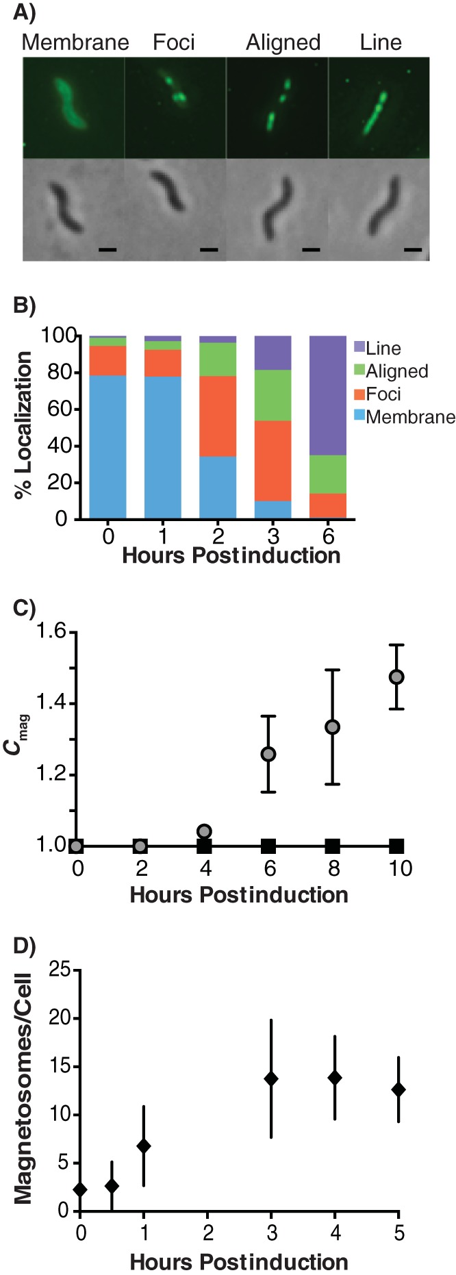
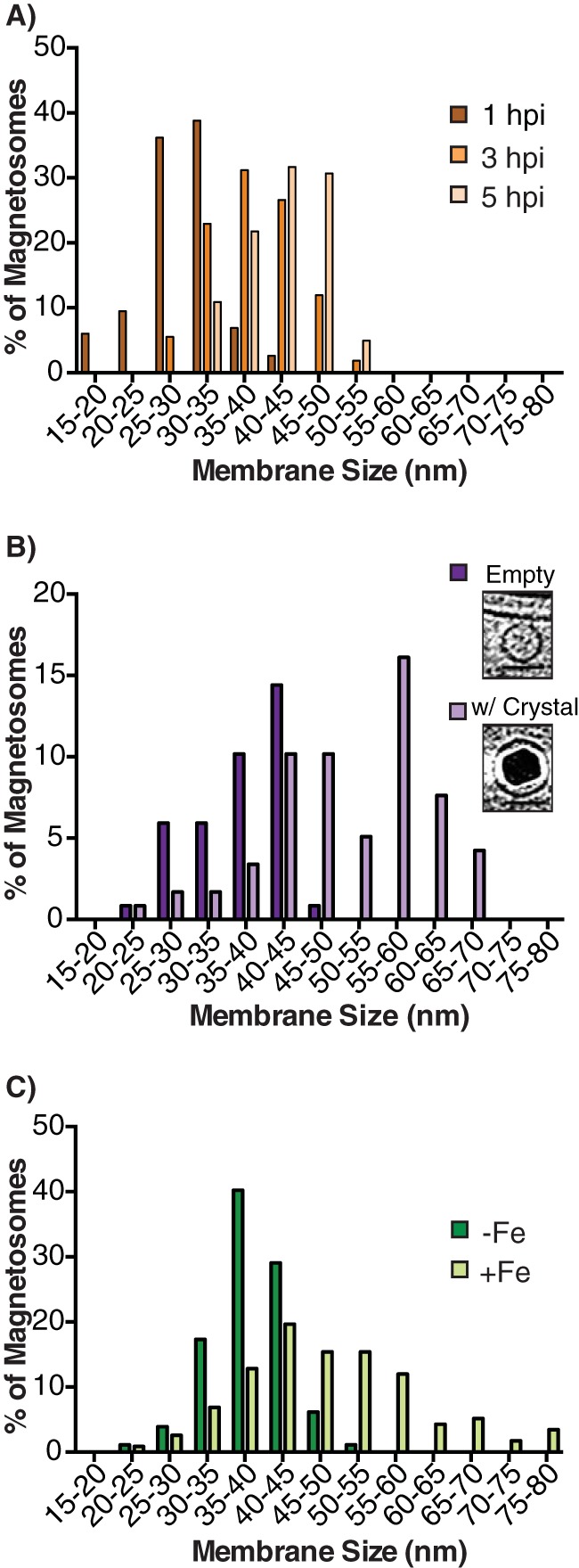
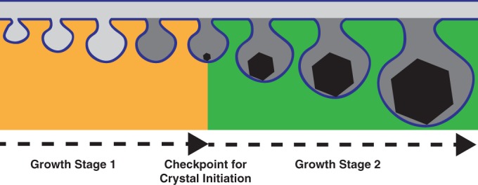
Magnetotactic bacteria produce chains of membrane-bound organelles that direct the biomineralization of magnetic nanoparticles. These magnetosome compartments are a model for studying the biogenesis and subcellular organization of bacterial organelles. Previous studies have suggested that discrete gene products build and assemble magnetosomes in a stepwise fashion. Here, using an inducible system, we show that the stages of magnetosome formation are highly dynamic and interconnected. During de novo formation, magnetosomes first organize into discontinuous chain fragments that are subsequently connected by the bacterial actin-like protein MamK. We also find that magnetosome membranes are not uniform in size and can grow in a biomineralization-dependent manner. In the absence of biomineralization, magnetosome membranes stall at a diameter of ~50 nm. Those that have initiated biomineralization then expand to significantly larger sizes and accommodate mature magnetic particles. We speculate that such a biomineralization-dependent checkpoint for membrane growth establishes the appropriate conditions within the magnetosome to ensure successful nucleation and growth of magnetic particles.

Importance: Magnetotactic bacteria make magnetic nanoparticles inside membrane-bound organelles called magnetosomes; however, it is unclear how the magnetosome membrane controls the biomineralization that occurs within this bacterial organelle. We placed magnetosome formation under inducible control in Magnetospirillum magneticum AMB-1 and used electron cryo-tomography to capture magnetosomes in their near-native state as they form de novo. An inducible system provided the key evidence that magnetosome membranes grow continuously unless they have not properly initiated biomineralization. Our finding that the size of a bacterial organelle impacts its biochemical function is a fundamental advance that impacts our perception of organelle formation and can inform future attempts aimed at creating designer magnetic particles.


Figures

FIG 1
FIG 1
Magnetosome formation can be placed under genetically inducible control to follow de novo magnetosome formation and organization. (A) GFP-MmsF can be classified into 4 distinct localization patterns: membrane localized, unaligned foci, aligned foci, and a solid line (scale bars, 500 nm). (B) Mean localization patterns of GFP-MmsF for three independent 6-h induction time courses. (C) Magnetite crystal formation is assessed by the degree of magnetic response (Cmag) of triplicate cell culture induced over a 10-h time course. (D) Quantification of the mean number of magnetosomes per cell that could be visualized by electron crytomography (ECT) over a 5-h time course. Seven to 10 tomograms of the induced cell population were reconstructed for each time point.
FIG 2
FIG 2
MamK is required for chain continuity but not long-range alignment. (A and B) Representative ECT images of long-range alignment of the magnetosome chain at 3 hpi and 5 hpi for QInd and QIndΔK, respectively (scale bars, 100 nm). The inset is a higher magnification of the same cell to show where gaps occur in the chain (scale bars, 50 nm). Gaps are denoted by double-sided orange arrows. A yellow arrowhead denotes the same magnetosome at each magnification for reference. Purple arrowheads point to individual magnetosomes in the chain. (C) Quantification of the number of gaps found in magnetosome chains of induced QInd and QIndΔK at 3 hpi and 5 hpi. The gap percentage is the percentage of edge-to-edge distances between adjacent and aligned magnetosomes that are >75 nm in length. The total numbers of edge-to-edge distances measured (n) for each time point are as follows: QInd 3 hpi, n = 74; QInd 5 hpi, n = 99; QIndΔK 3 hpi, n = 94; and QIndΔK 5 hpi, n = 97. (D) Cartoon depicting magnetosome chain organization in QInd versus QIndΔK. At 3 hpi, both strains exhibit long-range but discontinuous magnetosome alignment. At 5 hpi, the QInd strain has filled these gaps, whereas QIndΔK does not fill the chain discontinuities.
FIG 3
FIG 3
Magnetosome membranes grow in diameter in a manner that is dependent on their biomineralization state. (A) Distribution of magnetosome size at three points in the induction time course of QInd: 1 hpi (n = 116), 3 hpi (n = 110), and 5 hpi (n = 101). (B) Distribution of magnetosome size in wild-type AMB-1 (n = 117). Shown are representative images of an empty magnetosome versus a magnetosome with crystal (scale bars, 50 nm). (C) Magnetosome membrane size distribution in wild-type AMB-1 grown in either iron-rich (+Fe; n = 117) or iron-poor (−Fe; n = 172) medium. Membrane size is an average of 3 independent diameter measurements of the same magnetosome at the tomographic slice where it is largest and most visible. The number of magnetosomes measured is n.
FIG 4
FIG 4
Crystal growth does not physically expand the magnetosome membrane. (A) Representative ECT images of magnetosomes of different sizes in wild-type AMB-1 (scale bars, 50 nm). (B) Scatterplot and regression analysis of membrane size versus crystal size for magnetosomes that harbor crystals in wild-type AMB-1 (n = 72 magnetosomes). The long axis (crystal length) is reported as crystal size. (C) Representative ECT images of magnetosomes of different sizes in the ΔmmsF mutant (scale bars, 50 nm). (D) Scatterplot of membrane size versus crystal size for magnetosomes that harbor crystals in the ΔmmsF mutant (n = 140 magnetosomes). The long axis (crystal length) is reported as crystal size. (E) Distribution of magnetosome size in the ΔmmsF mutant (n = 243) compared to wild-type AMB-1 (n = 117).
FIG 5
FIG 5
Magnetosome membrane growth is a two-step growth mechanism dependent on biomineralization. The magnetosome membrane compartment is remodeled in two growth stages. In the first stage (orange), the inner membrane is remodeled to form the magnetosome compartment. The magnetosome lumen is most likely similar to that of the periplasm (light gray). Membrane size is restricted until conditions inside the magnetosome membrane are optimal for biomineralization (dark gray). Crystal initiation triggers a second growth stage (green) to accommodate a growing crystal.

References

    1. Federovitch CM, Ron D, Hampton RY. 2005. The dynamic ER: experimental approaches and current questions. Curr Opin Cell Biol 17:409–414. doi:10.1016/j.ceb.2005.06.010. - DOI - PubMed
    1. Egner A, Jakobs S, Hell SW. 2002. Fast 100-nm resolution three-dimensional microscope reveals structural plasticity of mitochondria in live yeast. Proc Natl Acad Sci U S A 99:3370–3375. doi:10.1073/pnas.052545099. - DOI - PMC - PubMed
    1. Huotari J, Helenius A. 2011. Endosome maturation. EMBO J 30:3481–3500. doi:10.1038/emboj.2011.286. - DOI - PMC - PubMed
    1. Voeltz GK, Barr FA. 2013. Cell organelles. Curr Opin Cell Biol 25:403–405. doi:10.1016/j.ceb.2013.06.001. - DOI - PubMed
    1. Murat D, Byrne M, Komeili A. 2010. Cell biology of prokaryotic organelles. Cold Spring Harb Perspect Biol 2:a000422. doi:10.1101/cshperspect.a000422. - DOI - PMC - PubMed

Publication types

MeSH terms