. 2016 Jun;30(6):2135-50.
doi: 10.1096/fj.201500173. Epub 2016 Feb 16.

The matricellular protein CCN1 enhances TGF-β1/SMAD3-dependent profibrotic signaling in fibroblasts and contributes to fibrogenic responses to lung injury

Affiliations

The matricellular protein CCN1 enhances TGF-β1/SMAD3-dependent profibrotic signaling in fibroblasts and contributes to fibrogenic responses to lung injury

Ashish R Kurundkar et al. FASEB J. 2016 Jun.

Abstract

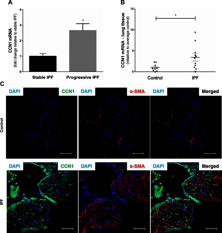
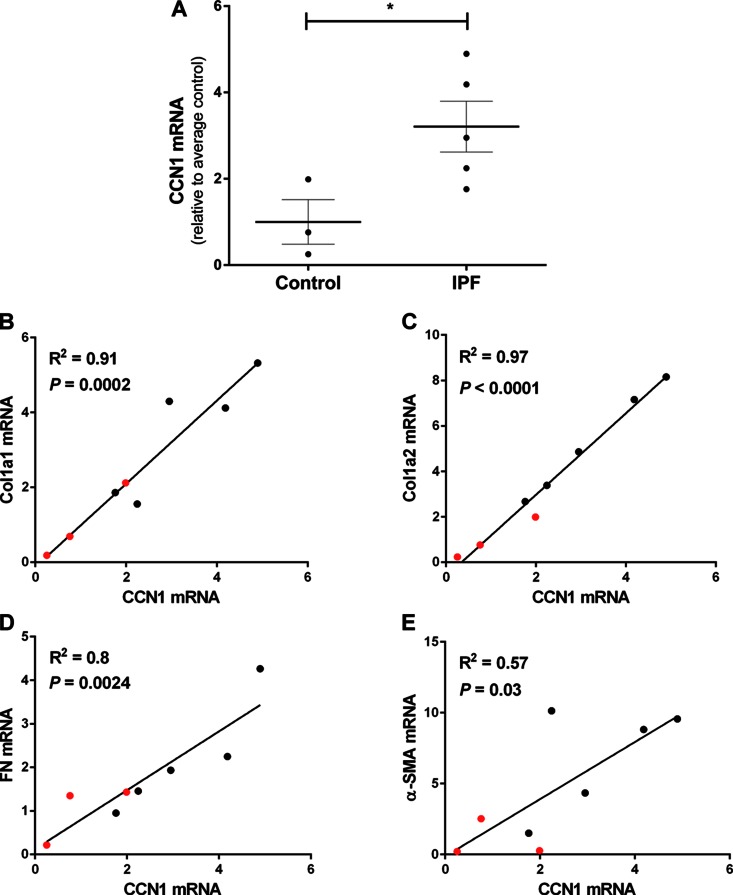
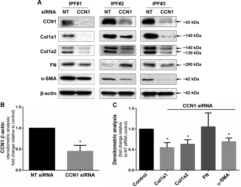
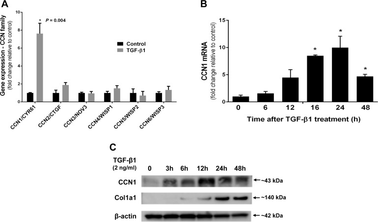
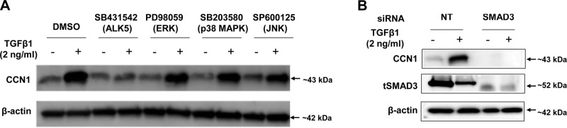
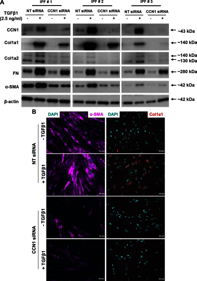
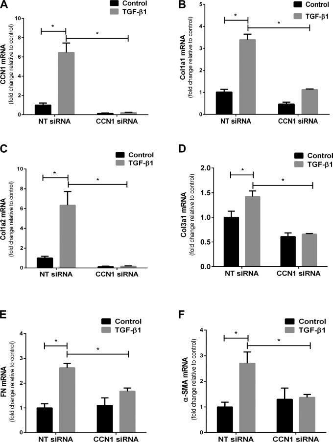
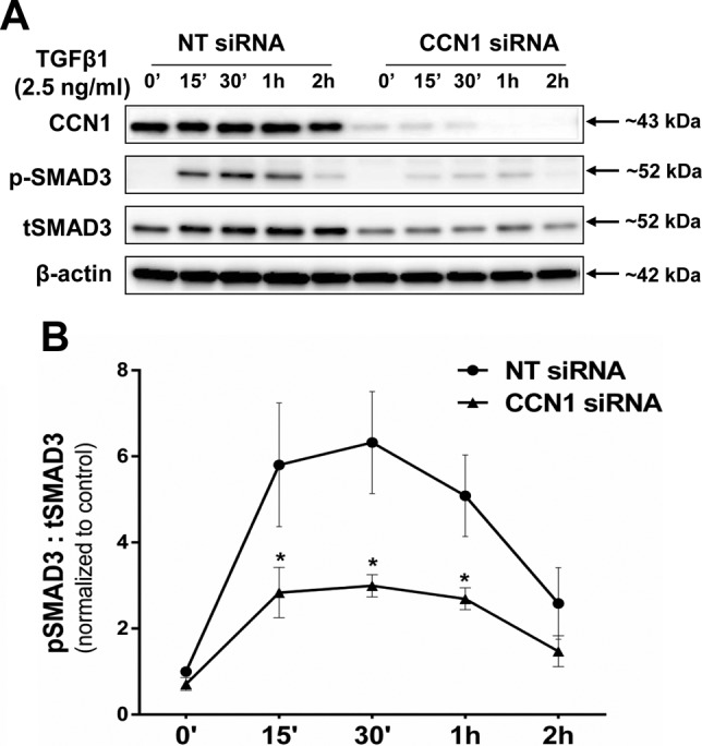
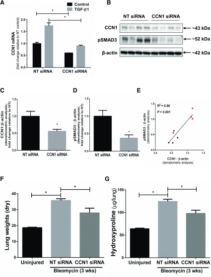
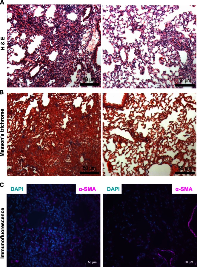
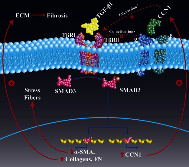
Matricellular proteins mediate pleiotropic effects during tissue injury and repair. CCN1 is a matricellular protein that has been implicated in angiogenesis, inflammation, and wound repair. In this study, we identified CCN1 as a gene that is differentially up-regulated in alveolar mesenchymal cells of human subjects with rapidly progressive idiopathic pulmonary fibrosis (IPF). Elevated levels of CCN1 mRNA were confirmed in lung tissues of IPF subjects undergoing lung transplantation, and CCN1 protein was predominantly localized to fibroblastic foci. CCN1 expression in ex vivo IPF lung fibroblasts correlated with gene expression of the extracellular matrix proteins, collagen (Col)1a1, Col1a2, and fibronectin as well as the myofibroblast marker, α-smooth muscle actin. RNA interference (RNAi)-mediated knockdown of CCN1 down-regulated the constitutive expression of these profibrotic genes in IPF fibroblasts. TGF-β1, a known mediator of tissue fibrogenesis, induces gene and protein expression of CCN1 via a mothers against decapentaplegic homolog 3 (SMAD3)-dependent mechanism. Importantly, endogenous CCN1 potentiates TGF-β1-induced SMAD3 activation and induction of profibrotic genes, supporting a positive feedback loop leading to myofibroblast activation. In vivo RNAi-mediated silencing of CCN1 attenuates fibrogenic responses to bleomycin-induced lung injury. These studies support previously unrecognized, cooperative interaction between the CCN1 matricellular protein and canonical TGF-β1/SMAD3 signaling that promotes lung fibrosis.-Kurundkar, A. R., Kurundkar, D., Rangarajan, S., Locy, M. L., Zhou, Y., Liu, R.-M., Zmijewski, J., Thannickal, V. J. The matricellular protein CCN1 enhances TGF-β1/SMAD3-dependent profibrotic signaling in fibroblasts and contributes to fibrogenic responses to lung injury.

Keywords: idiopathic pulmonary fibrosis; matrix remodeling; myofibroblasts; wound repair.


Figures

Figure 1.
Figure 1.
CCN1 expression in IPF. A) Fold change in mRNA of CCN1 in alveolar mesenchymal cells from stable (n = 3) vs. progressive (n = 3) IPF. B) Relative mRNA expression of CCN1 in lung tissue from controls (n = 9) and IPF (n = 11) normalized to average expression of controls. C) Immunofluorescence staining of IPF lung tissue for CCN1 (green) and α-SMA expression (red; ×40 magnification). Two-tailed unpaired Student’s t test was performed between groups. *P < 0.05. Scale bars, 50 µm.
Figure 2.
Figure 2.
Expression of CCN1 in IPF fibroblasts correlates with profibrotic gene expression. Control fibroblasts (n = 3) and IPF fibroblasts (n = 5) isolated from lung explants were serum starved overnight at 70–80% confluence. RNA was isolated and gene expression analyzed by RT-PCR. A) CCN1 mRNA expression relative to average of controls. Two-tailed unpaired Student’s t test was performed between groups. *P < 0.05. BE) Linear regression analysis was done to compare the mRNA levels of CCN1 to mRNA levels of Col1a1, Col1a2, FN, and α-SMA. Correlation graphs with mRNA levels CCN1 vs. Col1a1 (B), Col1a2 (C), FN (D), and α-SMA (E). Values represents means of 2 technical duplicates relative to average control and are color-coded as controls in red and IPF in black. R2 is coefficient of determination.
Figure 3.
Figure 3.
Silencing of CCN1 down-regulates constitutive expression of profibrotic proteins in IPF fibroblasts. IPF fibroblasts were subjected to NT and CCN1 siRNA (final concentration = 100 nM) overnight, then medium was changed to 10% FBS medium for 24 h, and then medium was changed to serum free for 3 d. A) Representative Western blots of showing expression of CCN1, Col1a1, Col1a2, FN, α-SMA, and β-actin with CCN1 knockdown in 3 IPF cell lines. B, C) Densitometric analyses of CCN1 expression (B) and Col1a1, Col1a2, FN, and α-SMA expression (C) relative to β-action on 4 IPF fibroblasts. Data represent means ± sem (n = 4). Two-way ANOVA was performed between NT and CCN1 siRNA groups for densitometric analyses. *P < 0.05.
Figure 4.
Figure 4.
Silencing of CCN1 down-regulates constitutive expression of profibrotic genes in IPF fibroblasts. IPF fibroblasts were subjected to NT and CCN1 siRNA (final concentration = 100 nM) for overnight, then medium was changed to 10% FBS medium for 24 h, and then medium was changed to serum free for 3 d. Graphs represent fold change in mRNA expression of CCN1 (A),Col1a1 (B), Col1a2 (C), Col3a1 (D), α-SMA (E), and FN (F) relative to NT siRNA-treated fibroblasts. n.s., not significant. Values represent means ± sem (n = 3). Two-tailed unpaired Student's t test was performed between groups. *P < 0.05.
Figure 5.
Figure 5.
TGF-β1 up-regulates CCN1 in lung fibroblasts. A) Relative mRNA expression of genes of CCN family in IMR-90 fibroblasts treated with or without TGF-β1 (2 ng/ml), as analyzed by whole-genome Affymetrix microarray for 24 h. B) RT-PCR showing fold change in mRNA levels of CCN1 compared with control at indicated intervals in IMR-90 fibroblasts treated with TGF-β1 (2.5 ng/ml) after serum starved for 24 h. C) Western blot showing expression of CCN1 and Col1a1 in primary adult lung fibroblasts treated with TGF-β1 (2 ng/ml) for indicated periods. *P < 0.05.
Figure 6.
Figure 6.
TGF-β1 mediated CCN1 up-regulation is ALK5/SMAD3-dependent in lung fibroblasts. A) Primary adult lung fibroblasts were pretreated with or without chemical inhibitors type 1 TGF-β receptor (TβR1/ALK5; SB431542, 1 µM), ERK (PD98059, 10 µM), MAPK (SB203580, 6 µM), and cJNK (SP600125, 100 nM) for 45 min and then treated with or without TGF-β1 (2 ng/ml) for 24 h. B) Primary adult lung fibroblasts were transfected with NT and SMAD3 siRNA (100 nM) for 2 d, then serum starved for 24 h and treated with TGF-β1 (2 ng/ml) for 24 h. Expression of CCN1 and tSMAD3 was evaluated by immunoblotting.
Figure 7.
Figure 7.
CCN1 knockdown impairs TGF-β1-induced expression of profibrotic proteins in IPF fibroblasts. IPF fibroblasts (n = 5) were subjected to NT and CCN1 siRNA (final concentration = 100 nM) for 2 d, then medium was changed to serum free for 24 h, and then fibroblasts were treated with TGF-β1 (2.5 ng/ml) for 24 h. A) Representative Western blot (n = 3) showing expression of CCN1, Col1a1, Col1a2, FN, and α-SMA. B) Immunofluorescence staining showing expression of α-SMA (magenta) and Col1a1 (red; ×20 magnification). Scale bars, 50 µm.
Figure 8.
Figure 8.
CCN1 knockdown impairs TGF-β1-induced expression of profibrotic genes in IPF fibroblasts. IPF fibroblasts (n = 3) were subjected to NT and CCN1 siRNA (final concentration = 100 nM) for 2 d, then medium was changed to serum free for 24 h, and then fibroblasts were treated with TGF-β1 (2.5 ng/ml) for 24 h. Graphs represent fold change in mRNA expression of CCN1 (A), Col1a1 (B), Col1a2 (C), Col3a1 (D), FN (E), and α-SMA (F). Data represent means ± sem (n = 3) of 1 IPF cell line. Similar results were seen in 2 additional IPF cell lines. Two-way ANOVA was performed between groups. *P < 0.05.
Figure 9.
Figure 9.
CCN1 knockdown inhibits SMAD3 phosphorylation in IPF fibroblasts. IPF fibroblasts (n = 3) were subjected to NT and CCN1 siRNA (final concentration = 100 nM) for 2 d, then medium was changed to serum free for 24 h, and then fibroblasts were treated with TGF-β1 (2.5 ng/ml) for indicated time periods. A) Representative Western blot of independent experiments on 3 different IPF fibroblasts showing expression of CCN1, phospho-SMAD3 (Ser423/425), and total SMAD3 (tSMAD3). B) Densitometric analyses of pSMAD3:tSMAD3 in IPF fibroblasts (n = 3). Two-way ANOVA was performed between groups. *P < 0.05.
Figure 10.
Figure 10.
In vivo RNAi-mediated silencing of CCN1 attenuates fibrogenic response to bleomycin injury. A) Mouse lung fibroblasts were transfected with NT and CCN1 siRNA (final concentration = 100 nM) for 2 d, then were serum free for 24 h, and then were treated with TGF-β1 (2.5 ng/ml) for 24 h. Graph shows the mRNA expression of CCN1 relative to NT control. Data represent means ± sem (n = 3). Two-way ANOVA analyses were performed between groups. BG) C57BL/6 mice were subjected to acute lung injury by airway (intratracheal) administration of bleomycin or saline/control on d 0. NT siRNA or CCN1 siRNA (50 µg/mouse) was administered by intranasal delivery every other day for 2 wk, starting from d 8 to 20 postinjury. BD) Whole lung harvested at the 3 wk following bleomycin injury and were analyzed for expression of CCN1 and pSMAD3 by Western blotting (B) and densitometric analyses (C, D). Unpaired Student’s t test was performed for densitometric analyses between 2 groups. E) Correlation graph of protein expression of CCN1 and pSMAD3 in lung tissue of NT and CCN1 siRNA-treated mice post–bleomycin injury. Linear regression analysis was done to compare the protein expression of CCN1 and pSMAD3 in lung tissue in NT/CCN1 siRNA-treated mice post–bleomycin injury (R2 = 0.86; P = 0.001; n = 4 in each group). Values represent densitometric analyses of CCN1 and pSMAD3 (normalized to β-actin; relative to NT average) and are color-coded as NT siRNA-treated in red and CCN1 siRNA-treated in black. F, G) Severity of fibrosis was assessed by measuring dry lung weights (F) and quantitative hydroxyproline assay (G). Data are expressed as increase in micrograms of hydroxyproline/whole lung. Values represent means ± sem; n = 4–6 per group. Two-way ANOVA was performed between groups. *P < 0.05.
Figure 11.
Figure 11.
In vivo RNAi-mediated silencing of CCN1 attenuates fibrogenic response to bleomycin injury. Lung tissue sections from NT/CCN1 siRNA-treated mice (n = 2 each group) were used for immunohistochemistry, including hematoxylin and eosin staining (A), Masson’s trichrome staining (B), and immunofluorescence staining for α-SMA (magenta) (C). Scale bars, 50 µm.
Figure 12.
Figure 12.
Schematic of proposed model of CCN1 and TGF-β1 interactions in profibrotic signaling. Our studies indicate that endogenous CCN1 is required for constitutive and TGF-β1-induced SMAD3 activation and profibrogenic gene expression. TGF-β1 induces CCN1 expression via a SMAD3-dependent pathway, supporting a positive feedback loop. Abrogation of CCN1 during the active fibrogenic phase of lung injury repair protects from fibrosis in an in vivo model of lung fibrosis. The precise mechanism by which CCN1 cooperates with TGF-β receptors and/or integrins to activate SMAD3 requires further investigation.

References

    1. Bornstein P., Sage E. H. (2002) Matricellular proteins: extracellular modulators of cell function. Curr. Opin. Cell Biol. 14, 608–616 - PubMed
    1. Murphy-Ullrich J. E., Sage E. H. (2014) Revisiting the matricellular concept. Matrix Biol. 37, 1–14 - PMC - PubMed
    1. Murphy-Ullrich J. E. (2001) The de-adhesive activity of matricellular proteins: is intermediate cell adhesion an adaptive state? J. Clin. Invest. 107, 785–790 - PMC - PubMed
    1. Kyriakides T. R., Bornstein P. (2003) Matricellular proteins as modulators of wound healing and the foreign body response. Thromb. Haemost. 90, 986–992 - PubMed
    1. Kubota S., Takigawa M. (2007) CCN family proteins and angiogenesis: from embryo to adulthood. Angiogenesis 10, 1–11 - PubMed

MeSH terms

Substances

LinkOut - more resources