. 2016 Feb 16;9(415):ra18.
doi: 10.1126/scisignal.aac4374.

ONC201 kills solid tumor cells by triggering an integrated stress response dependent on ATF4 activation by specific eIF2α kinases

Affiliations

ONC201 kills solid tumor cells by triggering an integrated stress response dependent on ATF4 activation by specific eIF2α kinases

C Leah B Kline et al. Sci Signal. .

Abstract

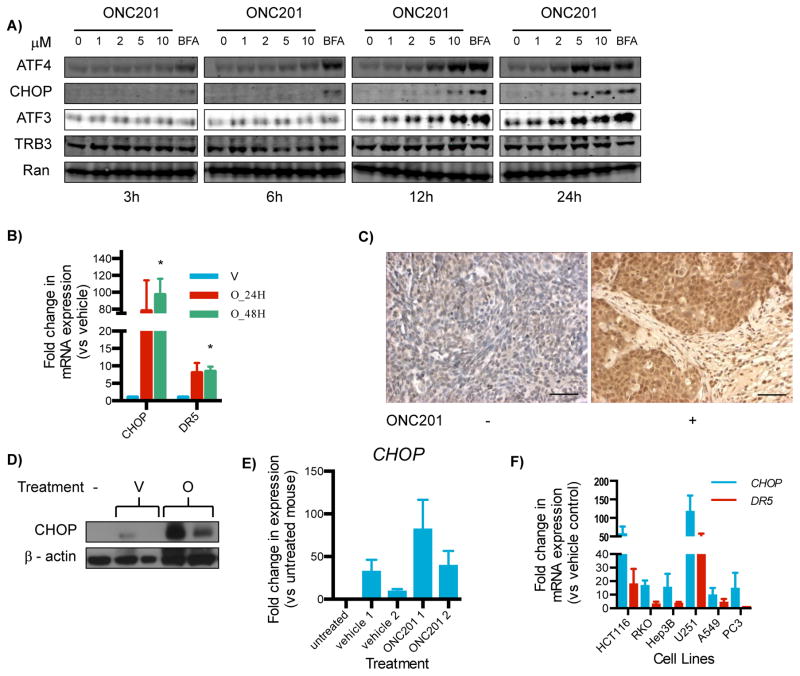
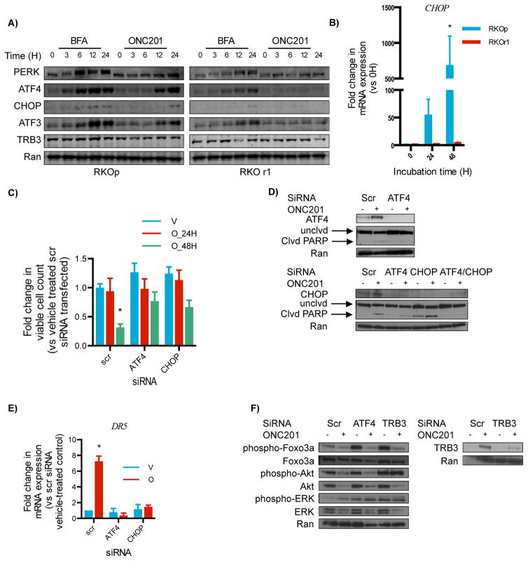
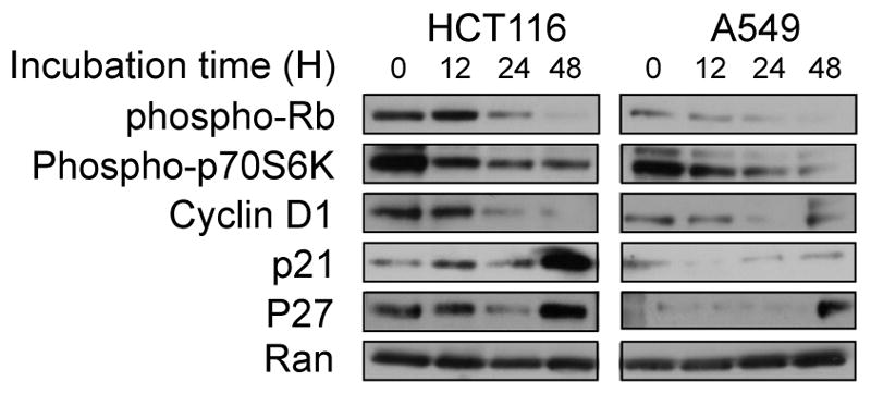
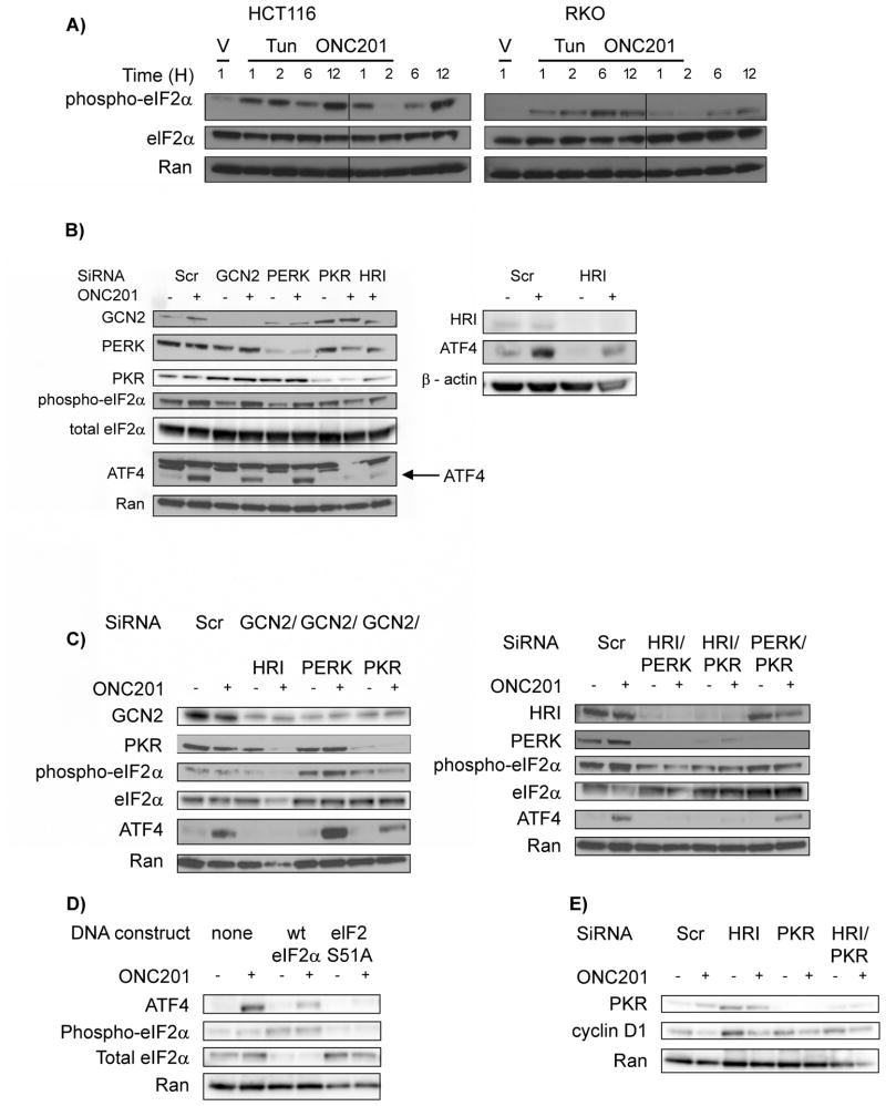
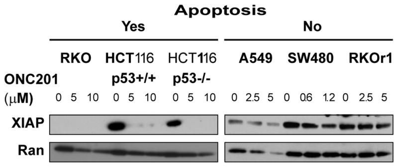
ONC201 (also called TIC10) is a small molecule that inactivates the cell proliferation- and cell survival-promoting kinases Akt and ERK and induces cell death through the proapoptotic protein TRAIL. ONC201 is currently in early-phase clinical testing for various malignancies. We found through gene expression and protein analyses that ONC201 triggered an increase in TRAIL abundance and cell death through an integrated stress response (ISR) involving the transcription factor ATF4, the transactivator CHOP, and the TRAIL receptor DR5. ATF4 was not activated in ONC201-resistant cancer cells, and in ONC201-sensitive cells, knockdown of ATF4 or CHOP partially abrogated ONC201-induced cytotoxicity and diminished the ONC201-stimulated increase in DR5 abundance. The activation of ATF4 in response to ONC201 required the kinases HRI and PKR, which phosphorylate and activate the translation initiation factor eIF2α. ONC201 rapidly triggered cell cycle arrest, which was associated with decreased abundance of cyclin D1, decreased activity of the kinase complex mTORC1, and dephosphorylation of the retinoblastoma (Rb) protein. The abundance of X-linked inhibitor of apoptosis protein (XIAP) negatively correlated with the extent of apoptosis in response to ONC201. These effects of ONC201 were independent of whether cancer cells had normal or mutant p53. Thus, ONC201 induces cell death through the coordinated induction of TRAIL by an ISR pathway.


Conflict of interest statement

Competing interests: J.E.A. and W.S.E-D. hold a patent on ONC201 and are shareholders of Oncoceutics, Inc. W.S.E-D. is a co-founder of Oncoceutics, Inc. Pennsylvania State University has licensed ONC201/TIC10 to Oncoceutics, Inc. for clinical development. W.S.E-D. is fully compliant with institutional disclosure requirements and conflict of interest rules.

Figures

Fig. 1
Fig. 1. ONC201’s broad-spectrum activity can be attributed to its anti-proliferative and pro-apoptotic effects
(A) EC50 of ONC201 was assessed by Cell Titer Glo assay in 23 cell lines treated with ONC201 for 72 hours. (B) Apoptosis measured by Sub-G1 analysis in cells treated with ONC201 for 72 hours. (C) Cell cycle profile analyses in HCT 116 and A549 cells treated with ONC201 for 24 hours. Data is representative of two biological replicates. (D) BrdU incorporation assays in HCT116 and A549 cells treated with ONC201 for 48 hours. (E) Viable cell counts assessed by trypan blue exclusion in cells treated with ONC201 for up to 72 hours. *p < 0.05, Student’s t-test with Holm-Sidak correction. (F) Proliferative capacity of cells after 72 hours in the presence of ONC201, assessed by clonogenic assays. Bars that are not visible indicate a proliferative fraction of 0. Concentrations of ONC201 used for each cell line are listed in table S1. Data in (A), (B), (D), (E) and (F) are means ± SEM from three biological replicates.
Fig. 2
Fig. 2. ONC201 activates the integrated stress response
(A)Western blotting analysis for ATF4, CHOP, ATF3, and TRB3 on lysates from HCT116 cells cultured with ONC201 (0–10 μM) for 3 to 24 hours. Brefeldin A (BFA, an ER stressor) served as a positive control. Blots are representative of 2 experiments. (B) qRT-PCR for the expression of CHOP and DR5 relative to GAPDH in HCT116 cells treated with ONC201 (O; 10 μM) or vehicle (V) for 24 or 48 hours. (C) Immunohistochemical analysis for CHOP in HCT116-derived subcutaneous xenografts from mice that received either vehicle (−) or ONC201 (+; 25 mg/kg). Results are representative of tissues from two vehicle-treated mice and six mice intraperitoneally or intravenously injected with ONC201 (3 mice each). Scale bar = 10 μm (D) Western blotting analysis for CHOP in lysates from colorectal cancer stem cell-like cell-derived xenografts (45) in athymic nude mice extracted 72 hours after treatment [vehicle (V) or ONC201 (O) (50 mg/kg, i.p.)]. Tumor lysates from untreated mice (−) served as controls. (E) Densitometric analyses of the bands in the Western blots in (D). (F) qRT-PCR for the fold change in expression of CHOP and DR5 relative to GAPDH in different cell types cultured with ONC201 (compared to vehicle) for 72 hours. Data in (B), (E) and (F) are means ± SEM of three biological replicates. *p < 0.05, Student’s t-test with Holm-Sidak correction, comparing ONC201-treated versus vehicle-treated cells.
Fig. 3
Fig. 3. ATF4 is required for ONC201-induced cytotoxicity
(A) Western blotting analysis for the indicated proteins in wild-type RKO (RKOp) and ONC201-resistant RKO (RKOr1) cells treated with 10 μM brefeldin A (an ER stressor, positive control) or 5 μM ONC201. (B) qRT-PCR for the fold-change in expression of CHOP relative to GAPDH in RKOp and RKOr1 cells treated with ONC201 (5 μM, 24–48 hours) compared with the 0 time point (H, hours). (C) Viability of HCT116 cells assessed by trypan blue exclusion assay after ATF4 and CHOP knockdown and treatment with vehicle (V) or ONC201 (O) (10μM, 24 and 48 hours). (D) Western blotting analysis for ATF4, CHOP, and uncleaved (unclvd) and cleaved (clvd) PARP in HCT116 cells transfected with ATF4 or CHOP siRNA. (E) qPCR of DR5 expression relative to GAPDH in HCT116 cells transfected with ATF4 or CHOP siRNA and subsequently treated with vehicle (V) or ONC201 (O) (10 μM, 48 hours). (F) Western blotting for the indicated proteins in HCT116 cells transfected with ATF4 or TRB3 siRNA and subsequently treated with ONC201 (10 μM, 72 hours upper set, 48 hours lower set). Blots in (A), (D), and (F) are representative of 2 experiments. Data in (B), (C) and (E) are means ± SEM of 3 biological replicates. *p < 0.05, Student’s t-test with Holm-Sidak correction.
Fig. 4
Fig. 4. ONC201 induces a decrease in cyclin D1 abundance
Western blotting analyses for the indicated proteins were performed in HCT116 or A549 cells treated with ONC201 (10 or 5 μM, respectively) for up to 48 hours. Blots are representative of two experiments.
Fig. 5
Fig. 5. ONC201 activates the ATF4 pathway through the eIF2α kinases HRI and PKR
(A) Western blotting analysis for phosphorylated and total eIF2α was performed in the indicated cell types treated with ONC201. (B to D) Western blotting analysis for the indicated proteins in lysates from HCT116 cells that were transfected with: siRNA against one (B) or two eIF2α kinases (C) as indicated for 24 hours, and subsequently treated with ONC201 (10 μM) for 12 hours. (D) Western blotting analysis for indicated proteins in lysates from HCT116 cells that were transfected with plasmid constructs for wild-type (wt) and a nonphosphorylatable eIF2α mutant (S51A) for 48 hours, and subsequently treated with ONC201 for 12 hours. (E) Western blotting analysis for cyclin D1 in HCT116 cells transfected with the indicated siRNA(s) and treated with ONC201 for 24 hours. Blots in (A) to (E) are representative of at least two independent experiments.
Fig. 6
Fig. 6. The effect of ONC201 on XIAP amount correlates with fate of cells treated with ONC201
Western blotting analysis for XIAP in cells treated with the indicated concentrations of ONC201 for 72 hours. Blots are representative of at least two independent experiments.

Comment in

  • ONC201: Stressing tumors to death.
    Endo Greer Y, Lipkowitz S. Endo Greer Y, et al. Sci Signal. 2016 Feb 16;9(415):fs1. doi: 10.1126/scisignal.aad7955. Sci Signal. 2016. PMID: 26884598 Free PMC article.

References

    1. Ashkenazi A, et al. Safety and antitumor activity of recombinant soluble Apo2 ligand. The Journal of clinical investigation. 1999;104:155–162. - PMC - PubMed
    1. Lemke J, von Karstedt S, Zinngrebe J, Walczak H. Getting TRAIL back on track for cancer therapy. Cell death and differentiation. 2014;21:1350–1364. - PMC - PubMed
    1. Merchant MS, et al. Phase I trial and pharmacokinetic study of lexatumumab in pediatric patients with solid tumors. Journal of clinical oncology: official journal of the American Society of Clinical Oncology. 2012;30:4141–4147. - PMC - PubMed
    1. Wagner KW, et al. Death-receptor O-glycosylation controls tumor-cell sensitivity to the proapoptotic ligand Apo2L/TRAIL. Nature medicine. 2007;13:1070–1077. - PubMed
    1. Lu M, et al. Cell death. Opposing unfolded-protein-response signals converge on death receptor 5 to control apoptosis. Science. 2014;345:98–101. - PMC - PubMed

Publication types

MeSH terms