. 2016 May;59(5):1030-9.
doi: 10.1007/s00125-016-3885-8. Epub 2016 Feb 17.

ANT1-mediated fatty acid-induced uncoupling as a target for improving myocellular insulin sensitivity

Affiliations

ANT1-mediated fatty acid-induced uncoupling as a target for improving myocellular insulin sensitivity

Lauren M Sparks et al. Diabetologia. 2016 May.

Abstract

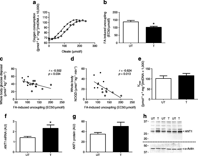
Aims/hypothesis: Dissipating energy via mitochondrial uncoupling has been suggested to contribute to enhanced insulin sensitivity. We hypothesised that skeletal muscle mitochondria of endurance-trained athletes have increased sensitivity for fatty acid (FA)-induced uncoupling, which is driven by the mitochondrial protein adenine nucleotide translocase 1 (ANT1).

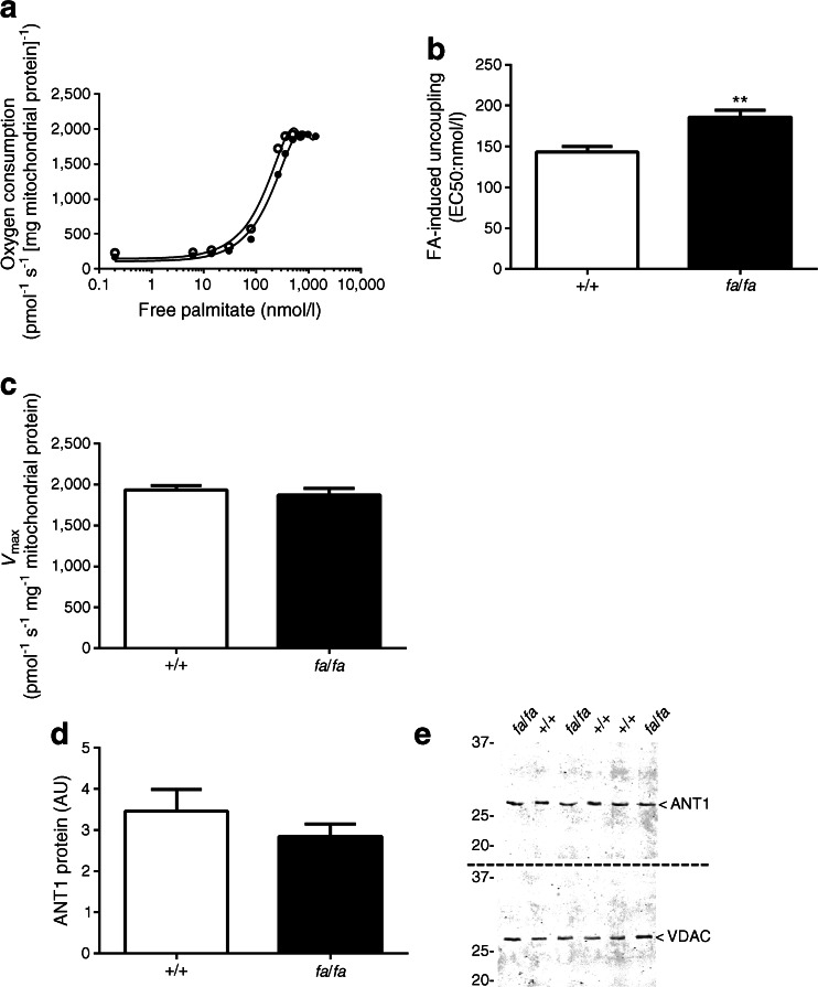
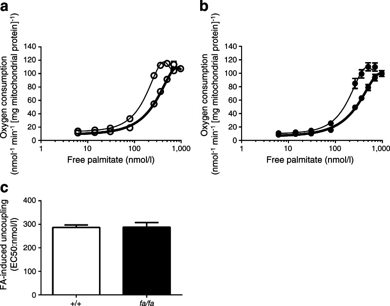
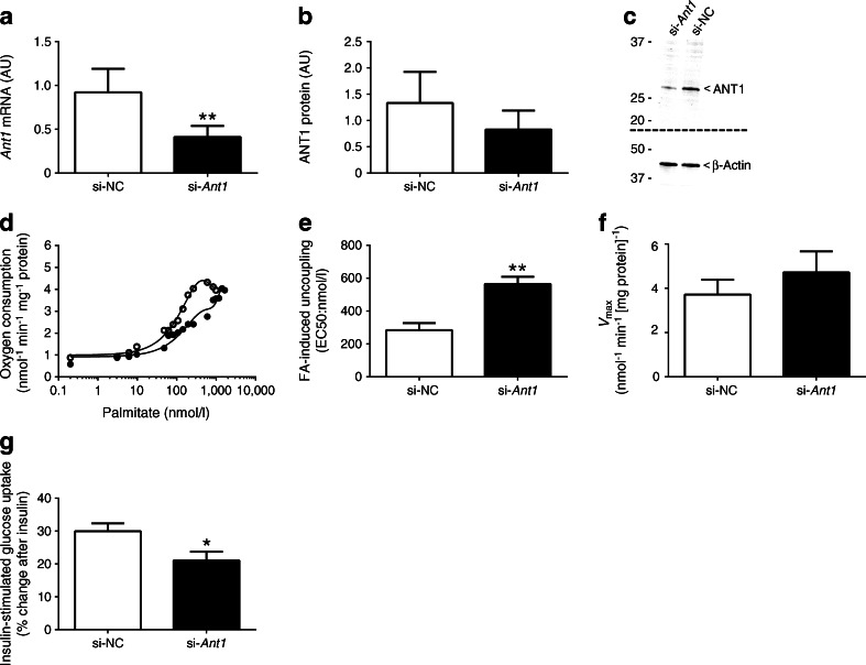
Methods: Capacity for FA-induced uncoupling was measured in endurance-trained male athletes (T) and sedentary young men (UT) in an observational study and also in isolated skeletal muscle mitochondria from Zucker diabetic fatty (ZDF) rats and C2C12 myotubes following small interfering RNA (siRNA)-mediated gene silencing of ANT1. Thus, fuelled by glutamate/succinate (fibres) or pyruvate (mitochondria and myotubes) and in the presence of oligomycin to block ATP synthesis, increasing levels of oleate (fibres) or palmitate (mitochondria and myotubes) were automatically titrated while respiration was monitored. Insulin sensitivity was measured by hyperinsulinaemic-euglycaemic clamp in humans and via insulin-stimulated glucose uptake in myotubes.

Results: Skeletal muscle from the T group displayed increased sensitivity to FA-induced uncoupling (p = 0.011) compared with muscle from the UT group, and this was associated with elevated insulin sensitivity (p = 0.034). ANT1 expression was increased in T (p = 0.013). Mitochondria from ZDF rats displayed decreased sensitivity for FA-induced uncoupling (p = 0.008). This difference disappeared in the presence of the adenine nucleotide translocator inhibitor carboxyatractyloside. Partial knockdown of ANT1 in C2C12 myotubes decreased sensitivity to the FA-induced uncoupling (p = 0.008) and insulin-stimulated glucose uptake (p = 0.025) compared with controls.

Conclusions/interpretation: Increased sensitivity to FA-induced uncoupling is associated with enhanced insulin sensitivity and is affected by ANT1 activity in skeletal muscle. FA-induced mitochondrial uncoupling may help to preserve insulin sensitivity in the face of a high supply of FAs.

Trial registration: www.trialregister.nl NTR2002.

Keywords: ANT1; Fatty acid-induced uncoupling; Skeletal muscle insulin sensitivity.


Figures

Fig. 1
Fig. 1
(a) Typical experiment assessing FA-induced (oleate) uncoupling in muscle fibres of healthy UT and T. (b) Sensitivity to FA-induced uncoupling (EC50) is shown in UT and T. (c, d) FA-induced uncoupling correlated with insulin sensitivity (whole-body glucose disposal) (c) and insulin-stimulated NOGD (d). (e) Maximal oleate-induced uncoupling (V max) in permeabilised muscle fibres from UT and T. (f, g) ANT1 mRNA expression (f) and ANT1 protein abundance (g) in skeletal muscle tissue of UT and T (shown in arbitrary units [AU]). (h) Representative blots for protein abundance. Two separate gels and blots were used for ANT1 and α-actin as indicated by the dashed line. GAPDH was used as a reference gene for mRNA expression. α-Actin was used to quantify equal protein loading during the western blot analysis. White circles and bars, UT (n = 10); black circles and bars, T (n = 9). Data are presented as mean ± SEM. *p < 0.05, T vs UT
Fig. 2
Fig. 2
(a) Typical experiment assessing FA-induced (palmitate) uncoupling in isolated skeletal muscle mitochondria from wild-type control (+/+) and ZDF (fa/fa) rats. (b) Sensitivity to FA-induced uncoupling (EC50) in isolated skeletal muscle mitochondria from ZDF rats compared with wild-type control rats (n = 4 per group). (c, d) Maximal palmitate-induced uncoupling (V max) (c) and ANT1 protein levels (shown in arbitrary units [AU]) (d) in isolated skeletal muscle mitochondria from wild-type controls and ZDF rats (n = 8 per group). (e) Representative blots for protein abundance. Two separate gels and blots were used for ANT1 and VDAC as indicated by the dashed line. VDAC was used to quantify equal protein loading during the western blot analysis. White circles and bars, wild-type control rats; black circles and bars, ZDF rats. Free palmitate concentrations were plotted on a logarithmic scale for the representative trace of EC50 in (a). Data are presented as mean ± SEM. **p < 0.01, T vs UT
Fig. 3
Fig. 3
(a, b) Sensitivity to FA-induced uncoupling (see Fig. 2a) in the absence (thin line) or presence (thick line) of the ANT inhibitor CATR, in wild-type control (+/+) rats (white circles) (a) and ZDF (fa/fa) rats (black circles) (b) was determined. (c) EC50 values in the wild-type control (white bar) and ZDF rats (black bar) in the presence of CATR. Free palmitate concentrations were plotted on a logarithmic scale in (a) and (b) (n = 4 per group). Data are presented as mean ± SEM
Fig. 4
Fig. 4
(a, b) Abundance of Ant1 mRNA (a) and levels of ANT1 protein (b) after transfection with negative control siRNA (si-NC) or Ant1 siRNA (si-Ant1) in differentiated C2C12 myotubes (n = 4 per group) (shown in arbitrary units [AU]). (c) Representative blots for protein abundance. Two separate gels and blots were used for ANT1 and β-actin as indicated by the dashed line. (d) Typical experiment assessing FA-induced (palmitate) uncoupling in differentiated, permeabilised C2C12 myotubes after transfection with either si-NC or si-Ant1. (e) Sensitivity to FA-induced uncoupling (EC50) in si-NC- and si-Ant1-transfected C2C12 myotubes (n = 3 per group). (f) Maximal palmitate-induced uncoupling (V max) in si-NC- and si-Ant1-transfected C2C12 myotubes (n = 3 per group). (g) Percentage change in insulin-stimulated glucose uptake in si-NC- and si-Ant1-transfected C2C12 myotubes (n = 9 per group). Palmitate concentrations were plotted on a logarithmic scale for the representative trace of EC50 in (d). mRNA expression data were normalised to the reference gene Rn18s. α-Actin was used to verify equal protein loading during the western blot analyses. White circles and bars, si-NC; black circles and bars, si-Ant1. Data are presented as mean ± SEM. *p<0.05 and **p<0.01 for si-Ant1 vs si-NC

References

    1. Meex RC, Schrauwen-Hinderling VB, Moonen-Kornips E, et al. Restoration of muscle mitochondrial function and metabolic flexibility in type 2 diabetes by exercise training is paralleled by increased myocellular fat storage and improved insulin sensitivity. Diabetes. 2010;59:572–579. doi: 10.2337/db09-1322. - DOI - PMC - PubMed
    1. Phielix E, Meex R, Moonen-Kornips E, Hesselink MK, Schrauwen P. Exercise training increases mitochondrial content and ex vivo mitochondrial function similarly in patients with type 2 diabetes and in control individuals. Diabetologia. 2010;53:1714–1721. doi: 10.1007/s00125-010-1764-2. - DOI - PMC - PubMed
    1. Nielsen J, Mogensen M, Vind BF, et al. Increased subsarcolemmal lipids in type 2 diabetes: effect of training on localization of lipids, mitochondria, and glycogen in sedentary human skeletal muscle. Am J Physiol Endocrinol Metab. 2010;298:E706–713. doi: 10.1152/ajpendo.00692.2009. - DOI - PubMed
    1. Phielix E, Meex R, Ouwens DM, et al. High oxidative capacity due to chronic exercise training attenuates lipid-induced insulin resistance. Diabetes. 2012;61:2472–2478. doi: 10.2337/db11-1832. - DOI - PMC - PubMed
    1. Befroy DE, Petersen KF, Dufour S, Mason GF, Rothman DL, Shulman GI. Increased substrate oxidation and mitochondrial uncoupling in skeletal muscle of endurance-trained individuals. Proc Natl Acad Sci U S A. 2008;105:16701–16706. doi: 10.1073/pnas.0808889105. - DOI - PMC - PubMed

Publication types

MeSH terms

LinkOut - more resources