Review
. 2016 Oct;263(10):1893-902.
doi: 10.1007/s00415-016-8060-0. Epub 2016 Feb 17.

Spring cleaning: time to rethink imaging research lines in MS?

Affiliations
Review

Spring cleaning: time to rethink imaging research lines in MS?

Martina Absinta et al. J Neurol. 2016 Oct.

Abstract

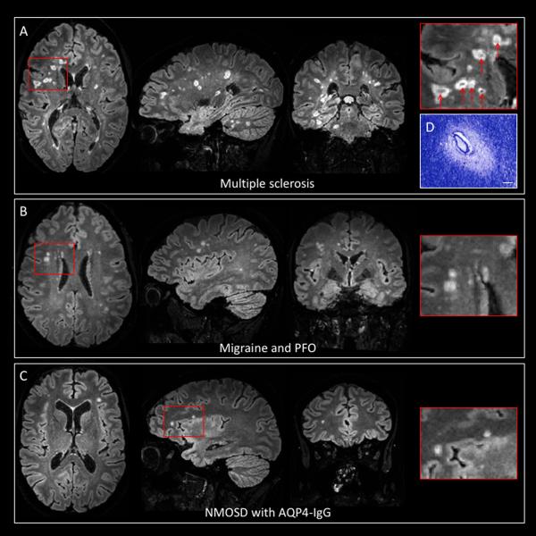
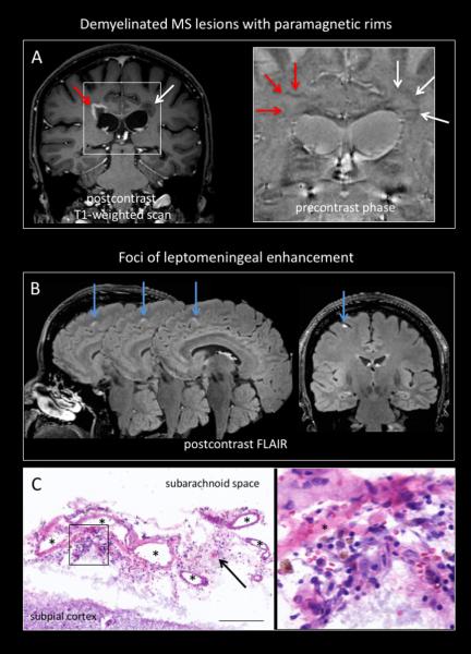
Together with recently advanced MRI technological capability, new needs and updated questions are emerging in imaging research in multiple sclerosis (MS), especially with respect to the identification of novel in vivo biomarkers of MS-relevant pathological processes. Expected benefits will involve approaches to diagnosis and clinical classification. In detail, three main points of discussion are addressed in this review: (1) new imaging biomarkers (centrifugal/centripetal lesion enhancement, central vein, paramagnetic rims at the lesion edge, subpial cortical demyelination); (2) thinking about high-resolution MR from a pathological perspective (from postmortem to in vivo staging); and (3) the clinical utility of quantitative MRI. In this context, research efforts should increasingly be focused on the direct in vivo visualization of "hidden" inflammation, beyond what can be detected with conventional gadolinium-based methods, as well as remyelination and repair, since these are likely to represent critical pathological processes and potential therapeutic targets. Concluding remarks concern the limitations, challenges, and ultimately clinical role of non-conventional MRI techniques.

Keywords: Biomarkers; Multiple sclerosis; Neuroimaging.


Figures

Figure 1
Figure 1. Vasculoscentric vs non-vasculocentric lesion appearance
Precontrast 3T FLAIR* images [magnified views in red boxes, for sequence details see Sati et al., Radiology 2012] in three different neurological conditions showing discrete white matter lesions: (A) 33-year-old woman with relapsing-remitting MS; (B) 53-year-old woman with migraine and patent foramen ovale (PFO); (C) 54-year-old woman with neuromyelitis optica spectrum disorder (NMOSD) with serum aquaporin-4 immunoglobulin G antibodies (AQP4-IgG). A prominent central vein is clearly discernable in the majority of demyelinated MS lesions in vivo (red arrows, A), whereas MS-mimicking lesions in microembolic/ischemic conditions (B) and NMOSD (C) do not present this morphological feature. (D) Pathological insert showing the vasculocentric development of a demyelinated MS lesion in a 59-year-old man with progressive MS (Luxol fast blue-periodic acid Schiff staining [LFB-PAS], scale bar 100 μm).
Figure 2
Figure 2. MS-related “hidden” inflammation
(A) Two periventricular lesions with paramagnetic rims on precontrast 3T phase images in a 31-year-old woman with relapsing-remitting MS. The rim on phase images reflects the presence of paramagnetic substances, (possibly inflammation-related) at the lesion edge. Lesion 1. Active lesion with peripheral leakage of gadolinium (centripetal pattern) and paramagnetic rim (red arrows); Lesion 2. Chronic lesion with paramagnetic rim (white arrows). (B) Multiple foci of leptomeningeal enhancement (cyan arrows) on postcontrast FLAIR images in a 42-year-old woman with relapsing-remitting MS. (C) Perivascular inflammatory infiltrate in the leptomeninges (black arrows and magnified box) where leptomeningeal enhancement was found in vivo [10 μm-thick Hematoxylin & Eosin (H&E) representative section; asterisks indicate meningeal venules; scale bar 200 μm]. From Absinta et al., Neurology 2015, Jul 7;85(1):18–28, doi:10.1212/WNL.0000000000001587 with permission.

References

    1. Kutzelnigg A, Lassmann H. Pathology of multiple sclerosis and related inflammatory demyelinating diseases. Handb Clin Neurol. 2014;122:15–58. - PubMed
    1. Polman CH, et al. Diagnostic criteria for multiple sclerosis: 2010 Revisions to the McDonald criteria. Ann Neurol. 2011;69(2):292–302. - PMC - PubMed
    1. Filippi M, et al. Insights from magnetic resonance imaging. Handb Clin Neurol. 2014;122:115–49. - PubMed
    1. Filippi M, et al. Association between pathological and MRI findings in multiple sclerosis. Lancet Neurol. 2012;11(4):349–60. - PubMed
    1. Lublin FD, et al. Defining the clinical course of multiple sclerosis: the 2013 revisions. Neurology. 2014;83(3):278–86. - PMC - PubMed

MeSH terms

LinkOut - more resources