

Nuclear medicine imaging of posttraumatic osteomyelitis
- PMID: 26886235
- PMCID: PMC4969346
- DOI: 10.1007/s00068-016-0647-8
Nuclear medicine imaging of posttraumatic osteomyelitis
Abstract
Introduction: Early recognition of a possible infection and therefore a prompt and accurate diagnostic strategy is essential for a successful treatment of posttraumatic osteomyelitis (PTO). However, at this moment there is no single routine test available that can detect osteomyelitis beyond doubt and the performed diagnostic tests mostly depend on personal experience, available techniques and financial aspects. Nuclear medicine techniques focus on imaging pathophysiological changes which usually precede anatomical changes. Together with recent development in hybrid camera systems, leading to better spatial resolution and quantification possibilities, this provides new opportunities and possibilities for nuclear medicine modalities to play an important role in diagnosing PTO.
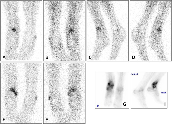
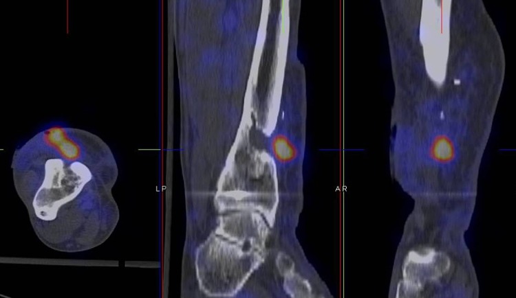
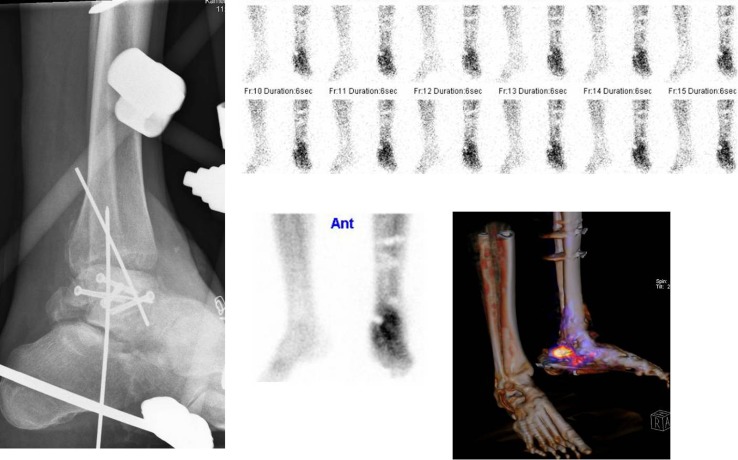
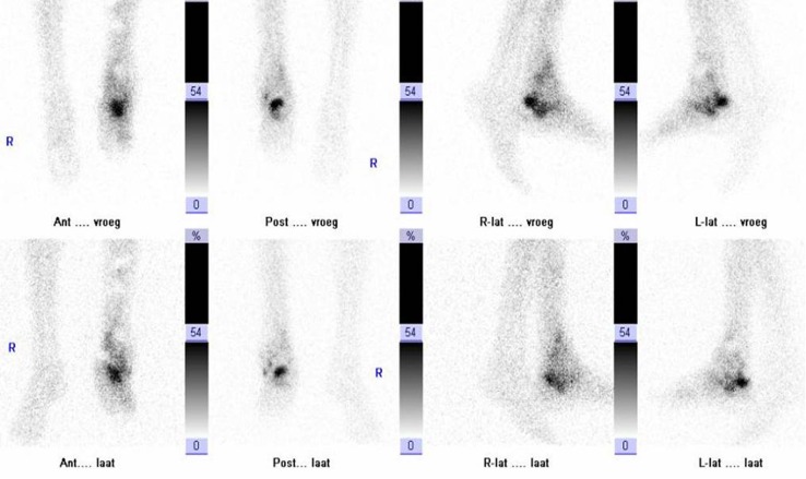
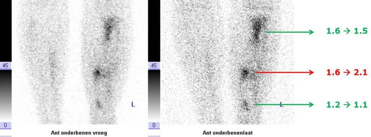
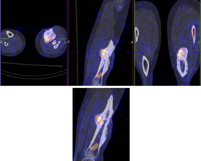
Aim: In this overview paper the techniques and available literature results for PTO are discussed for the three most commonly used nuclear medicine techniques: the three phase bone scan (with SPECT-CT), white blood cell scintigraphy (also called leukocyte scan) with SPECT-CT and (18)F-fluorodeoxyglucose (FDG)-PET/CT. Emphasis is on how these techniques are able to answer the diagnostic questions from the clinicians (trauma and orthopaedic surgeons) and which technique should be used to answer a specific question. Furthermore, three illustrative cases from clinical practice are described.
Keywords: Bone scan; FDG-PET; Nuclear medicine; Posttraumatic osteomyelitis; White blood cell scan.
Figures
References
-
- Ochsner PE, Borens O, Bodler P-M, Broger I, Clauss M, Eich G et al. Infections of the musculoskeletal system; swiss orthopaedics and Swiss society for infectious diseases. 2014.
-
- Lerner RK, Esterhai JL, Jr, Polomano RC, Cheatle MD, Heppenstall RB. Quality of life assessment of patients with posttraumatic fracture nonunion, chronic refractory osteomyelitis, and lower-extremity amputation. Clin Orthop Relat Res. 1993;295:28–36. - PubMed
-
- Glaudemans AW, Galli F, Pacilio M, Signore A. Leukocyte and bacteria imaging in prosthetic joint infection. Eur Cell Mater. 2013;25:61–77. - PubMed
Publication types
MeSH terms
LinkOut - more resources
Full Text Sources
Other Literature Sources
Medical
