Bedside Lung Ultrasound During Acute Chest Syndrome in Sickle Cell Disease
- PMID: 26886600
- PMCID: PMC4998600
- DOI: 10.1097/MD.0000000000002553
Bedside Lung Ultrasound During Acute Chest Syndrome in Sickle Cell Disease
Abstract
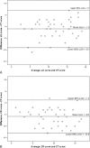
Lung ultrasound (LU) is increasingly used to assess pleural and lung disease in intensive care unit (ICU) and emergency unit at the bedside. We assessed the performance of bedside chest radiograph (CR) and LU during severe acute chest syndrome (ACS), using computed tomography (CT) as the reference standard. We prospectively explored 44 ACS episodes (in 41 patients) admitted to the medical ICU. Three imaging findings were evaluated (consolidation, ground-glass opacities, and pleural effusion). A score was used to quantify and compare loss of lung aeration with each technique and assess its association with outcome. A total number of 496, 507, and 519 lung regions could be assessed by CT scan, bedside CR, and bedside LU, respectively. Consolidations were the most common pattern and prevailed in lung bases (especially postero-inferior regions). The agreement with CT scan patterns was significantly higher for LU as compared to CR (κ coefficients of 0.45 ± 0.03 vs 0.30 ± 0.03, P < 0.01 for the parenchyma, and 0.73 ± 0.08 vs 0.06 ± 0.09, P < 0.001 for pleural effusion). The Bland and Altman analysis showed a nonfixed bias of -1.0 (P = 0.12) between LU score and CT score whereas CR score underestimated CT score with a fixed bias of -5.8 (P < 0.001). The specificity for the detection of consolidated regions or pleural effusion (using CT scan as the reference standard) was high for LU and CR, whereas the sensitivity was high for LU but low for CR. As compared to others, ACS patients with an LU score above the median value of 11 had a larger volume of transfused and exsanguinated blood, greater oxygen requirements, more need for mechanical ventilation, and a longer ICU length of stay. LU outperformed CR for the diagnosis of consolidations and pleural effusion during ACS. Higher values of LU score identified patients at risk of worse outcome.
Conflict of interest statement
The authors have no funding and conflicts of interest to disclose.
Figures


References
-
- Weatherall D, Hofman K, Rodgers G, et al. A case for developing North-South partnerships for research in sickle cell disease. Blood 2005; 105:921–923. - PubMed
-
- Rees DC, Williams TN, Gladwin MT. Sickle-cell disease. Lancet 2010; 376:2018–2031. - PubMed
-
- Vichinsky EP, Neumayr LD, Earles AN, et al. Causes and outcomes of the acute chest syndrome in sickle cell disease. National Acute Chest Syndrome Study Group. N Engl J Med 2000; 342:1855–1865. - PubMed
-
- Platt OS, Brambilla DJ, Rosse WF, et al. Mortality in sickle cell disease. Life expectancy and risk factors for early death. N Engl J Med 1994; 330:1639–1644. - PubMed
Publication types
MeSH terms
LinkOut - more resources
Full Text Sources
Other Literature Sources
Medical
Research Materials
Miscellaneous

