

Association Between Changes in Mammographic Image Features and Risk for Near-Term Breast Cancer Development
- PMID: 26886970
- PMCID: PMC4938728
- DOI: 10.1109/TMI.2016.2527619
Association Between Changes in Mammographic Image Features and Risk for Near-Term Breast Cancer Development
Abstract
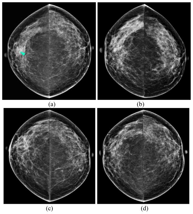
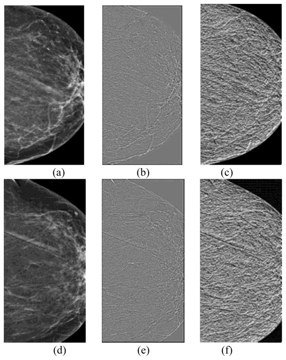
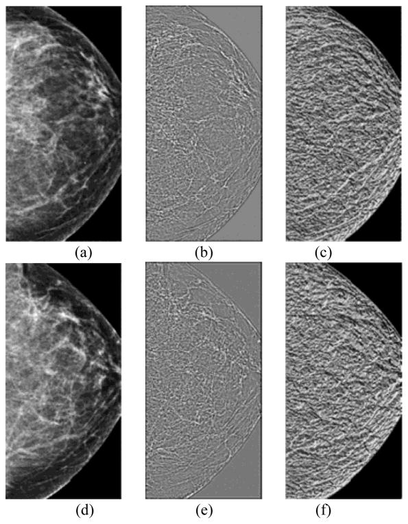
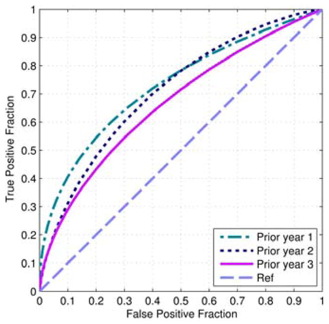
The purpose of this study is to develop and test a new computerized model for predicting near-term breast cancer risk based on quantitative assessment of bilateral mammographic image feature variations in a series of negative full-field digital mammography (FFDM) images. The retrospective dataset included series of four sequential FFDM examinations of 335 women. The last examination in each series ("current") and the three most recent "prior" examinations were obtained. All "prior" examinations were interpreted as negative during the original clinical image reading, while in the "current" examinations 159 cancers were detected and pathologically verified and 176 cases remained cancer-free. From each image, we initially computed 158 mammographic density, structural similarity, and texture based image features. The absolute subtraction value between the left and right breasts was selected to represent each feature. We then built three support vector machine (SVM) based risk models, which were trained and tested using a leave-one-case-out based cross-validation method. The actual features used in each SVM model were selected using a nested stepwise regression analysis method. The computed areas under receiver operating characteristic curves monotonically increased from 0.666±0.029 to 0.730±0.027 as the time-lag between the "prior" (3 to 1) and "current" examinations decreases. The maximum adjusted odds ratios were 5.63, 7.43, and 11.1 for the three "prior" (3 to 1) sets of examinations, respectively. This study demonstrated a positive association between the risk scores generated by a bilateral mammographic feature difference based risk model and an increasing trend of the near-term risk for having mammography-detected breast cancer.
Figures
Similar articles
-
Assessment of global and local region-based bilateral mammographic feature asymmetry to predict short-term breast cancer risk.Phys Med Biol. 2018 Jan 9;63(2):025004. doi: 10.1088/1361-6560/aaa096. Phys Med Biol. 2018. PMID: 29226849 Free PMC article.
-
Assessment of a Four-View Mammographic Image Feature Based Fusion Model to Predict Near-Term Breast Cancer Risk.Ann Biomed Eng. 2015 Oct;43(10):2416-28. doi: 10.1007/s10439-015-1316-5. Epub 2015 Apr 8. Ann Biomed Eng. 2015. PMID: 25851469 Free PMC article.
-
A new near-term breast cancer risk prediction scheme based on the quantitative analysis of ipsilateral view mammograms.Comput Methods Programs Biomed. 2018 Mar;155:29-38. doi: 10.1016/j.cmpb.2017.11.019. Epub 2017 Nov 29. Comput Methods Programs Biomed. 2018. PMID: 29512502
-
Using multiscale texture and density features for near-term breast cancer risk analysis.Med Phys. 2015 Jun;42(6):2853-62. doi: 10.1118/1.4919772. Med Phys. 2015. PMID: 26127038 Free PMC article.
-
Impact of full field digital mammography on the classification and mammographic characteristics of interval breast cancers.Eur J Radiol. 2015 Jun;84(6):1056-61. doi: 10.1016/j.ejrad.2015.03.007. Epub 2015 Mar 14. Eur J Radiol. 2015. PMID: 25816990 Review.
Cited by
-
Clinical Significance of Combined Density and Deep-Learning-Based Texture Analysis for Stratifying the Risk of Short-Term and Long-Term Breast Cancer in Screening.Diagnostics (Basel). 2024 Aug 21;14(16):1823. doi: 10.3390/diagnostics14161823. Diagnostics (Basel). 2024. PMID: 39202310 Free PMC article.
-
Assessment of global and local region-based bilateral mammographic feature asymmetry to predict short-term breast cancer risk.Phys Med Biol. 2018 Jan 9;63(2):025004. doi: 10.1088/1361-6560/aaa096. Phys Med Biol. 2018. PMID: 29226849 Free PMC article.
-
Prediction of breast cancer risk using a machine learning approach embedded with a locality preserving projection algorithm.Phys Med Biol. 2018 Jan 30;63(3):035020. doi: 10.1088/1361-6560/aaa1ca. Phys Med Biol. 2018. PMID: 29239858 Free PMC article.
-
Automated volumetric breast density measures: differential change between breasts in women with and without breast cancer.Breast Cancer Res. 2019 Oct 28;21(1):118. doi: 10.1186/s13058-019-1198-9. Breast Cancer Res. 2019. PMID: 31660981 Free PMC article.
-
Classification of Breast Masses Using a Computer-Aided Diagnosis Scheme of Contrast Enhanced Digital Mammograms.Ann Biomed Eng. 2018 Sep;46(9):1419-1431. doi: 10.1007/s10439-018-2044-4. Epub 2018 May 10. Ann Biomed Eng. 2018. PMID: 29748869 Free PMC article.
References
MeSH terms
Grants and funding
LinkOut - more resources
Full Text Sources
Other Literature Sources
Medical
