. 2016 Feb 18:6:16836.
doi: 10.1038/srep16836.

A Modular Assembly Platform for Rapid Generation of DNA Constructs

Affiliations

A Modular Assembly Platform for Rapid Generation of DNA Constructs

Elliot H Akama-Garren et al. Sci Rep. .

Abstract

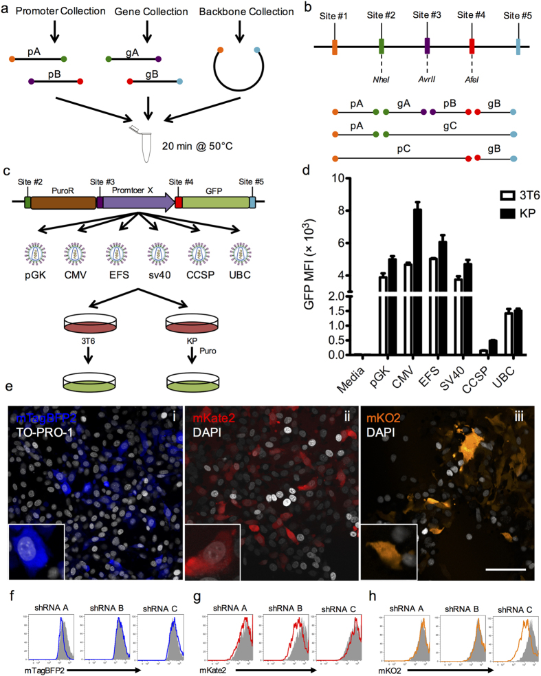
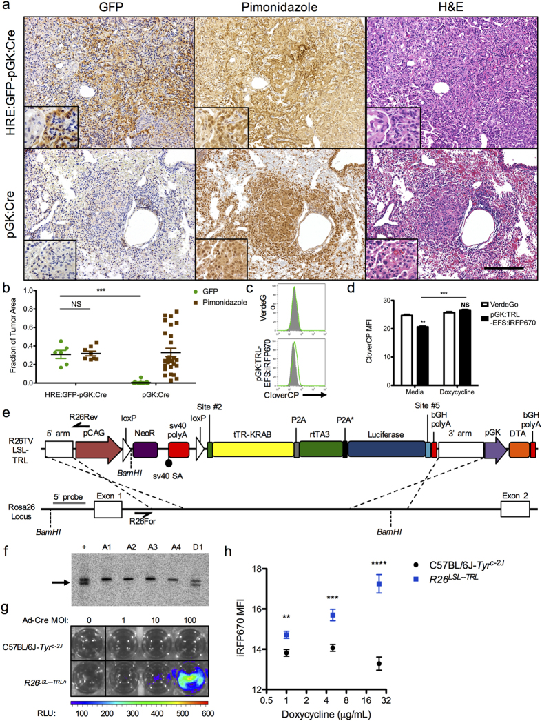
Traditional cloning methods have limitations on the number of DNA fragments that can be simultaneously manipulated, which dramatically slows the pace of molecular assembly. Here we describe GMAP, a Gibson assembly-based modular assembly platform consisting of a collection of promoters and genes, which allows for one-step production of DNA constructs. GMAP facilitates rapid assembly of expression and viral constructs using modular genetic components, as well as increasingly complicated genetic tools using contextually relevant genomic elements. Our data demonstrate the applicability of GMAP toward the validation of synthetic promoters, identification of potent RNAi constructs, establishment of inducible lentiviral systems, tumor initiation in genetically engineered mouse models, and gene-targeting for the generation of knock-in mice. GMAP represents a recombinant DNA technology designed for widespread circulation and easy adaptation for other uses, such as synthetic biology, genetic screens, and CRISPR-Cas9.


Figures

Figure 1
Figure 1. Gibson assembly-based modular assembly platform (GMAP).
(A) Promoters, genes, and backbones from established GMAP-compatible collections are used in a one step isothermal assembly reaction to produce DNA constructs on demand. (B) Schematic of possible orderings of genes and promoters. Promoter A (pA) is flanked by sites 1 and 2, promoter B (pB) is flanked by sites 3 and 4, promoter C (pC) is flanked by sites 1 and 3, gene A (gA) is flanked by sites 2 and 3, gene B (gB) is flanked by sites 4 and 5, and gene C (gC) is flanked by sites 2 and 5. (C) Schematic diagram of experiment using GMAP retroviral backbone. Retroviruses expressing GFP driven by different promoters were assembled using GMAP and used to transduce murine 3T6 fibroblasts or murine lung cancer (KP) cells. PuroR, puromycin resistance; pGK, human phosphoglycerate kinase 1 promoter; CMV, cytomegalovirus immediate-early promoter; EFS, elongation factor 1α promoter; SV40, simian virus early 40 promoter; CCSP, clara cell secretory protein promoter; UBC, human Ubiquitin C promoter. (D) Bar graph shows flow cytometry measurements of median fluorescence intensity (MFI) of GFP from 3T6 and KP cells transduced with GMAP-generated retrovirus. Data are representative of at least three independent experiments. (E) GMAP-generated lentiviruses expressing mTagBFP2-AUTR (i), mKate2-BUTR (ii), or mKO2-CUTR(iii) sensor cassettes were assembled and used to transduce a Cre reporter cell line (3TB). 3TB cells were selected with hygromycin and visualized by confocal microscopy. Insets show higher magnification. Scale bar, 100 μm. (F-H) Histograms from 3TB cells expressing mTagBFP2-AUTR (F), mKate2-BUTR (G), or mKO2-CUTR (H) transfected with three inducible hairpin constructs targeting the A, B, or C 3’UTRs assembled using GMAP. After transfection 3TB cells were selected with blasticidin, treated with doxycycline, and knockdown was assessed by flow cytometry analysis on GFP+ cells. Grey histograms represent cells lines transfected with an inducible shRNA targeting luciferase. Data are representative of at least three independent experiments.
Figure 2
Figure 2. In vivo applications of GMAP using lentiviral and Rosa26 targeting constructs.
(A) K-rasLSL−G12D/+;p53fl/fl mice were infected with GMAP-generated hypoxia response element (HRE):GFP-pGK:Cre or pGK:Cre lentivirus. After 30 weeks tumors were assessed for GFP expression, hypoxia as determined by pimonidazole staining, and hematoxylin and eosin (H&E). Insets show higher magnification. Data are representative of at least three independent experiments. Scale bar, 200 μm. (B) Column scatter plot shows quantification of immunohistochemistry shown in (A). Data are presented as fraction of tumor area that expressed GFP or pimonidazole staining. NS, p > 0.05; ***p = 5.82 × 10−12. (C) FACS histogram of KP cells engineered to express CloverCP in the presence of doxycycline and rtTA (VerdeGo) that were transduced with a GMAP-generated pGK:tTR-KRAB-rtTA3-Luc (TRL)-EFS:iRFP670 lentivirus and treated with media (grey histogram) or doxycycline (green line). Data are representative of at least three independent replicates. (D) Bar graph shows quantification of flow cytometry shown in (C). NS, p > 0.05; **p = 7.68 × 10−4; ***p = 2.46 × 10−4. (E) Map of a GMAP-generated Rosa26 targeting vector (R26TV) and scheme for knock-in of a Cre-dependent tTR-KRAB-rtTA3-Luc construct into the Rosa26 locus. LSL, loxP-Stop-loxP; NeoR, neomycin resistance; bGH polyA, bovine growth hormone polyadenylation signal; DTA, diphtheria toxin fragment A. Relevant primer binding sites (black half arrow) for PCR screening and restriction sites and probe (grey line) for Southern analysis are shown. (F) Southern analysis of ES cell DNA. C57BL/6J-Tyrc-2J ES cells were electroporated with AsiSI-linearized R26TV and subclone genomic DNA was digested with BamHI and probed, yielding a 5.8 kb product for the wild type Rosa26 locus and a 4.8 kb product for the targeted Rosa26 allele. A1-D1, clone number; +, positive control; arrow, targeted product. (G) Bioluminescence imaging of C57BL/6J-Tyrc-2J or R26LSL-TRL (clone D1) ES cells transduced with increasing multiplicity of infection (MOI) CMV-Cre adenovirus (Ad-Cre). Data are representative of at least three independent replicates. (H) Plot shows flow cytometry measurement of iRFP670 expression in C57BL/6J-Tyrc-2J or R26LSL-TRL ES cells transduced with a GMAP-generated tetracycline response element promoter (pTRE):iRFP670-EFS:Cre-2A-GFP lentivirus and treated with doxycycline. Data are representative of at least three independent replicates. **p = 3.71 × 10−3; ***p = 6.51 × 10−4; ****p = 3.60 × 10−5.

References

    1. Lodish H. et al. Molecular Cell Biology. (Freeman W. H., 2000).
    1. Li M. Z. & Elledge S. J. Harnessing homologous recombination in vitro to generate recombinant DNA via SLIC. Nat. Methods 4, 251–256 (2007). - PubMed
    1. Aslanidis C. & de Jong P. J. Ligation-independent cloning of PCR products (LIC-PCR). Nucleic Acids Res. 18, 6069–6074 (1990). - PMC - PubMed
    1. Scior A., Preissler S., Koch M. & Deuerling E. Directed PCR-free engineering of highly repetitive DNA sequences. BMC Biotechnol. 11, 87 (2011). - PMC - PubMed
    1. Werner S., Engler C., Weber E., Gruetzner R. & Marillonnet S. Fast track assembly of multigene constructs using Golden Gate cloning and the MoClo system. Bioeng. Bugs 3, 38–43 (2012). - PubMed

Publication types