. 2016 Apr 8;291(15):7888-901.
doi: 10.1074/jbc.M115.703124. Epub 2016 Feb 17.

Path to Collagenolysis: COLLAGEN V TRIPLE-HELIX MODEL BOUND PRODUCTIVELY AND IN ENCOUNTERS BY MATRIX METALLOPROTEINASE-12

Affiliations

Path to Collagenolysis: COLLAGEN V TRIPLE-HELIX MODEL BOUND PRODUCTIVELY AND IN ENCOUNTERS BY MATRIX METALLOPROTEINASE-12

Stephen H Prior et al. J Biol Chem. .

Abstract

Collagenolysis is essential in extracellular matrix homeostasis, but its structural basis has long been shrouded in mystery. We have developed a novel docking strategy guided by paramagnetic NMR that positions a triple-helical collagen V mimic (synthesized with nitroxide spin labels) in the active site of the catalytic domain of matrix metalloproteinase-12 (MMP-12 or macrophage metalloelastase) primed for catalysis. The collagenolytically productive complex forms by utilizing seven distinct subsites that traverse the entire length of the active site. These subsites bury ∼1,080 Å(2)of surface area, over half of which is contributed by the trailing strand of the synthetic collagen V mimic, which also appears to ligate the catalytic zinc through the glycine carbonyl oxygen of its scissile G∼VV triplet. Notably, the middle strand also occupies the full length of the active site where it contributes extensive interfacial contacts with five subsites. This work identifies, for the first time, the productive and specific interactions of a collagen triple helix with an MMP catalytic site. The results uniquely demonstrate that the active site of the MMPs is wide enough to accommodate two strands from collagen triple helices. Paramagnetic relaxation enhancements also reveal an extensive array of encounter complexes that form over a large part of the catalytic domain. These transient complexes could possibly facilitate the formation of collagenolytically active complexes via directional Brownian tumbling.

Keywords: collagen; docking; encounter complex; matrix metalloproteinase (MMP); nuclear magnetic resonance (NMR); paramagnetic relaxation enhancement (PRE); protein-protein interaction.


Figures

FIGURE 1.
FIGURE 1.
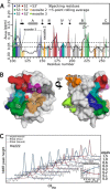
The largest shifts of peaks in NMR spectra of MMP-12 cat introduced by the α1(V)436–450 triple-helical peptide map to the catalytic cleft. A, shifts of amide NMR peaks are plotted versus sequence with a light gray line for individual values and a black line for the rolling average of five points. The shifts of the amide NMR peaks are defined as the radius of a triangle defined by the shifts of the 1H and 15N NMR peaks as ΔωHN = [ΔωH2 + (ΔωN/5)2]1/2 where the larger 15N scale is normalized to the 1H scale. The peak shifts are concentrated at residues at subsites within the cleft (solid colors) or residues packing with them (hatched colors). Exosite 2 (orange; Ile-152 to Gly-155) is the only area outside the catalytic cleft showing significant perturbation. Locations of the β-strands in the sequence are marked with arrows, and the helices are marked with cylinders. B, MMP-12 cat subsites exhibiting significant peak shifts span the length of the catalytic cleft. The colored surface corresponds to the colored areas in A. The gray sphere is the catalytic zinc. Red and white spheres mark the active water. C, spectral changes of His-222, which coordinates the catalytic zinc, strongly suggest binding nearby. Titration with the unlabeled triple-helical peptide shifts the His-222 amide peak (solid line) from that of the free form (fitted blue dashed line) to that of the bound form (fitted red dashed line). The inset shows the line widths of both free and bound peaks as a function of the concentration of α1(V)436–450 THP increasing to 1.5-fold molar excess. The broadening of the line shapes during partial occupancy of the active sites followed by the peak sharpening upon saturation to full occupancy is characteristic of intermediate-slow exchange. r.m.s. error of each fit of a Lorentzian line shape is indicated in the inset.
FIGURE 2.
FIGURE 2.
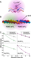
The collagen V-mimicking triple-helical peptide was synthesized with a nitroxide-containing amino acid to introduce strongly distance-dependent PREs to MMP-12 cat. A, the modified triple-helical peptide shows the nitroxide-containing TOAC residue with sticks and the leading, middle, and trailing chains in red, yellow, and blue, respectively. The radially decreasing violet color symbolizes the strong distance dependence of PRE that decreases in proportion to 1/r6 where r is the distance between unpaired electron and the proton monitored by NMR. B, peptide synthesis placed TOAC at either the P5 or P8′ position in the homotrimer. This introduces paramagnetic relaxation radiating up to 25 Å from each nitroxide group. The paramagnetic relaxation emanating from the TOAC at P5 is symbolized by green, and that from TOAC at P8′ is symbolized by violet. C, relaxation patterns indicate that Gly-105 at the unprimed end of the catalytic cleft is near a nitroxide at P5, whereas Gly-179 at the primed end of the cleft is near a nitroxide at P8′. The relaxation in the left panels was measured by the widely used two-point method (35, 45). In the right panels, the new multipoint approach (39–41) was used. Its exponential decays confirm suppression of J coupling. Error bars indicate S.D. of spectral noise. The unpaired electron in TOAC was reduced, and its paramagnetic relaxation was removed by incubation with 6 mm ascorbate, establishing the diamagnetic reference state.
FIGURE 3.
FIGURE 3.
Circular dichroism spectroscopy demonstrates the formation of triple helix and thermal stability of the TOAC-substituted triple-helical peptides used in this study. CD spectra of the triple-helical peptides with TOAC substitution at the P5 position (A) or the P8′ position (B) are shown. The ratio of the peak near 210 nm to the peak near 230 nm describes the degree of triple helix formation, which in this case is 8:1 for the unprimed substrate and 12.3:1 for the primed substrate, indicating the triple helix to be intact. Each inset shows the thermal melting curve for each triple-helical peptide. The Tm values are 28 °C with P5 substitution and 16 °C for P8′ substitution. NMR was performed at a temperature below the Tm of the substrate for triple-helical integrity during the experiment. Above each spectrum is a schematic representation showing the location of the TOAC residue within the sequence. mdeg, millidegrees.
FIGURE 4.
FIGURE 4.
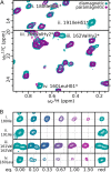
Paramagnetic NMR line broadenings introduced by triple-helical peptide with TOAC at P8′ are extreme and remote from the active site. A, i–iv, 13C heteronuclear multiple quantum coherence of Ile-Leu-Val-labeled, free MMP-12 cat (cyan) at 800 MHz reveals several methyl peaks broadened away by addition of α1(V) THP with TOAC on the primed side of the scissile bond (purple). Ile-180 is in the catalytic cleft. Ile-191, Val-162, and Leu-160 cluster on the β-sheet, suggesting alternative modes of binding. Each asterisk (*) symbolizes all three degenerate methyl protons. B, i–iv, concentration dependence of paramagnetic broadening. The last column was recorded after reduction with 6 mm ascorbate, which restored NMR peaks broadened by the spin-labeled triple-helical peptide.
FIGURE 5.
FIGURE 5.
Logic flow of the Monte Carlo algorithm q_test.py used to identify ensembles of binding poses in accord with the PREs. The structural coordinates tried were drawn randomly from a library of thousands of unique binding poses calculated with HADDOCK. The algorithm depends upon the optimization of the three thresholds x, y, and z. The threshold x defines the improvement needed to justify addition of a model to the ensemble. Lowering this cutoff results in more accurate ensembles but at the expense of significantly longer run time. The cutoff y defines the exit condition. Increasing y tests a larger number of ensembles but at the expense of longer run time. The threshold z defines the level of statistical value as the Akaike information criterion (AIC) above which ensemble members are deemed not to add worthwhile information. Raising z results in smaller ensembles at the expense of potentially higher Q-factors.
FIGURE 6.
FIGURE 6.
Statistics of parsimonious ensembles generated by the metaheuristic algorithm q_test.py. A, minimal ensembles generated showed a typical Gaussian distribution of between six and 10 members with an ensemble size of 8 being most frequent. B, certain clusters of structures were highly represented among the 1000 best ensembles (when ranked by Q-factor). Three clusters (clusters 1, 2, and 3) appeared in over 80% of ensembles. C, occurrence of clusters among the 1000 best minimal ensembles. 65 clusters were represented in at least 1% of ensembles of which only three (clusters 1, 2, and 3) occurred in more than 80%.
FIGURE 7.
FIGURE 7.
Productive complex of a collagen triple helix in the active site of an MMP revealed by paramagnetic NMR. A, structure of the collagen V-derived triple helix primed for hydrolysis reveals that both the middle (yellow surface) and trailing (blue surface) chains fit in the active site. The leading strand (red surface) makes little contact with the enzyme. The region of the large dashed box is expanded in walleyed stereo in B. The small dashed boxes show approximate locations of TOAC residues, each containing a spin label. B, middle and trailing strands manage to fit in the active site through efficient subsite usage, positioning the trailing chain of the Michaelis complex for initial hydrolysis. Active water (red and white spheres) and the carbonyl oxygen of the Gly at the P1 position (blue sticks of Gly-88) coordinate the active site zinc (black sphere). This positions the carbonyl carbon of Gly at P1 in the trailing chain for nucleophilic attack by the water. Seven distinct subsites (labeled in the left image) are occupied by side chains (labeled with magenta in the right image) from the middle and trailing chains. Dots are plotted for peptide atoms in apparent contact with the enzyme. Two bad contacts outside subsite S3′ are colored blue-green. Green dashes mark a hydrogen bond inferred between Lys-241 and Gln at P5′. The exosite marked was identified in Refs. and .
FIGURE 8.
FIGURE 8.
Ensembles of structures containing remote encounter complexes account for the distribution of paramagnetic broadenings from TOAC-substituted triple-helical miniproteins (A–C). The productively bound member of the ensembles is symbolized by the black arrow. The two major remote binding poses (red, yellow, and blue schematic) are members of most of the ensembles. An ensemble of ensembles (mesh representing 20% occupancy) representing a cohort of lowly populated or mobile binding poses is required to explain the majority of the remaining PREs.
FIGURE 9.
FIGURE 9.
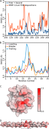
Structural distortions in the complex upon binding appear small, localized, and within the experimental range of variation. A, per-residue Cα r.m.s.d. of crystallographic and NMR solution structures of MMP-12 cat in the Protein Data Bank (PDB). The blue line shows the structural changes needed for MMP-12 to accommodate two chains of the triple helix. The red line corresponds to the natural diversity of all MMP-12 structures in the Protein Data Bank (currently 18 structures). All the significant distortion needed for MMP-12 cat to accommodate the triple helix (r.m.s.d. > 1.0 Å; residues 178–182, 225–230, and 237–242) maps to flexible loops and falls within the natural range of structural diversity. B, per-residue structural changes are plotted for the α1(V)436–450 triple-helical peptide as it binds to MMP-12 cat. The largest change occurs in the trailing strand (blue line) in the scissile triplet. Only Glu-20 of the leading and middle strands (red and orange lines) exhibits significant distortion, which accompanies displacement of the scissile triplet of the trailing strand. C, per-residue structural changes in MMP-12 as it binds the triple-helical miniprotein plotted as r.m.s.d. of free versus bound forms (i.e. pre- and postenergy minimization). Much of the distortion is confined to the flexible S1′ specificity loop, i.e. the “lower lip” of the active site. D, the information of B is plotted onto the surface of the homology model of the α1(V)436–450 triple-helical peptide. The boxed region shows the approximate location of the scissile region.
FIGURE 10.
FIGURE 10.
Ensembles of structures are needed to account for the many PREs measured. Theoretical PREs (colored symbols) are compared with measured PREs (gray) introduced by a 1.5-fold excess of the triple-helical peptide with TOAC substitution either at P5 (A) or at P8′ (B). The PREs from triple-helical peptide to amide protons are marked with gray columns for measurements and squares for back-calculations from the productive model in red, from the three frequent binding poses (clusters 1–3) in blue, and the ensemble of parsimonious ensembles in green. Triangles mark PREs from P8′ to methyl groups measured (gray) or back-calculated (color). Theoretical PREs calculated from the productive mode of binding (red) explain broadening in the active site (e.g. residues 102–105 and 221–230 in A and 202–220 in B) but fail to explain other areas experiencing significant broadening (e.g. residues 140–165 in B). Addition of the two frequently sampled remote poses explains the PREs of several other areas (blue). However, an ensemble of ensembles is required to model (green) the widely observed PREs with the comparatively high quality of a Q-factor of 0.21. Error bars indicate S.D. of spectral noise.
FIGURE 11.
FIGURE 11.
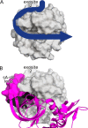
Remote binding to exosite 2 and a hydrophobic channel topping the β-sheet might impart a hypothetical advantage to reload another triple helix into the active site. A, proposed route of diffusion that might reload productive binding (blue arrow) after molecular recognition near exosite 2. B, tissue inhibitor of metalloproteinases-2 (magenta; Protein Data Bank code 2E2D) reaches its long sA-sB loop (magenta surface) around the S-shaped loop to fill the channel over the β-sheet to approach exosite 2. This occupies the hypothetical path of diffusion of the triple helix.

References

    1. Fields G. B. (1991) A model for interstitial collagen catabolism by mammalian collagenases. J. Theor. Biol. 153, 585–602 - PubMed
    1. Wolf K., Wu Y. I., Liu Y., Geiger J., Tam E., Overall C., Stack M. S., and Friedl P. (2007) Multi-step pericellular proteolysis controls the transition from individual to collective cancer cell invasion. Nat. Cell Biol. 9, 893–904 - PubMed
    1. Chun T. H., Sabeh F., Ota I., Murphy H., McDonagh K. T., Holmbeck K., Birkedal-Hansen H., Allen E. D., and Weiss S. J. (2004) MT1-MMP-dependent neovessel formation within the confines of the three-dimensional extracellular matrix. J. Cell Biol. 167, 757–767 - PMC - PubMed
    1. Rundhaug J. E. (2005) Matrix metalloproteinases and angiogenesis. J. Cell. Mol. Med. 9, 267–285 - PMC - PubMed
    1. Libby P. (2008) The molecular mechanisms of the thrombotic complications of atherosclerosis. J. Intern. Med. 263, 517–527 - PMC - PubMed

Publication types

LinkOut - more resources