. 2016 Feb 18:7:10708.
doi: 10.1038/ncomms10708.

Cryo-EM structures of the eukaryotic replicative helicase bound to a translocation substrate

Affiliations

Cryo-EM structures of the eukaryotic replicative helicase bound to a translocation substrate

Ferdos Abid Ali et al. Nat Commun. .

Abstract

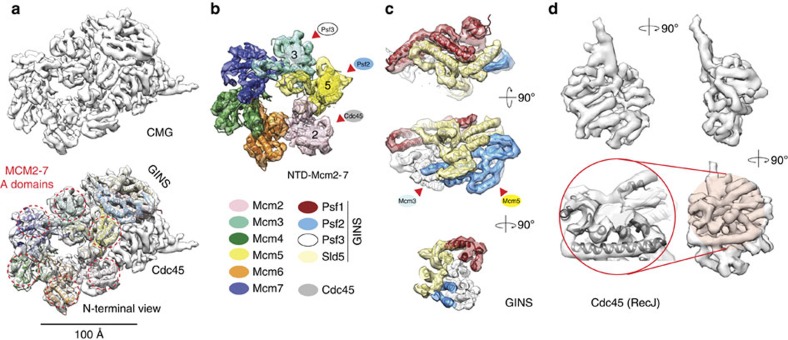
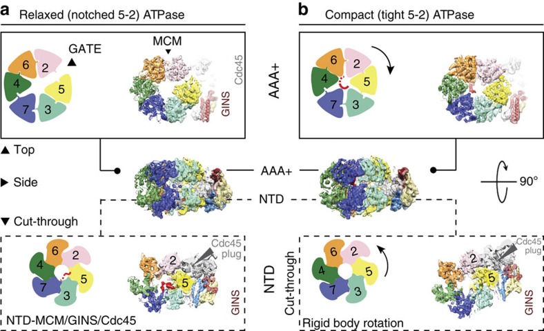
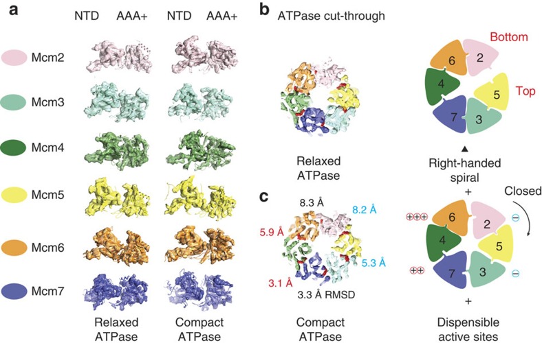
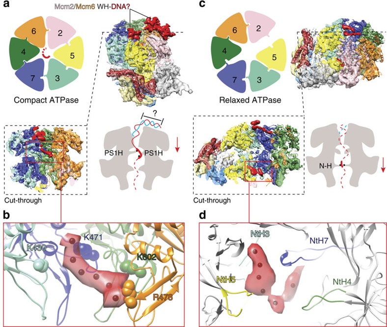
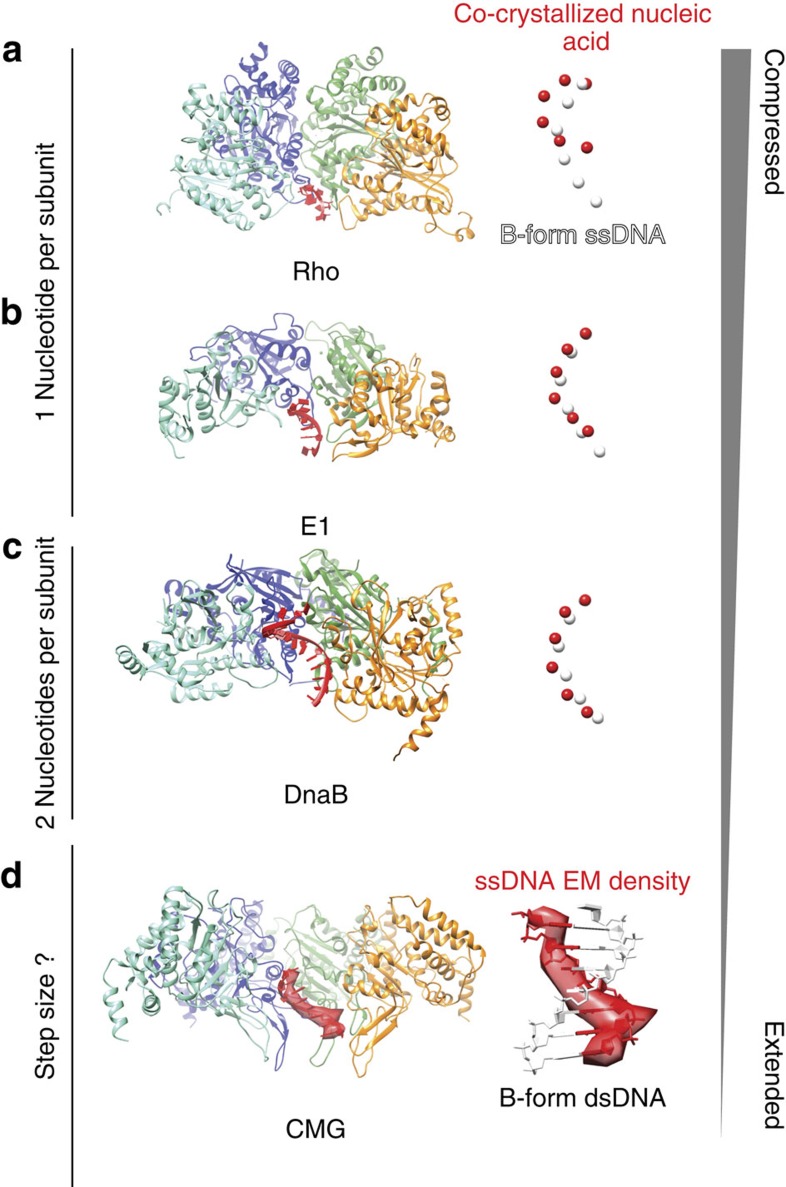
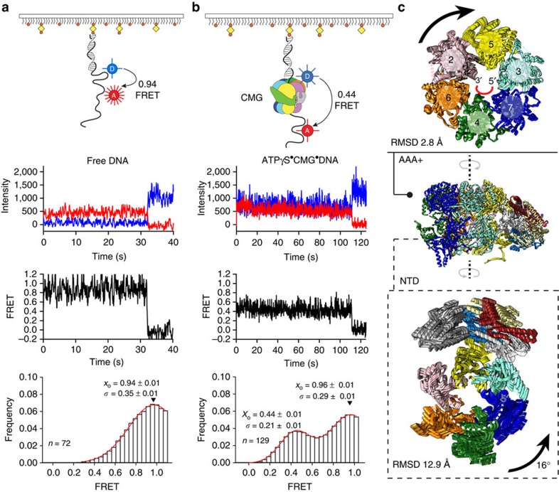
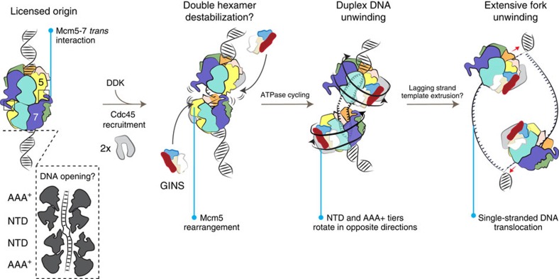
The Cdc45-MCM-GINS (CMG) helicase unwinds DNA during the elongation step of eukaryotic genome duplication and this process depends on the MCM ATPase function. Whether CMG translocation occurs on single- or double-stranded DNA and how ATP hydrolysis drives DNA unwinding remain open questions. Here we use cryo-electron microscopy to describe two subnanometre resolution structures of the CMG helicase trapped on a DNA fork. In the predominant state, the ring-shaped C-terminal ATPase of MCM is compact and contacts single-stranded DNA, via a set of pre-sensor 1 hairpins that spiral around the translocation substrate. In the second state, the ATPase module is relaxed and apparently substrate free, while DNA intimately contacts the downstream amino-terminal tier of the MCM motor ring. These results, supported by single-molecule FRET measurements, lead us to suggest a replication fork unwinding mechanism whereby the N-terminal and AAA+ tiers of the MCM work in concert to translocate on single-stranded DNA.


Figures

Figure 1
Figure 1. CMG helicase structure at subnanometre resolution.
(a) Resolution density (7.4 Å) map of the CMG viewed from the MCM N-terminal face, without or with docked MCM and GINS atomic structures. (b) Detailed view of the MCM N-terminal DNA-interacting collar. Psf3 contacts Mcm3, Psf2 contacts Mcm5 and Cdc45 contacts Mcm2. (c) Detailed view of the GINS assembly with docked human atomic structure (PDB entry 2Q9Q). (d) Density assigned to Cdc45. The Cdc45 core matches the secondary structure elements of the bacterial RecJ exonuclease (PDB entry 1IR6).
Figure 2
Figure 2. Two configurations in the ATPγS–CMG–DNA complex.
(a) AAA+, side and cut-through view of the CMG in a relaxed ATPase configuration. Cdc45 topologically locks the Mcm5-2 gate. (b) AAA+, side and cut-through N-terminal view of the CMG in a compact ATPase configuration. DNA density surmounting the AAA+ domain has been removed for visualization purposes (also see Fig. 4).
Figure 3
Figure 3. The MCM ATPase centres.
(a) Segmented density and docked atomic structures of the six Mcm2-7 protomers in the two conformers. The NTD and AAA+ domains tilt and rock with respect to one another. (b) ATPase sites in the relaxed ATPase conformer. (c) ATPase sites in the compact ATPase conformer. One side of the MCM ring is more static and the corresponding active sites are dispensable for viability (−means Walker A mutation kills DNA unwinding, +means tolerated mutation).
Figure 4
Figure 4. DNA-bound form of the CMG helicase.
(a) The compact ATPase form contains rod-shaped, bent density features surmounting the ATPase face tentatively assigned to duplex DNA engaged by the MCM winged helix (WH) C-terminal extensions. An MCM slice through the side view reveals an extended density feature, which we assign to single-stranded DNA, traversing the AAA+ pore. (b) Single-stranded DNA contacts conserved positively charged residues on the AAA+ PS1 hairpins that have a key role in DNA unwinding. (c) The relaxed ATPase form contains a thin density feature surmounting the AAA+ ring, which we assign to the flexible C-terminal MCM WH extensions. Although the AAA+ tier appears substrate free, a well-resolved elongated density threads through the MCM N-terminal collar and (d) contacts a set of N-terminal hairpins important for DNA binding and helicase activity.
Figure 5
Figure 5. Comparison of the ATPγS–CMG–DNA structure with available helicase–nucleic acid assemblies.
(a) The Rho termination factor (PDB entry 3ICE) contacts and compresses single-stranded RNA. The RNA structure is compared with one single DNA strand extracted from a B-form double helix. Spheres represent the centre of mass of each nucleotide. (b) The Papillomavirus E1 replicative helicase (PDB entry 2GXA) contacts and compresses single-stranded DNA. (c) The bacterial DnaB replicative helicase contacts and slightly compresses single-stranded DNA (approximating A-form DNA, PDB entry 4ESV). (d) The MCM motor of the CMG harbours a more extended form of single-stranded DNA, which appears to match B-form DNA. The DNA density matches the structure of one single strand in the double helix. This supports the notion that the CMG is a single-stranded DNA translocase.
Figure 6
Figure 6. DNA engagement and deformation by the CMG helicase.
Single-molecule FRET analysis of (a) isolated DNA and (b) DNA+ATPγS CMG. Top to bottom: cartoon schematic of the single-molecule FRET experiment indicating FRET between Cy3 (Donor, blue sphere) and Cy5 (Acceptor, red sphere, 7-nt separation) attached to biotinylated 3′-tailed duplex DNA, immobilized on a neutravidin-coated biotin-PEG surface; donor (blue) and acceptor (red) intensity trajectories are anti-correlated until single-step photobleaching of the acceptor. FRET trajectories (black) between Cy3 and Cy5 exhibit a sharp drop to zero FRET when the acceptor photobleaches; histogram of FRET values collected from all molecules with a Gaussian fit are shown in red, where x0 is the mean FRET and σ is the distribution width. ATPase controlled ring rotation and DNA stretching by the CMG helicase. (c) The AAA+ tier of the Mcm2-7 motor rotates clockwise as Mcm2 moves towards the Mcm5 protomer, to close a gap in the motor domain. The DNA-interacting NTD rotates anticlockwise as a rigid body, together with GINS and Cdc45.
Figure 7
Figure 7. Origin activation and replication fork unwinding by the CMG helicase.
Origin licensing involves the loading of a head-to-head double hexameric ring that encircles duplex DNA, which might become partially deformed. Cdc45 is loaded onto the double hexamer, in a process that requires DDK phosphorylation of MCM. DDK phosphorylation might cause a rearrangement in Mcm5 and disrupt the (yeast specific) Mcm5-7 trans interaction. This rearrangement would expose a GINS interacting element in Mcm5. ATP hydrolysis by the MCM promotes the relative rotation of the NTD and AAA+ tiers of the helicase, in a movement that might promote duplex DNA underwinding. Following a poorly understood lagging-strand extrusion process, the CMG helicase extensively unwinds the replication fork, by translocating on single-stranded DNA.

References

    1. Remus D. & Diffley J. F. Eukaryotic DNA replication control: lock and load, then fire. Curr. Opin. Cell Biol. 21, 771–777 (2009) . - PubMed
    1. Remus D. et al. Concerted loading of Mcm2-7 double hexamers around DNA during DNA replication origin licensing. Cell 139, 719–730 (2009) . - PMC - PubMed
    1. Evrin C. et al. A double-hexameric MCM2-7 complex is loaded onto origin DNA during licensing of eukaryotic DNA replication. Proc. Natl Acad. Sci. USA 106, 20240–20245 (2009) . - PMC - PubMed
    1. Li N. et al. Structure of the eukaryotic MCM complex at 3.8A. Nature 524, 186–191 (2009) (2015) . - PubMed
    1. Yeeles J. T., Deegan T. D., Janska A., Early A. & Diffley J. F. Regulated eukaryotic DNA replication origin firing with purified proteins. Nature 519, 431–435 (2015) . - PMC - PubMed

Publication types