. 2016 May 1;115(5):2593-607.
doi: 10.1152/jn.01073.2015. Epub 2016 Feb 17.

Testing the hypothesis of neurodegeneracy in respiratory network function with a priori transected arterially perfused brain stem preparation of rat

Affiliations

Testing the hypothesis of neurodegeneracy in respiratory network function with a priori transected arterially perfused brain stem preparation of rat

Sarah E Jones et al. J Neurophysiol. .

Abstract

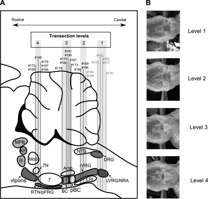
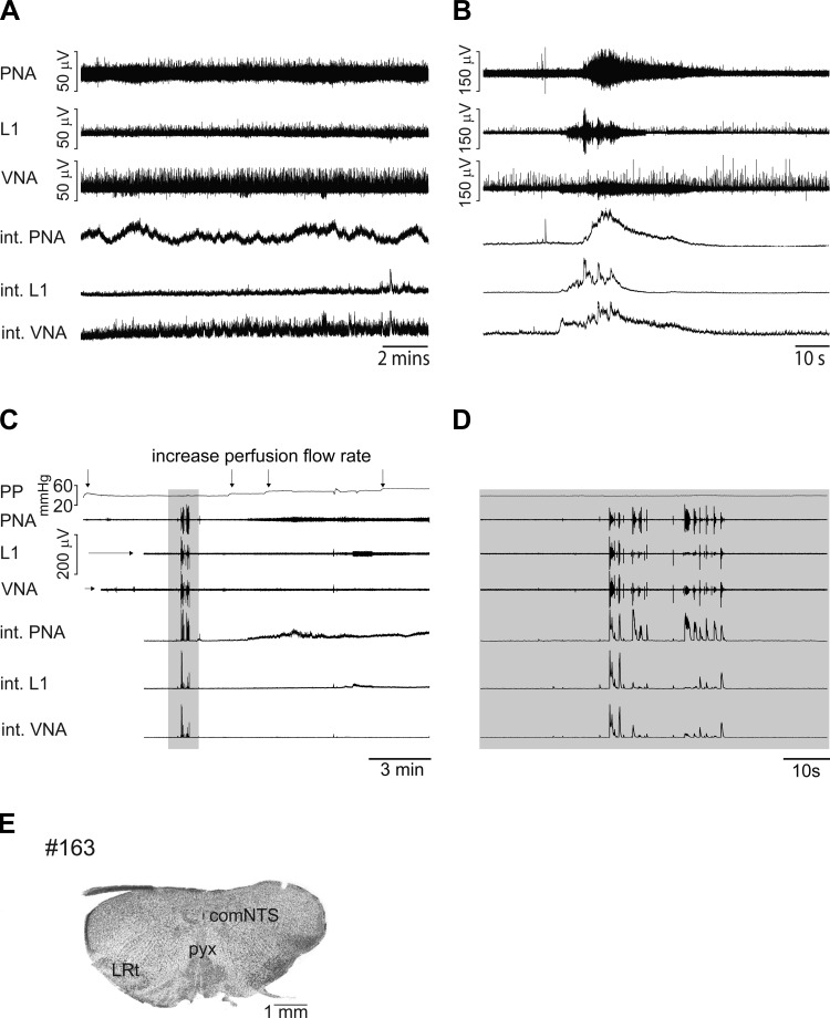
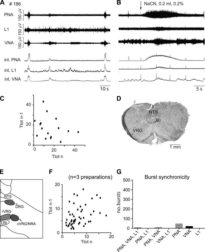
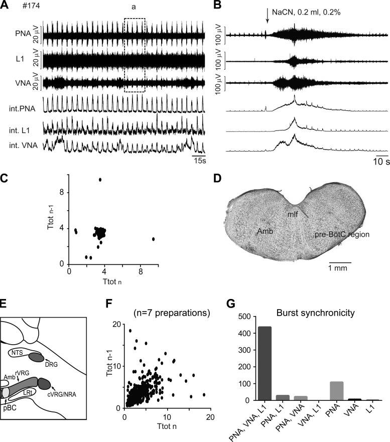
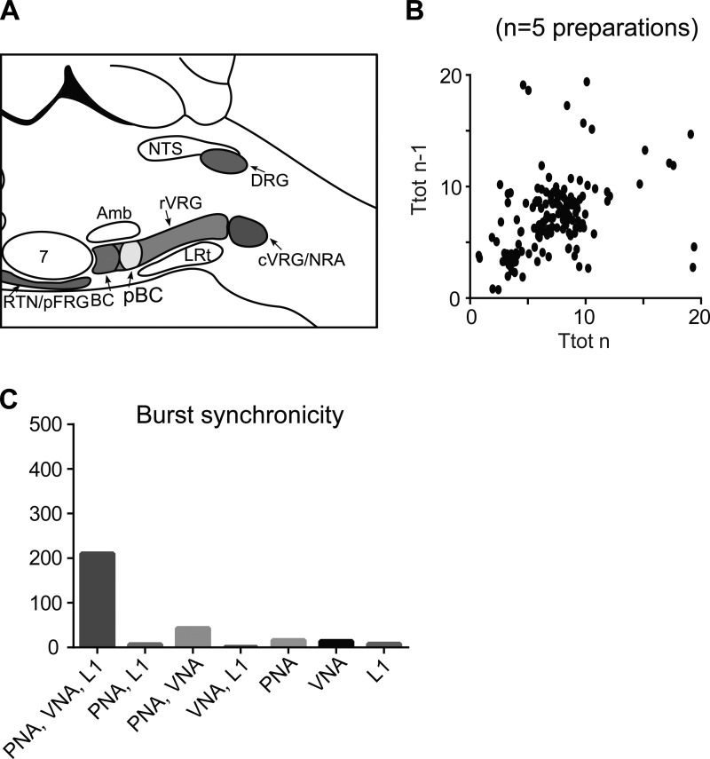
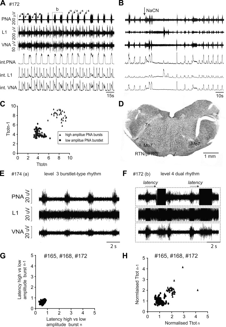
Degeneracy of respiratory network function would imply that anatomically discrete aspects of the brain stem are capable of producing respiratory rhythm. To test this theory we a priori transected brain stem preparations before reperfusion and reoxygenation at 4 rostrocaudal levels: 1.5 mm caudal to obex (n = 5), at obex (n = 5), and 1.5 (n = 7) and 3 mm (n = 6) rostral to obex. The respiratory activity of these preparations was assessed via recordings of phrenic and vagal nerves and lumbar spinal expiratory motor output. Preparations with a priori transection at level of the caudal brain stem did not produce stable rhythmic respiratory bursting, even when the arterial chemoreceptors were stimulated with sodium cyanide (NaCN). Reperfusion of brain stems that preserved the pre-Bötzinger complex (pre-BötC) showed spontaneous and sustained rhythmic respiratory bursting at low phrenic nerve activity (PNA) amplitude that occurred simultaneously in all respiratory motor outputs. We refer to this rhythm as the pre-BötC burstlet-type rhythm. Conserving circuitry up to the pontomedullary junction consistently produced robust high-amplitude PNA at lower burst rates, whereas sequential motor patterning across the respiratory motor outputs remained absent. Some of the rostrally transected preparations expressed both burstlet-type and regular PNA amplitude rhythms. Further analysis showed that the burstlet-type rhythm and high-amplitude PNA had 1:2 quantal relation, with burstlets appearing to trigger high-amplitude bursts. We conclude that no degenerate rhythmogenic circuits are located in the caudal medulla oblongata and confirm the pre-BötC as the primary rhythmogenic kernel. The absence of sequential motor patterning in a priori transected preparations suggests that pontine circuits govern respiratory pattern formation.

Keywords: breathing; medulla oblongata; neural circuits; respiratory pattern; respiratory rhythm generation.


Figures

Fig. 1.
Fig. 1.
A: original recordings of phrenic (PNA), vagal (VNA), and lumbar iliohypogastric nerve (L1) activity from the intact in situ perfused brain stem preparation, including the physiological response to sodium cyanide (NaCN) bolus injection of the intact preparation (B). integ., Integrated burst amplitude. C: enlarged single burst illustrates nerve discharge during phases of inspiration (I), postinspiration (PI), and expiration (E) in the intact preparation. Arrows indicate, for each nerve, the onset of nerve discharge within the respiratory cycle. D: schematic illustration of pontomedullary brain stem circuitry remaining intact following precollicular decerebration. E: Poincaré plots of respiratory cycle length (Ttot) averaged over 5 min from 9 intact preparations. Amb, nucleus ambiguus; BC, Bötzinger complex; c-rVRG, caudal and rostral ventral respiratory group; DRG, dorsal respiratory group; I5, intermediate reticular nucleus; LRt, lateral reticular nucleus; Mo5, motor trigeminal nucleus; MPB, medial parabrachial nucleus; NRA, nucleus retroambiguus; NTS, nucleus tractus solitarii; pBC, pre-Bötzinger complex; vlpons, ventrolateral pons; 7N, facial nerve.
Fig. 2.
Fig. 2.
A: illustration of the anatomical location of the a priori transection at 4 distinct levels (level 1, 1.5 mm caudal to obex; level 2, obex level and rostral to obex; level 3, 1.5 mm rostral to obex; and level 4, 3 mm rostral to obex) in a schematic drawing of a sagittal section through the pontomedullary brain stem. Examples of respiratory activity and histology of individual preparations (#) are illustrated in subsequent figures. B: photographs showing examples of a priori transected brain stems.
Fig. 3.
Fig. 3.
A: original recordings of phrenic (PNA), vagal (VNA), and lumbar iliohypogastric nerve (L1) activity from an a priori transected brain stem preparation at level 1 (1.5 mm caudal to obex). B: respiratory response to carotid body stimulation via intra-arterial bolus injection of sodium cyanide (NaCN). C: recording of transient and robust respiratory motor bursting (shaded area) during early stages of the reperfusion protocol. D: expanded view of initial motor bursting. E: photograph of the first intact coronal section cut from the a priori transected brain stem preparation. comNTS, commissural nucleus of the nucleus tractus solitarii; LRt, lateral reticular nucleus; pyx: pyramidal decussation.
Fig. 4.
Fig. 4.
A: original recordings of phrenic (PNA), vagal (VNA), and lumbar iliohypogastric nerve (L1) activity from a priori transected brain stem preparation #186 at level 2 (obex). B: respiratory response to carotid body stimulation via intra-arterial bolus injection of sodium cyanide (NaCN). C: Poincaré plots of respiratory cycle length (Ttot) during the most rhythmic 5-min period of preparation #186. D: photograph of the first intact coronal section cut from the a priori transected brain stem preparation. E: schematic illustration of the transection level. F: Poincaré plots of Ttot from weakly rhythmic preparations that were a priori transected at level 2. G: bar diagram illustrating the synchronicity of bursting activity across the 3 recorded motor outputs. c-rVRG, caudal and rostral ventral respiratory group; DRG, dorsal respiratory group; LRt: lateral reticular nucleus; NRA, nucleus retroambiguus; NTS, nucleus tractus solitarii; XII, hypoglossal motor nucleus.
Fig. 5.
Fig. 5.
A: original recordings of phrenic (PNA), vagal (VNA), and lumbar iliohypogastric nerve (L1) activity from a priori transected brain stem preparation #174 at level 3 (1.5 mm rostral to obex). Note that recording area in box a is used for comparison with the activity from more rostrally transected preparations (see Fig. 7). B: respiratory response to carotid body stimulation via intra-arterial bolus injection of sodium cyanide (NaCN). C: Poincaré plots of respiratory cycle length (Ttot) from a rhythmic 5-min period of preparation #174. D: photograph of the first intact coronal section cut from the a priori transected brain stem preparation. E: schematic illustration of the transection level. F: Poincaré plots of Ttot from all rhythmic preparations a priori transected at level 3. G: bar diagram illustrating the synchronicity of bursting activity across the 3 recorded motor outputs. Amb, nucleus ambiguus; c-rVRG, caudal and rostral ventral respiratory group; DRG, dorsal respiratory group; LRt, lateral reticular nucleus; NRA, nucleus retroambiguus; mlf, medial longitudinal fasciculus; NTS, nucleus tractus solitarii; pre-BötC/pBC, pre-Bötzinger complex; XII, hypoglossal motor nucleus.
Fig. 6.
Fig. 6.
Analysis of high-amplitude bursts observed after transections at level 4. A: schematic illustration of the transection level. B: Poincaré plots of Ttot for high-amplitude phrenic nerve activity (PNA) of preparation a priori transected at level 4. C: bar diagram illustrating the synchronicity of burst activity across the 3 recorded motor outputs. Amb, nucleus ambiguus; BC, Bötzinger complex; c-rVRG, caudal and rostral ventral respiratory group; DRG, dorsal respiratory group; LRt, lateral reticular nucleus; NRA, nucleus retroambiguus; NTS, nucleus tractus solitarii; RTN/pFRG, retrotrapezoid nucleus/parafacial respiratory group.
Fig. 7.
Fig. 7.
Original recordings of phrenic (PNA), vagal (VNA), and lumbar iliohypogastric nerve (L1) activity from a priori transected brain stem preparation #172 at level 4 (3 mm rostral to obex). Note the simultaneous expression of 2 distinct bursting rhythms with high (triangles) and low (circles) amplitudes in this preparation. B: respiratory response to carotid body stimulation via intra-arterial bolus injection of sodium cyanide (NaCN). C: Poincaré plots of respiratory cycle length (Ttot) of high- and low-amplitude bursts during 5-min recording period of preparation #172. D: photograph of the first intact coronal section cut from the a priori transected brain stem preparation. E and F: recordings show the similarity of low-amplitude burstlet-type rhythm observed after transection at level 3 (E) and level 4 (F). G: Poincaré plots of the latency between onset of burstlet-type activity and high-amplitude burst from all preparations that expressed dual rhythmic activity. H: Poincaré plots of Ttot from all rhythmic preparations that expressed dual bursting rhythms after a priori transection at level 4. 7n, facial nerve; Mo7, facial motor neurons; RTN/pFRG, retrotrapezoid nucleus/parafacial respiratory group.

References

    1. Abbott SB, Stornetta RL, Coates MB, Guyenet PG. Phox2b-expressing neurons of the parafacial region regulate breathing rate, inspiration, and expiration in conscious rats. J Neurosci 31: 16410–16422, 2011. - PMC - PubMed
    1. Abdala AP, Rybak IA, Smith JC, Paton JF. Abdominal expiratory activity in the rat brainstem-spinal cord in situ: patterns, origins and implications for respiratory rhythm generation. J Physiol 587: 3539–3559, 2009. - PMC - PubMed
    1. Alheid GF, Milsom WK, McCrimmon DR. Pontine influences on breathing: an overview. Respir Physiol Neurobiol 143: 105–114, 2004. - PubMed
    1. Aoki M, Mori S, Kawahara K, Watanabe H, Ebata N. Generation of spontaneous respiratory rhythm in high spinal cats. Brain Res 202: 51–63, 1980. - PubMed
    1. Baghdadwala MI, Duchcherer M, Trask WM, Gray PA, Wilson RJ. Diving into the mammalian swamp of respiratory rhythm generation with the bullfrog. Respir Physiol Neurobiol 14: 37–51, 2016. - PubMed

LinkOut - more resources