. 2016 Feb 18:6:21747.
doi: 10.1038/srep21747.

TRPV-1-mediated elimination of residual iPS cells in bioengineered cardiac cell sheet tissues

Affiliations

TRPV-1-mediated elimination of residual iPS cells in bioengineered cardiac cell sheet tissues

Katsuhisa Matsuura et al. Sci Rep. .

Abstract

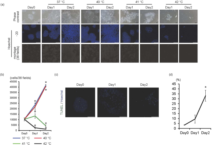
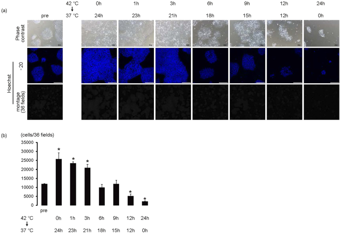
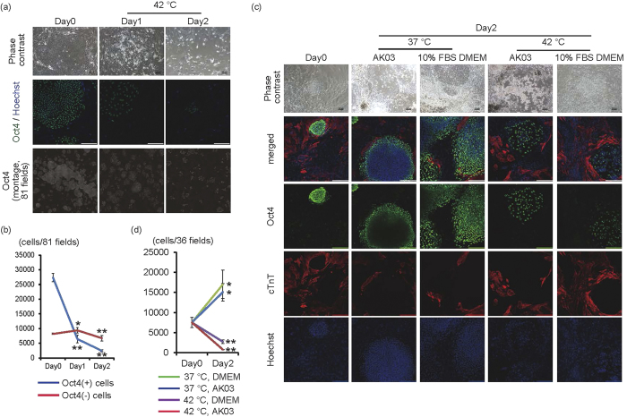
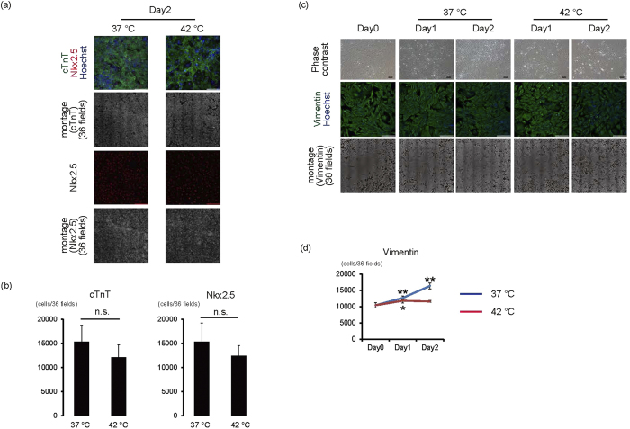
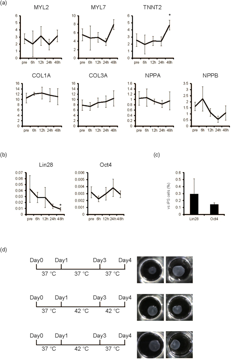
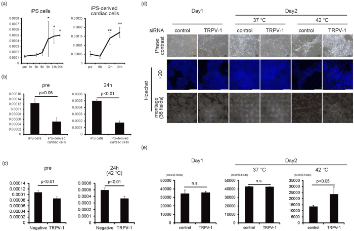
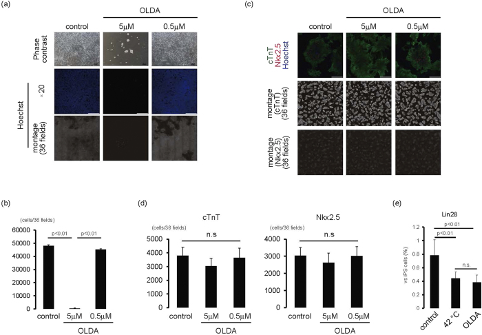
The development of a suitable strategy for eliminating remaining undifferentiated cells is indispensable for the use of human-induced pluripotent stem (iPS) cell-derived cells in regenerative medicine. Here, we show for the first time that TRPV-1 activation through transient culture at 42 °C in combination with agonists is a simple and useful strategy to eliminate iPS cells from bioengineered cardiac cell sheet tissues. When human iPS cells were cultured at 42 °C, almost all cells disappeared by 48 hours through apoptosis. However, iPS cell-derived cardiomyocytes and fibroblasts maintained transcriptional and protein expression levels, and cardiac cell sheets were fabricated after reducing the temperature. TRPV-1 expression in iPS cells was upregulated at 42 °C, and iPS cell death at 42 °C was TRPV-1-dependent. Furthermore, TRPV-1 activation through thermal or agonist treatment eliminated iPS cells in cardiac tissues for a final concentration of 0.4% iPS cell contamination. These findings suggest that the difference in tolerance to TRPV-1 activation between iPS cells and iPS cell-derived cardiac cells could be exploited to eliminate remaining iPS cells in bioengineered cell sheet tissues, which will further reduce the risk of tumour formation.


Conflict of interest statement

There is potential competing interest. Teruo Okano is a founder and a member of the board of CellSeed Inc., which has licenses for certain cell sheet-related technologies and patents from Tokyo Women’s Medical University. Teruo Okano and Tatsuya Shimizu are stake holders of CellSeed Inc. Tokyo Women’s Medical University receives research funds from CellSeed Inc. Teruo Okano, Tatsuya Shimizu, and Katsuhisa Matsuura are inventors of bioreactor systems.

Figures

Figure 1
Figure 1. 42 °C is the critical temperature for feeder-less human iPS cells.
(a) Human iPS cells cultured on laminin E8 fragment were cultured at 37 °C, 40 °C, 41 °C, or 42 °C for 1 or 2 days. Upper panels are representative of phase-contrast images. Bars, 100 μm. Middle panels, Hoechst staining images. Bars, 200 μm. Lower panels, montage images of 36 fields (6 × 6) of Hoechst staining images (original magnification of each field is ×20). Multiple images from the same sample were acquired using the same microscope settings. (b) The cell number at each condition was calculated and shown in the graph (n = 3). *p < 0.01 vs. day 0. (c) Representative images of TUNEL staining (green). Nuclei were stained with Hoechst. (d) Percentage of TUNEL(+) cells was calculated and shown in the graph (n = 3). *p < 0.01 vs. day 0.
Figure 2
Figure 2. Over 12-hour culture at 42 °C is necessary for eliminating iPS cells.
iPS cells on laminin E8 fragment were cultured for 1 hour, 3 hours, 6 hours, 9 hours, or 12 hours at 42 °C and subsequently cultured at 37 °C until 24 hours. (a) Upper panels are representative of phase-contrast images. Bars, 100 μm. Middle panels, Hoechst staining images. Bars, 200 μm. Lower, montage images of 36 fields (6 × 6) of Hoechst staining images (original magnification of each field is ×20). Multiple images from the same sample were acquired using the same microscope settings. (b) Cell number at each condition was calculated and shown in the graph (n = 3–4). *p < 0.01 vs. pre.
Figure 3
Figure 3. Effects of 42 °C culture on iPS cells in co-culture with other cells.
(a) Human iPS cells cultured on MEF were cultured at 42 °C for 1 or 2 days. Upper panels are representative of phase-contrast images. Bars, 100 μm. Middle panels are representative images of Oct4 (green) and Hoechst (blue) staining. Bars, 200 μm. Lower, montage images of 81 fields (9 × 9) of Oct4 staining images (original magnification of each field is ×20). Multiple images from the same sample were acquired using the same microscope settings. (b) The Oct4(+) and Oct4(-) cell number in 81 fields at each time point was calculated and shown in the graph (n = 3). (c) Co-culture experiments between iPS cells and iPS cell-derived cardiac cells. Two days after starting co-culture of cell aggregates of iPS cells with iPS cell-derived cells, cells were cultured at 37 °C or 42 °C for 2 days in AK03 or 10% FBS DMEM. Upper panels are representative of phase-contrast images. Bars, 100 μm. Lower panels are representative of immunofluorescent images (Oct4; green, cTnT; red). Nuclei were stained with Hoechst (blue). Bars, 200 μm. (d) The Oct4(+) cell number in 36 fields at each time point was calculated and shown in the graph (n = 4). *p < 0.05 vs. day 0. **p < 0.01 vs. day 0.
Figure 4
Figure 4. Effects of 42 °C culture on iPS cell-derived cardiomyocytes and fibroblasts.
(a,b) iPS cell-derived purified cardiomyocytes were cultured at 37 °C or 42 °C for 2 days. (a) Representative images of cardiomyocytes. 1st lines, merged images of Nkx2.5 (red), cTnT (green), and Hoechst (blue). 2nd lines, montage images of 36 fields (6 × 6) of cTnT images (original magnification of each field is ×20). 3rd lines, Nkx2.5 staining. 4th lines, montage images of 36 fields (6 × 6) of Nkx2.5 images (original magnification of each field is ×20). Bars, 200 μm. Multiple images on cTnT and Nkx2.5 from the same sample were acquired using the same microscope settings. (b) The number of cTnT(+) and Nkx2.5(+) cells (n = 4) n.s., not significant. (c,d) iPS cell-derived fibroblasts after cardiac differentiation were cultured at 37 °C or 42 °C for 1 or 2 days. (c) Representative images of fibroblasts. 1st lines, phase-contrast images. Bars, 100 μm. 2nd lines, vimentin staining. Nuclei were stained with Hoechst. Bars, 200 μm. 3rd lines, montage images of 36 fields (6 × 6) of vimentin images (original magnification of each field is ×20). Multiple images on vimentin from the same sample were acquired using the same microscope settings. (d) Number of vimentin(+) cells (n = 4). *p < 0.05 vs. day 0. **p < 0.01 vs. day 0.
Figure 5
Figure 5. Elimination of remaining iPS cells in cardiac cell sheet preparation.
(a) The mRNA expression in cardiac cells after cardiac differentiation of human iPS cells from each time point at 42 °C (n = 3). Y-axis indicates relative gene expression compared with GAPDH. *p < 0.05 vs. pre. (b) The mRNA expression of Lin28 and Oct4 of cardiac cells after cardiac differentiation of human iPS cells from each time point at 42 °C (n = 3). Y-axis indicates relative gene expression compared with GAPDH. *p < 0.05 vs. pre. (c) The relative mRNA expression of Lin28 and Oct4 of iPS cell-derived cardiac cells at 48 hours in 42 °C (n = 3, iPS cell = 100). Y-axis indicates relative gene expression compared with undifferentiated iPS cells cultured on MEF. (d) Effects of 42 °C culture on cell sheet fabrication. Four days after starting culture of iPS-derived cardiac cells in temperature-responsive multi-well culture plates at 37 °C, monolayered cell sheets were fabricated following lowering of culture temperature (upper). Even if cells were cultured at 42 °C from day 1 to day 4 (middle), or from day 1 to day 3 (lower), cell sheets were fabricated following lowering of culture temperature at day 4. Left, culture scheme. Right, macroscopic view of monolayered cell sheets from the different experiments.
Figure 6
Figure 6. TRPV-1-mediated iPS cell elimination in 42 °C culture.
(a) The mRNA expression of TRPV-1 on feeder-less iPS cells (left, n = 3) and iPS-derived cardiac cells (right, n = 3). Y-axis indicates relative gene expression of TRPV-1 compared with β-actin. *p < 0.05 vs. pre. **p < 0.01 vs. pre. (b) Comparison of TRPV-1 mRNA expression between feeder-less iPS cells and iPS-derived cardiac cells at pre and 24 hours in 42 °C culture (n = 3). (c) The mRNA expression of TRPV-1 in iPS cells transfected with TRPV-1 siRNA or control siRNA (n = 4). (d) One day after transfection of TRPV-1 siRNA or control siRNA (day 1), iPS cells were cultured at 37 °C or 42 °C for 1 day (day 2). Upper panels are representative of phase-contrast images. Bars, 100 μm. Middle panels, Hoechst staining images. Bars, 200 μm. Lower panels, montage images of 36 fields (6 × 6) of Hoechst staining images (original magnification of each field is ×20). Multiple images from the same sample were acquired using the same microscope settings. (e) Cell number at each condition was calculated and shown in the graph (n = 3).
Figure 7
Figure 7. iPS elimination using TRPV-1 agonist.
(a,b) iPS cells on laminin E8 fragment were cultured with OLDA for 1 day. (a) Upper panels are representative of phase-contrast images. Bars, 100 μm. Middle panels, Hoechst staining images. Bars, 200 μm. Lower, montage images of 36 fields (6 × 6) of Hoechst staining images (original magnification of each field is ×20). Multiple images from the same sample were acquired using the same microscope settings. (b) Cell number at each condition was calculated and shown in the graph (n = 3). (c,d) iPS cell-derived cardiac cells were cultured with OLDA for 1 day. (c) Upper, merged images of cTnT (green), Nkx2.5 (red), and Hoechst (blue). Bars, 200 μm. Middle, montage images of 36 fields (6 × 6) of cTnT staining images (original magnification of each field is ×20). Lower, montage images of 36 fields (6 × 6) of Nkx2.5 staining images (original magnification of each field is ×20). Multiple images on cTnT and Nkx2.5 from the same sample were acquired using the same microscope settings. (d) Number of cTnT-positive (left) and Nkx2.5-positive (right) cells. n.s., not significant. (e) iPS cell-derived cardiac cells were cultured at 42 °C or with OLDA (5 μM) at 37 °C for 2 days. The mRNA expression of Lin28 of cardiac cells after cardiac differentiation of human iPS cells (n = 3). Y-axis indicates relative gene expression of Lin28 compared with undifferentiated iPS cells cultured on MEF.

References

    1. Nishida K. et al.. Corneal reconstruction with tissue-engineered cell sheets composed of autologous oral mucosal epithelium. N Engl J Med 351, 1187–1196 (2004). - PubMed
    1. Sawa Y. et al.. Tissue engineered myoblast sheets improved cardiac function sufficiently to discontinue LVAS in a patient with DCM: report of a case. Surg Today 42, 181–184 (2012). - PubMed
    1. Ohki T. et al.. Prevention of esophageal stricture after endoscopic submucosal dissection using tissue-engineered cell sheets. Gastroenterology 143, 582–588 e582 (2012). - PubMed
    1. Takahashi K. et al.. Induction of pluripotent stem cells from adult human fibroblasts by defined factors. Cell 131, 861–872 (2007). - PubMed
    1. Kuroda T. et al.. Highly sensitive in vitro methods for detection of residual undifferentiated cells in retinal pigment epithelial cells derived from human iPS cells. PLoS One 7, e37342 (2012). - PMC - PubMed

Publication types