. 2016 Feb 18:6:20833.
doi: 10.1038/srep20833.

Abnormal tau induces cognitive impairment through two different mechanisms: synaptic dysfunction and neuronal loss

Affiliations

Abnormal tau induces cognitive impairment through two different mechanisms: synaptic dysfunction and neuronal loss

J Di et al. Sci Rep. .

Abstract

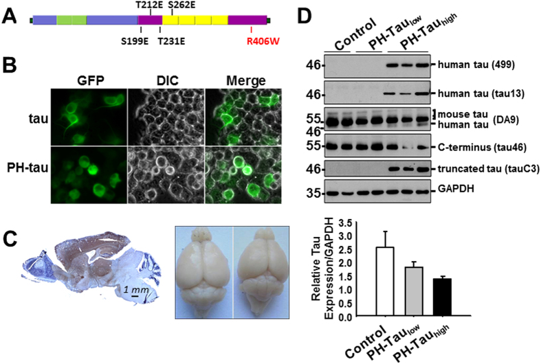
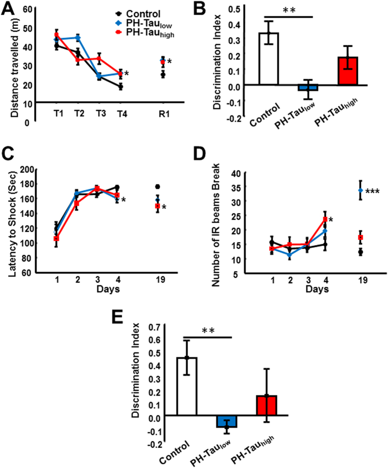
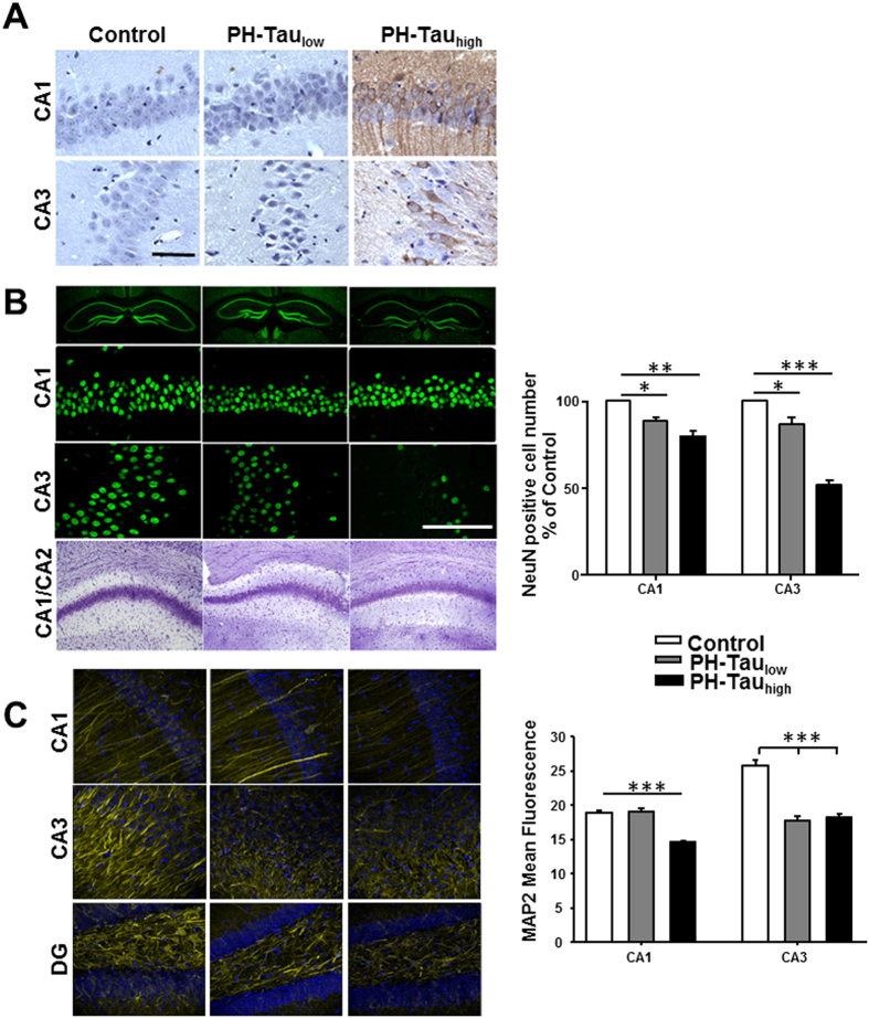
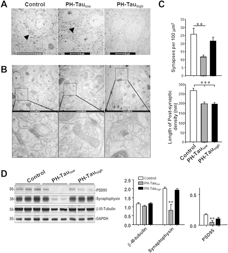
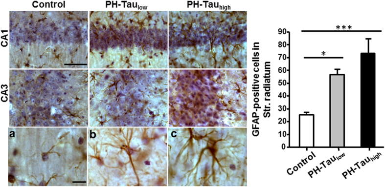
The hyperphosphorylated microtubule-associated protein tau is present in several neurodegenerative diseases, although the causal relationship remains elusive. Few mouse models used to study Alzheimer-like dementia target tau phosphorylation. We created an inducible pseudophosphorylated tau (Pathological Human Tau, PH-Tau) mouse model to study the effect of conformationally modified tau in vivo. Leaky expression resulted in two levels of PH-Tau: low basal level and higher upon induction (4% and 14% of the endogenous tau, respectively). Unexpectedly, low PH-Tau resulted in significant cognitive deficits, decrease in the number of synapses (seen by EM in the CA1 region), reduction of synaptic proteins, and localization to the nucleus. Induction of PH-Tau triggered neuronal death (60% in CA3), astrocytosis, and loss of the processes in CA1. These findings suggest, that phosphorylated tau is sufficient to induce neurodegeneration and that two different mechanisms can induce cognitive impairment depending on the levels of PH-Tau expression.


Figures

Figure 1
Figure 1. Generation of inducible transgenic mice expressing PH-Tau resulting in cognitive impairments in bigenic mice.
(A) Illustration of full-length tau with pseudophosphorylation and mutation sites marked. (B) Live imaging of N2A cells transfected with plasmids expressing tau and PH-Tau. Neurite formation is observed in the presence of WT tau, but the cells become rounded when PH-Tau is expressed. (C) Left: Sagittal paraffin section of PH-Tau expressing mouse stained with human-Tau antibody Tau-13 (brown) and counterstained with hematoxyline (blue). Right: Brains of control (left) and PH-Tauhigh (right) mice showing a loss of size when PH-Tau is expressed. (D) Tau proteins were examined in hippocampus homogenates. The graph at the right was determined by performing densitometry using the DA9 antibody.
Figure 2
Figure 2. Tau protein characterization.
(A) Hippocampal homogenates incubated at 37 °C for 1 h resulted in the appearance of a ~100 kDa species visualized with tau13 antibody in both bigenic mice. (B) Human and mouse tau proteins were measured in the sarkosyl fractionates of forebrain homogenates. C: Control, L: PH-Taulow, H: PH-Tauhigh, GAPDH as loading control. (C) Quantitation by densitometry showed sarkosyl insoluble tau increased significantly (***P < 0.001) in PH-Taulow mice vs control, and further increased (**P < 0.01) in PH-Tauhigh vs PH-Taulow mice (n = 3 per group).
Figure 3
Figure 3. Decreased cognitive behaviors observed in bigenic mice.
Bigenic mice (12-month old) were tested for behavior deficits in the (A) Morris Water Maze, (B,E) Novel Object Recognition (12-month old and 5-month old, respectively), and (C,D) Passive Avoidance tasks. Significant decreases in spatial memory and memory storage were observed. In the passive avoidance task there was an increase in anxiety. Significant differences are observed between control and PH-Taulow mice (*P < 0.5, **P < 0.01,***P < 0.001).
Figure 4
Figure 4. Neuronal loss and synaptic dysfunction in mice expressing PH-Tau.
(A) Sagittal paraffin sections of hippocampus stained with antibody tau13 recognizing human Tau. The nuclei are counterstained with hematoxylin. (B) Top three panels: Coronal slices of hippocampus stained with antibody NeuN recognizing nucleus of neurons. Bottom: Coronal sections of Nissl staining showed thinner layer of CA1 and CA2 area in bigenic mice compared to control. Scale bar = 50 μm. Right: Counts of the NeuN positive cells in both the CA1 and CA3 regions. (C) Coronal slices of hippocampus stained with MAP2 shows changes in protein levels in mice expressing PH-Tau. Right: Quantitation of the mean fluorescence observed using ImageJ analysis software. (*P < 0.05, **P < 0.01, ***P < 0.001).
Figure 5
Figure 5. Insufficient developmental dendrite spine and synapse loss in suppressed mice hippocampus.
(A) CA1 pyramidal neuron cell body layer, myelinated axons are shown in control and PH-Taulow mice (arrow head), but absent in PH-Tauhigh mice. (B) Synapses in CA1 stratum radiatum area, lower panel is the magnified images of square boxes, decreased post-synaptic density and enlarged pre-synaptic portion are shown in PH-Taulow mice. (C) Quantitation of the number of synapses in the CA1 stratum radiatum area, significant loss of synapses in PH-Taulow mice is observed. Decrease in the length of the post-synaptic density was observed in both PH-Taulow and PH-Tauhigh mice. (D) Representative Western blot of mouse hippocampus homogenate. The levels of synaptophysin, PSD95, and β-III-tubulin were measured by densitometry and normalized with the levels of GAPDH (*P < 0.05, **P < 0.01, ***P < 0.001).
Figure 6
Figure 6. Astrocytosis of bigenic mice.
Left: Coronal sections stained with GFAP antibody showed activation of astrocytes in CA1 and CA3 area of bigenic mice brains, with recognized morphological changes, including enlarged size and numerous cytoplasmic processes (b,c). Right: The numbers of CA1 stratum radiatum (str. radiatum) GFAP positive cells were analyzed by unbiased stereological estimates (*P < 0.05). Scale bar = 50 μm.

References

    1. Lee V. M., Goedert M. & Trojanowski J. Q. Neurodegenerative tauopathies. Annu Rev Neurosci 24, 1121–1159 (2001). - PubMed
    1. Iqbal K., Liu F., Gong C. X., Alonso A. D. & Grundke-Iqbal I. Mechanisms of tau-induced neurodegeneration. Acta Neuropathol 118, 53–69 (2009). - PMC - PubMed
    1. McKee A. C. et al. Chronic traumatic encephalopathy in athletes: progressive tauopathy after repetitive head injury. J Neuropathol Exp Neurol 68, 709–735 (2009). - PMC - PubMed
    1. Murray M. E. et al. Clinicopathologic and 11C-Pittsburgh compound B implications of Thal amyloid phase across the Alzheimer’s disease spectrum. Brain 138, 1370–1381 (2015). - PMC - PubMed
    1. Alonso A. D., Zaidi T., Novak M., Grundke-Iqbal I. & Iqbal K. Hyperphosphorylation induces self-assembly of tau into tangles of paired helical filaments/straight filaments. Proc Natl Acad Sci USA. 98, 6923–6928 (2001). - PMC - PubMed

Publication types

MeSH terms