. 2016 Feb 17;36(7):2131-47.
doi: 10.1523/JNEUROSCI.2921-15.2016.

Selective Disruption of Metabotropic Glutamate Receptor 5-Homer Interactions Mimics Phenotypes of Fragile X Syndrome in Mice

Affiliations

Selective Disruption of Metabotropic Glutamate Receptor 5-Homer Interactions Mimics Phenotypes of Fragile X Syndrome in Mice

Weirui Guo et al. J Neurosci. .

Abstract

Altered function of the Gq-coupled, Group 1 metabotropic glutamate receptors, specifically mGlu5, is implicated in multiple mouse models of autism and intellectual disability. mGlu5 dysfunction has been most well characterized in the fragile X syndrome mouse model, the Fmr1 knock-out (KO) mouse, where pharmacological and genetic reduction of mGlu5 reverses many phenotypes. mGlu5 is less associated with its scaffolding protein Homer in Fmr1 KO mice, and restoration of mGlu5-Homer interactions by genetic deletion of a short, dominant negative of Homer, H1a, rescues many phenotypes of Fmr1 KO mice. These results suggested that disruption of mGlu5-Homer leads to phenotypes of FXS. To test this idea, we examined mice with a knockin mutation of mGlu5 (F1128R; mGlu5(R/R)) that abrogates binding to Homer. Although FMRP levels were normal, mGlu5(R/R) mice mimicked multiple phenotypes of Fmr1 KO mice, including reduced mGlu5 association with the postsynaptic density, enhanced constitutive mGlu5 signaling to protein synthesis, deficits in agonist-induced translational control, protein synthesis-independent LTD, neocortical hyperexcitability, audiogenic seizures, and altered behaviors, including anxiety and sensorimotor gating. These results reveal new roles for the Homer scaffolds in regulation of mGlu5 function and implicate a specific molecular mechanism in a complex brain disease.

Significance statement: Abnormal function of the metabotropic, or Gq-coupled, glutamate receptor 5 (mGlu5) has been implicated in neurodevelopmental disorders, including a genetic cause of intellectual disability and autism called fragile X syndrome. In brains of a mouse model of fragile X, mGlu5 is less associated with its binding partner Homer, a scaffolding protein that regulates mGlu5 localization to synapses and its ability to activate biochemical signaling pathways. Here we show that a mouse expressing a mutant mGlu5 that cannot bind to Homer is sufficient to mimic many of the biochemical, neurophysiological, and behavioral symptoms observed in the fragile X mouse. This work provides strong evidence that Homer-mGlu5 binding contributes to symptoms associated with neurodevelopmental disorders.

Keywords: Homer; LTD; UP states; fragile X syndrome; mGluR5; protein synthesis.


Figures

Figure 1.
Figure 1.
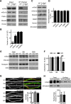
Selective disruption of mGlu5-Homer reduces PSD association, but not dendritic or surface expression of mGlu5. A, Representative Western blots from coimmunoprecipitations using a pan long-Homer antibody of cortical homogenates of WT and mGlu5R/R littermates. B, Quantified group data of the levels of mGlu5, mGlu1α, PIKE-L, and Shank3 that coimmunoprecipitate with Homer from mGlu5R/R cortical homogenates. Values are expressed as percentage (mean ± SEM) of WT littermates (n = 3–5 mice/genotype; one-sample t test with Bonferroni correction for multiple comparisons). C, D, Representative Western blots and quantified group data demonstrating that total levels of mGlu5, mGlu1α, Shank3, FMRP, and long-Homers are normal in cortical homogenates of mGlu5R/R mice. Values are normalized to β3-tubulin (n = 4–6 mice/genotype). E, F, Representative Western blots and quantified group data of mGlu5 levels in total, synaptoneurosome (SN) and PSD fractions of mGlu5F/R (Het) or mGlu5R/R littermate forebrains. Data are expressed as a percentage of WT levels. mGlu5 levels are normal in total and SN fractions but reduced in the PSD fractions of mGlu5R/R (n = 3 mice/genotype; two-way ANOVA, Sidak's post hoc). G, Representative immunofluorescence images of mGlu5 and β3-tubulin in dendrites of dissociated cortical neuron cultures from WT, Het, or mGlu5R/R mice. Quantified group data reveal no effect of the F>R mutation on dendritic mGlu5 levels (n = 45 dendrites [15–30 cells]/genotype from 2 independent cultures). H, Representative blots and quantified group data of total and surface (biotinylated) mGlu5 and β3 tubulin in acute hippocampal slices prepared from WT, Het, or mGlu5R/R mice (n = 4 mice/genotype). *p < 0.05. **p < 0.01.
Figure 2.
Figure 2.
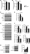
Selective disruption of mGlu5-Homer leads to constitutive mGlu5-driven signaling to ERK, translation initiation factors, and protein synthesis rates. A, Acute hippocampal slices from mGlu5R/R mice display elevated protein synthesis rates compared with WT littermates as measured by incorporation of 35S Met/Cys into total protein. Pretreatment with MPEP (10 μm), an mGlu5 NAM, equalizes protein synthesis rates between WT and mGlu5R/R slices (n = 14 slices/condition from 7 mice/genotype). B, In a separate set of experiments, inhibition of the MEK with U0126 (10 μm) rescues protein synthesis rates in mGlu5R/R slices to WT levels (n = 6 slices/condition from 3 mice/genotype). C, D, Representative Western blots and quantified group data reveal enhanced level translation initiation complexes in mGlu5R/R hippocampal slices, as measured by coimmunoprecipitation of eIF4E with eIF4G that are rescued by pretreatment with MPEP (n = 4 mice/genotype). E, F, Representative Western blots and quantified group data demonstrate elevated phosphorylation of translation initiation factors downstream of ERK (Mnk1; 4EBP, S65; eIF4E) in acute hippocampal slices of mGlu5R/R mice that are rescued by pretreatment with MPEP. P-ERK levels are unchanged in mGlu5R/R slices and unaffected by MPEP (n = 4 mice/genotype). G, Western blots of fresh hippocampal lysates from WT and mGlu5R/R littermates demonstrate elevated P-4EBP (S65) and P-eIF4E, but not P-ERK in vivo (n = 5–7 mice per genotype; one-sample t test). A–F, Two-way ANOVA; Sidak's post hoc tests. *p < 0.05. **p < 0.01. ***p < 0.001.
Figure 3.
Figure 3.
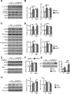
Selective disruption of mGlu5-Homer leads to constitutive mGlu5-driven signaling of the mTORC1 pathway. A, B, Representative Western blots and quantified group data demonstrate elevated mGlu5-dependent activity of the mTORC1 signaling pathway to translation factors in lysates of acute hippocampal slices of mGlu5R/R mice as measured by phosphorylation of mTORC1, 4EBP (T37/46), and S6K. Pretreatment with MPEP reduces P-mTORC1, P-4EBP(T37/46), and P-S6K in mGlu5R/R slices to WT levels n = 4 mice/genotype; mTORC1; paired t test; 4EBP and S6K; two-way ANOVA; Sidak's post hoc tests). *p < 0.05. **p < 0.01.
Figure 4.
Figure 4.
Selective disruption of mGlu5-Homer alters agonist-induced signaling to translation initiation and elongation. A, B, Representative Western blots and quantitative group data demonstrate enhanced basal activity of the mTORC1 pathway in acute hippocampal slices from mGlu5R/R compared with WT mice, as measured by P-mTORC1 and P-4EBP (T37/46). Treatment with the Group1 mGluR agonist, DHPG (D; 100 μm; 5 min) increases P-mTORC1 and P-4EBP levels in WT slices, but not mGlu5R/R slices (n = 4–6 mice/genotype). C, D, Enhanced basal activity of translation factors downstream of ERK is observed using Western blots of acute hippocampal slice lysates from mGlu5R/R compared with WT mice, as measured by P-Mnk1, P-eIF4E, and P-4EBP (S65). Although DHPG treatment (100 μm; 5 min) increases P-ERK in mGlu5R/R slices, it fails to increase P-Mnk1, P-eIF4E, and P-4EBP (S65) levels (n = 4–10 mice/genotype). E, F, Enhanced levels of eIF4E coimmunoprecipitation with eIF4G (eIF4E/4G) from lysates of acute hippocampal slices of mGlu5R/R mice, compared with WT mice. Treatment with DHPG increases 4E/4G levels in WT slices, but not in mGlu5R/R slices (n = 6 mice/genotype). G, DHPG treatment of acute hippocampal slices from mGlu5R/R mice results in greater P-EF2, compared with slices from WT mice (n = 15 mice/genotype). H, DHPG treatment results in rapid increases in MAP1b and APP levels in acute hippocampal slices from WT, but not mGlu5R/R mice (n = 9–14 mice/genotype). A–H, Two-way ANOVA; Sidak's post hoc tests. *p < 0.05. **p < 0.01. ***p < 0.001.
Figure 5.
Figure 5.
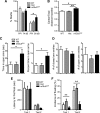
Disruption of mGlu5-Homer prevents mGlu1/5 agonist-induced, translation-dependent LTD. A, Brief DHPG (100 μm; 5 min) induces LTD of synaptic transmission in WT hippocampal slices (n = 14) that is reduced in slices of mGlu5R/R mice (n = 11; p < 0.05; t test). Plotted are group averages of fEPSP slope (mean ± SEM) normalized to pre-DHPG baseline as a function of time. Inset, Average of 10 fEPSPs taken during the baseline period (1) and 55–60 min after DHPG treatment (2). Calibration: 0.5 mV, 5 ms. B, C, Pretreatment with the protein synthesis inhibitor anisomycin (25 μm) reduced DHPG-induced LTD in WT mice (B; Veh; n = 11 slices; anisomycin; n = 9; p < 0.001; two-way ANOVA; Sidak's post hoc tests) but has no effect on LTD in mGlu5R/R mice (C; Veh; n = 11; anisomycin; n = 10; not significant). D, Low-frequency, 1 Hz, presynaptic stimulation of Schaffer collaterals induces robust LTD in slices from WT (n = 9 slices), but not mGlu5R/R mice (n = 14; p < 0.01; t test). E, There were no genotypic differences in the effect of mGlu5 blockade (MPEP; 10 μm; 1 h) on fEPSP slopes (WT; n = 6; mGlu5R/R; n = 7).
Figure 6.
Figure 6.
Disruption of mGlu5-Homer interactions is sufficient to mimic neocortical hyperexcitability and audiogenic seizures observed in Fmr1 KO mice. A, B, The mGlu5F>R mutation dose-dependently increases spontaneous UP state duration. A, Representative extracellular multiunit recordings of spontaneous, persistent activity or UP states from layer 4 of acute somatosensory neocortical slices from each genotype. Calibration: 50 μV, 1 s. B, Group data of average UP state duration in WT, Het, and mGlu5R/R littermates (WT, n = 29 slices; Het, n = 14; mGlu5R/R, n = 18; ANOVA; Tukey post hoc). C, The average amplitude (normalized to baseline) of UP states is increased in mGlu5R/R, but UP states occur less frequently (ANOVA; Tukey post hoc). D, The mGlu5F>R mutation increases the incidence and severity of audiogenic seizures as assessed by seizures score and by percentage mice that seized (described in Materials and Methods) (N = 27, N = 28, and N = 21 for WT, Het, and mGlu5R/R littermates, respectively). Score: ANOVA; Tukey multiple-comparison test; percentage seized: Fisher's exact test. *p < 0.05. **p < 0.01. ***p < 0.001.
Figure 7.
Figure 7.
mGlu5R/R mice have impaired sensorimotor gating, reduced nonsocial anxiety, and an antidepressant phenotype. A, PPI of startle amplitude, measured by the percent inhibition of the startle to 110 dB tone when preceded by a 90 dB prepulse, is reduced in mGlu5R/R mice compared with WT controls (N = 20, N = 16, and N = 13 for WT, Het, and mGlu5R/R littermates, respectively). Prepulse effect: F(1,43) = 117.48, p < 0.0001; genotype × prepulse: F(2,43) = 4.52, p = 0.02. Startle amplitude at 74 or 90 dB was not different between genotypes. Data are mean ± SEM percent inhibition of the 110 dB startle amplitude by the 74 dB and the 90 dB prepulse intensities. *p < 0.05 versus WT (LSD post hoc tests). **p < 0.01. B, Open field activity, measured as a ratio of the distance traveled in the center to total distance traveled in an open arena, was increased in mGlu5R/R mice compared with WT or Het mice (F(2,82) = 5.376, p = 0.006; Dunnet's post hoc tests) (N = 19, N = 44, and N = 22 for WT, Het, and mGlu5R/R littermates, respectively). C, mGlu5R/R mice spent more time in the open arms as measured by seconds or percentage of total time in the maze (N = 15, N = 22, and N = 17 for WT, Het, and mGlu5R/R littermates, respectively). Sex effect: F(1,48) = 6.14, p = 0.02; genotype effect: F(2,48) = 3.45, p = 0.04; interaction: p = 0.21. D, Number of entries into closed or open arms of an elevated plus maze were not different between genotypes. E, The latency to first float in the Porsolt Swim Test was not different among genotypes. F, The percentage of incidence of floating as scored in the first and second swim sessions was reduced in mGlu5R/R mice (N = 15, N = 22, and N = 17 for WT, HET, and mGlu5R/R littermates, respectively). Genotype effect: F(2,49) = 5.52, p = 0.007; no interactions with the genotype factor: p > 0.20.
Figure 8.
Figure 8.
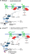
Working model of the role of Homer binding to mGlu5 in signaling pathways to translation initiation and elongation in WT (A) and mGlu5R/R (B) mice as considered in the Discussion.

Similar articles

Cited by

References

    1. Agmon A, Connors BW. Thalamocortical responses of mouse somatosensory (barrel) cortex in vitro. Neuroscience. 1991;41:365–379. doi: 10.1016/0306-4522(91)90333-J. - DOI - PubMed
    1. Ango F, Pin JP, Tu JC, Xiao B, Worley PF, Bockaert J, Fagni L. Dendritic and axonal targeting of type 5 metabotropic glutamate receptor is regulated by homer1 proteins and neuronal excitation. J Neurosci. 2000;20:8710–8716. - PMC - PubMed
    1. Ango F, Prézeau L, Muller T, Tu JC, Xiao B, Worley PF, Pin JP, Bockaert J, Fagni L. Agonist-independent activation of metabotropic glutamate receptors by the intracellular protein Homer. Nature. 2001;411:962–965. doi: 10.1038/35082096. - DOI - PubMed
    1. Ango F, Robbe D, Tu JC, Xiao B, Worley PF, Pin JP, Bockaert J, Fagni L. Homer-dependent cell surface expression of metabotropic glutamate receptor type 5 in neurons. Mol Cell Neurosci. 2002;20:323–329. doi: 10.1006/mcne.2002.1100. - DOI - PubMed
    1. Auerbach BD, Osterweil EK, Bear MF. Mutations causing syndromic autism define an axis of synaptic pathophysiology. Nature. 2011;480:63–68. doi: 10.1038/nature10658. - DOI - PMC - PubMed

Publication types

Substances