Review
. 2016 Mar;31(2):95-107.
doi: 10.1152/physiol.00034.2015.

Neural Control of Walking in People with Parkinsonism

Affiliations
Review

Neural Control of Walking in People with Parkinsonism

D S Peterson et al. Physiology (Bethesda). 2016 Mar.

Abstract

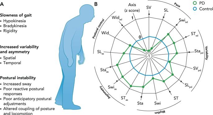
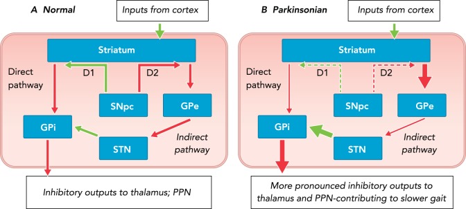
People with Parkinson's disease exhibit debilitating gait impairments, including gait slowness, increased step variability, and poor postural control. A widespread supraspinal locomotor network including the cortex, cerebellum, basal ganglia, and brain stem contributes to the control of human locomotion, and altered activity of these structures underlies gait dysfunction due to Parkinson's disease.


Conflict of interest statement

F. B. Horak and OHSU have an equity/interest in APDM, a company that may have a commercial interest in the results of the study. This potential conflict of interest has been reviewed and managed by the Research & Development Committee at the Portland VA Medical Center and OHSU. No other authors declare any conflict of interest.

Figures

FIGURE 1.
FIGURE 1.
Continuous gait disturbances A: continuous gait disturbances in people with PD. B: people with PD exhibit dysfunction in gait speed (pace/rhythm), variability and asymmetry, and postural control. This is depicted by a satellite plot showing deviations from control subjects (dotted line). SV, step velocity; SL, step length; Swi, swing time; ST, Step time; Sta, Stance time; Wid, Step width; sd, standard deviation (gait variability); as, asymmetry. *Differences between the control and PD group. Figure reproduced from Ref. with permission from Movement Disorders.
FIGURE 2.
FIGURE 2.
Rate model of basal ganglia dysfunction Rate model of basal ganglia dysfunction in normal (A) and parkinsonian (B) states. Over activity of the indirect and underactivity of the direct pathways result in more inhibitory output from the basal ganglia output structures (GPi) to the thalamus and the brain stem, and ultimately reduced amplitude of movements, including gait. Green arrows represent excitatory and red arrows inhibitory projections. Arrow thickness represents the relative firing rate of projections, and dashed arrows indicate the relative reduction of the SNpc D1 and D2 dopaminergic projections to the striatum. SNpc, substantia nigra pars reticulate; GPe, globus pallidus external segment; GPi, globas pallidus internal segment; STN, subthalamic nucleus.
FIGURE 4.
FIGURE 4.
The dysfunction in speed, variability and asymmetry, and postural control in PD are differently affected by levodopa Rightward (positive) values represent a positive impact of levodopa, whereas leftward (negative) values represent a negative impact of levodopa. Gait speed and size of limb movements are improved by levodopa (top). Postural sway can be worsened by levodopa (bottom). Although few variability or asymmetry variables were available in this particular analysis, asymmetry of arm swing and temporal coordination, such as double support time, were not consistently improved by levodopa. A value larger than 0.20 represents small, 0.50 moderate, and 0.80 large responsiveness. *Differences between the control and PD group. Figure reproduced from Ref. with permission from Movement Disorders.
FIGURE 3.
FIGURE 3.
Framework for supraspinal control of locomotion in people with PD Alterations in activity of the basal ganglia (1) and brain stem (4) contribute to gait slowness and increased postural instability, respectively, and increased cerebellar activity may partially compensate for these alterations (2). Increased volitional control (i.e., cortico-spinal) and reduced automatic control (3) may contribute to increased gait variability and asymmetry. See text box above for more information. PPN, pedunculopontine nucleus; MLR, mesencelphalic locomotor region; PMRF, pontomedulary reticular formation; SMA, supplementary motor area.

References

    1. Adkin AL, Bloem BR, Allum JH. Trunk sway measurements during stance and gait tasks in Parkinson's disease. Gait Posture 22: 240–249, 2005. - PubMed
    1. Albin RL, Young AB, Penney JB. The functional anatomy of basal ganglia disorders. Trends Neurosci 12: 366–375, 1989. - PubMed
    1. Baker R. Measuring Walking: A Handbook of Clinical Gait Analysis. Hoboken, NJ: Wiley, 2013.
    1. Baltadjieva R, Giladi N, Gruendlinger L, Peretz C, Hausdorff JM. Marked alterations in the gait timing and rhythmicity of patients with de novo Parkinson's disease. Eur J Neurosci 24: 1815–1820, 2006. - PubMed
    1. Blin O, Ferrandez AM, Pailhous J, Serratrice G. Dopa-sensitive and dopa-resistant gait parameters in Parkinson's disease. J Neurol Sci 103: 51–54, 1991. - PubMed

Publication types

LinkOut - more resources