

doi: 10.1128/JVI.03223-15.
Print 2016 May.
Structural Basis for Norovirus Inhibition by Human Milk Oligosaccharides
Affiliations
- PMID: 26889023
- PMCID: PMC4836343
- DOI: 10.1128/JVI.03223-15
Item in Clipboard
Structural Basis for Norovirus Inhibition by Human Milk Oligosaccharides
J Virol.
.
Abstract
Histo-blood group antigens (HBGAs) are important binding factors for norovirus infections. We show that two human milk oligosaccharides, 2'-fucosyllactose (2'FL) and 3-fucosyllactose (3FL), could block norovirus from binding to surrogate HBGA samples. We found that 2'FL and 3FL bound at the equivalent HBGA pockets on the norovirus capsid using X-ray crystallography. Our data revealed that 2'FL and 3FL structurally mimic HBGAs. These results suggest that 2'FL and 3FL might act as naturally occurring decoys in humans.
Copyright © 2016, American Society for Microbiology. All Rights Reserved.
Figures
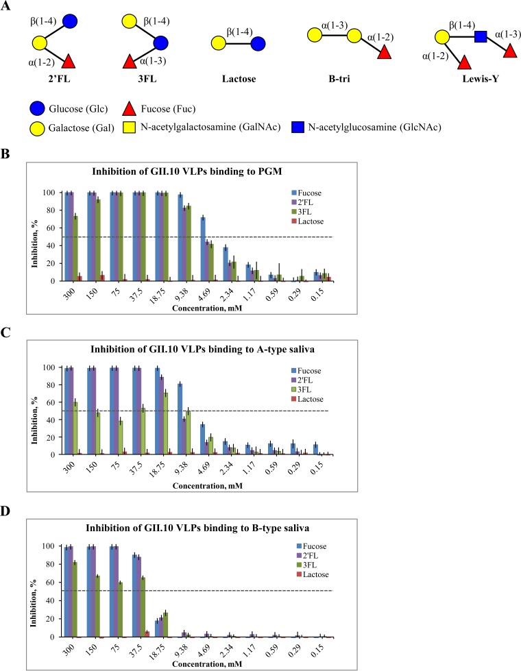
HMOs and blocking of binding of norovirus VLPs to HBGAs. (A) Schematic representation of HMOs and HBGAs. The 2′FL is an α-l -fucose-(1-2)-β-d -galactose-(1-4)-α-d -glucose; 3FL is an α-l -fucose-(1-3)-[β-d -galactose-(1-4)]-β-d -glucose; lactose is a β-d -galactose-(1-4)-α-d -glucose; B-trisaccharide (B-tri) is an α-l -fucose-(1-2)-α-d -galactose-(3-)-N-acetyl-α-d -galactosamine; and Lewis Y-tetrasaccharide (Lewis-Y) is an α-l -fucose-(1-2)-β-d -galactose-(1-4)-N-acetyl-β-d -glucosamine-(3-1)-α-l -fucose. (B) Inhibition of binding of GII.10 VLPs to PGM. (C) Inhibition of binding of GII.10 VLPs to A-type saliva. (D) Inhibition of binding of GII.10 VLPs to B-type saliva. All experiments were performed in triplicate (standard deviations are shown). The half-maximal inhibitory concentration (IC50) cutoff is shown as a dashed line.
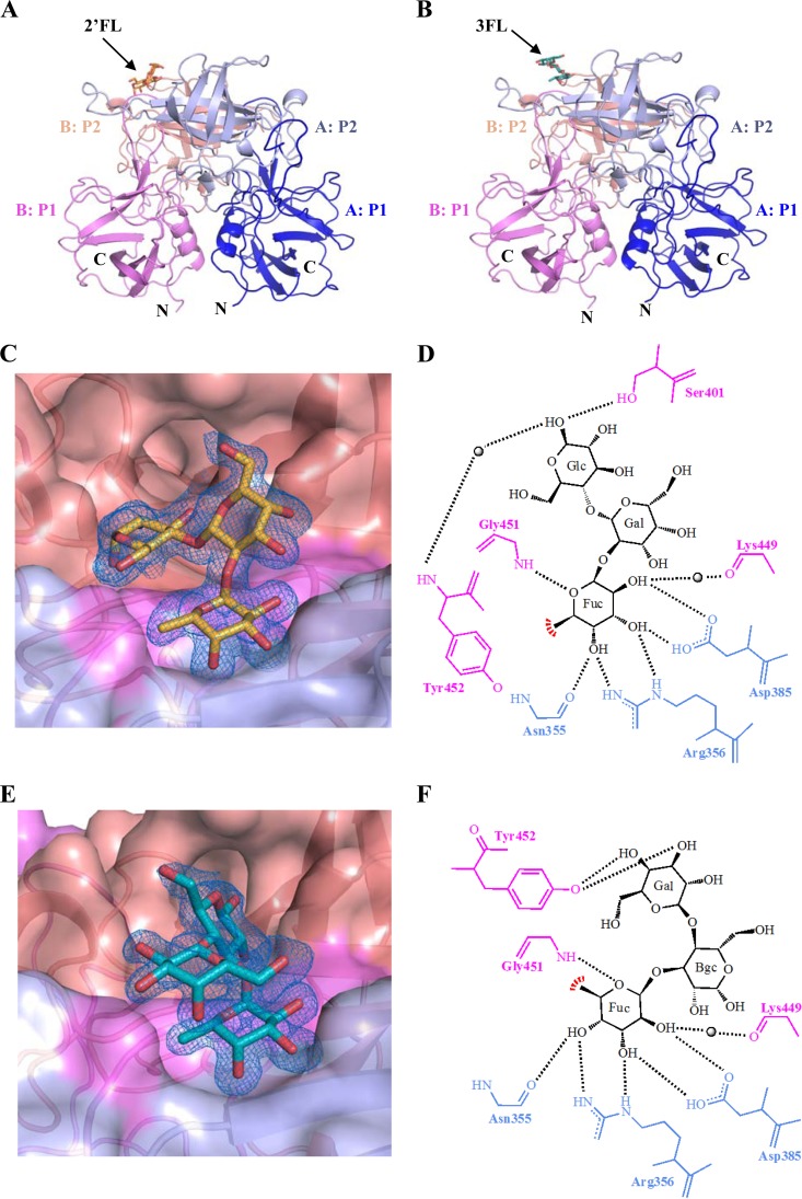
GII.10 P dimer binding interaction with HMOs. (A) The X-ray crystal structure of the GII.10 P domain dimer and 2′FL complex determined to 1.55 Å resolution and colored according to monomers (chain A and chain B) and P1 and P2 subdomains, i.e., chain A P1 (blue), chain A P2 (light blue), chain B P1 (violet), and chain B P2 (salmon). (B) The X-ray crystal structure of the GII.10 P domain dimer and 3FL complex determined to 1.35-Å resolution and colored as described for panel A. (C) A closeup surface and ribbon representation of the GII.10 and 2′FL (orange sticks) complex structure, showing a simulated annealing difference omit map (blue mesh) of 2′FL contoured at 2.0 σ. (D) The GII.10 P domain binding interaction with 2′FL showing α-fucose (Fuc), α-galactose (Gal), and β-glucose (Glc). The black lines represent the hydrogen bonds, the red line represents the hydrophobic interaction with the aromatic ring of Tyr452, and the black spheres represent water. Hydrogen bond distances were less than 3.3 Å. (E) A closeup surface and ribbon representation of the GII.10 and 3FL (deep teal sticks) complex structure, showing a simulated annealing difference omit map of 3FL contoured at 2.0 σ. (F) GII.10 P domain binding interaction with 3FL showing α-fucose (Fuc), β-galactose (Bgc), and β-glucose (Glc).
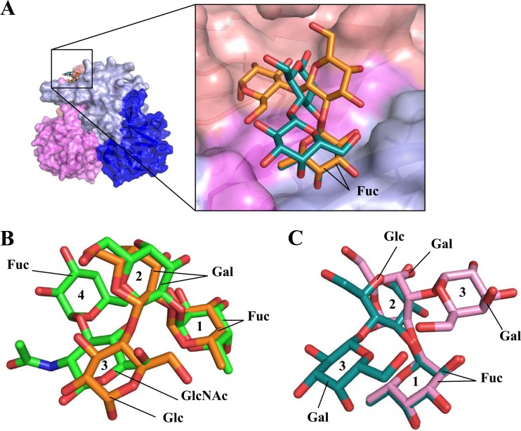
HMO and HBGA binding to GII.10 norovirus. (A) Surface representation of the GII.10 P domain in complex with 2′FL (orange sticks) and 3FL (deep-teal sticks). The fucose units of the HMO were positioned similarly on the P domain, whereas the other saccharide units were differently oriented. (B) Superposition of 2′FL and Lewis-Y tetrasaccharide (green sticks) showed that the 2′FL saccharide units essentially mimicked the orientations of first three saccharides of Lewis-Y. The saccharides are numbered (1, 2, 3, and 4) for viewing. (C) Superposition of 3FL and B-trisaccharide (pink sticks) indicated that the fucose units were similarly positioned, whereas the other saccharide units were orientated differently.
References
-
- Huang P, Farkas T, Marionneau S, Zhong W, Ruvoen-Clouet N, Morrow AL, Altaye M, Pickering LK, Newburg DS, LePendu J, Jiang X. 2003. Noroviruses bind to human ABO, Lewis, and secretor histo-blood group antigens: identification of 4 distinct strain-specific patterns. J Infect Dis 188:19–31. doi:10.1086/375742. - DOI - PubMed
-
- Huang P, Farkas T, Zhong W, Tan M, Thornton S, Morrow AL, Jiang X. 2005. Norovirus and histo-blood group antigens: demonstration of a wide spectrum of strain specificities and classification of two major binding groups among multiple binding patterns. J Virol 79:6714–6722. doi:10.1128/JVI.79.11.6714-6722.2005. - DOI - PMC - PubMed
-
- Harrington PR, Lindesmith L, Yount B, Moe CL, Baric RS. 2002. Binding of Norwalk virus-like particles to ABH histo-blood group antigens is blocked by antisera from infected human volunteers or experimentally vaccinated mice. J Virol 76:12335–12343. doi:10.1128/JVI.76.23.12335-12343.2002. - DOI - PMC - PubMed
Publication types
MeSH terms
Substances
LinkOut - more resources
Full Text Sources
Other Literature Sources
Medical
