. 2016 Apr 14;90(9):4481-4493.
doi: 10.1128/JVI.03206-15. Print 2016 May.

Range of CD4-Bound Conformations of HIV-1 gp120, as Defined Using Conditional CD4-Induced Antibodies

Affiliations

Range of CD4-Bound Conformations of HIV-1 gp120, as Defined Using Conditional CD4-Induced Antibodies

Gilad Kaplan et al. J Virol. .

Abstract

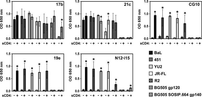
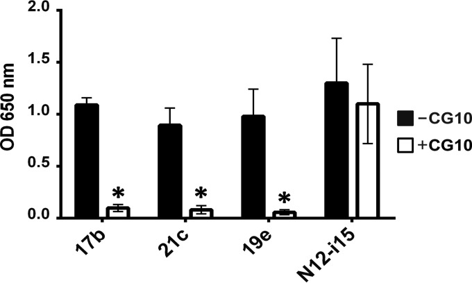
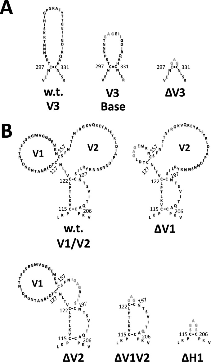
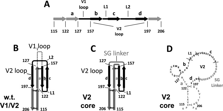
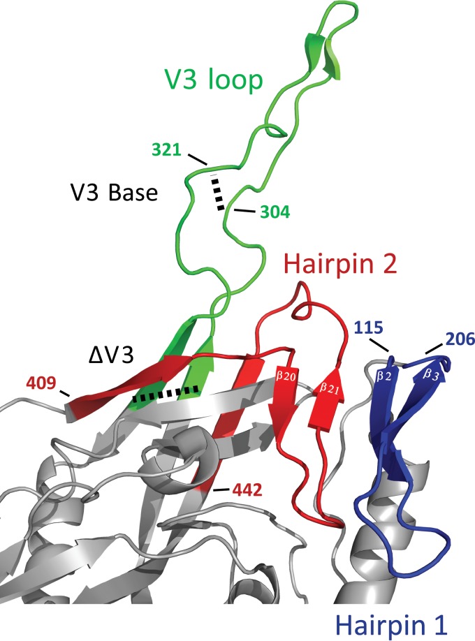
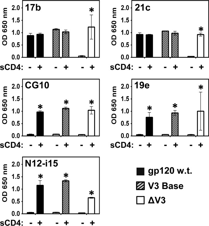
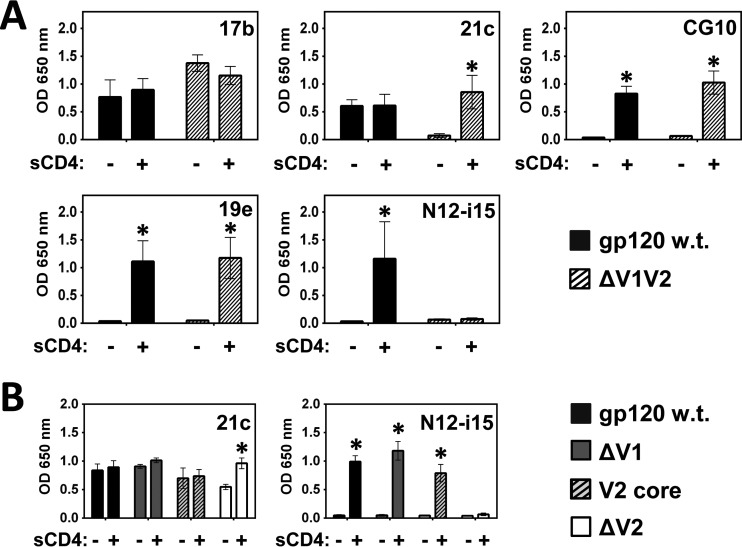
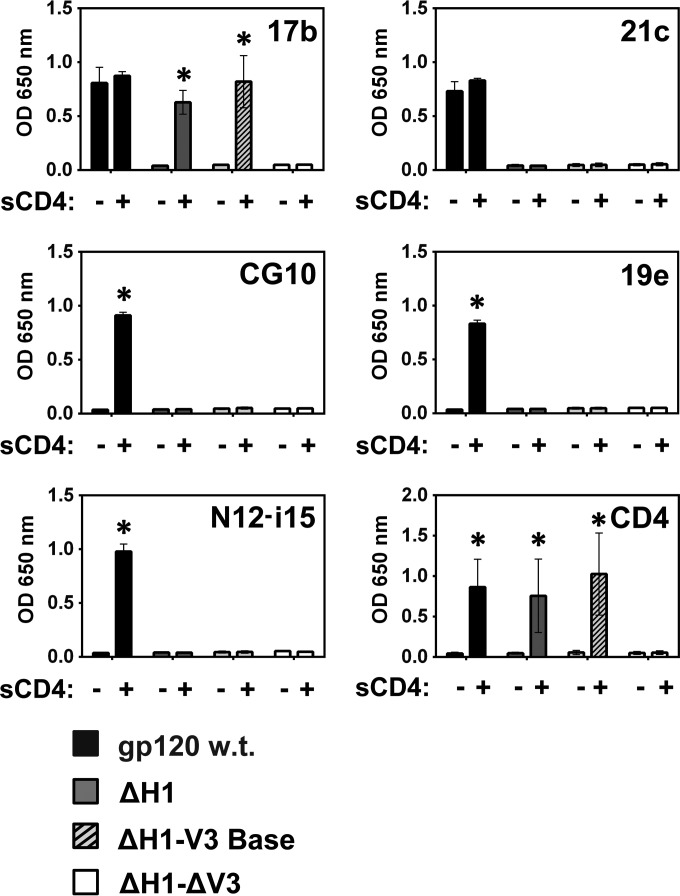
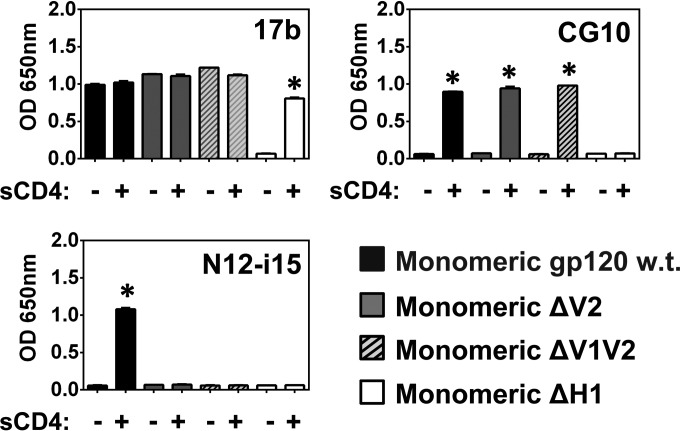
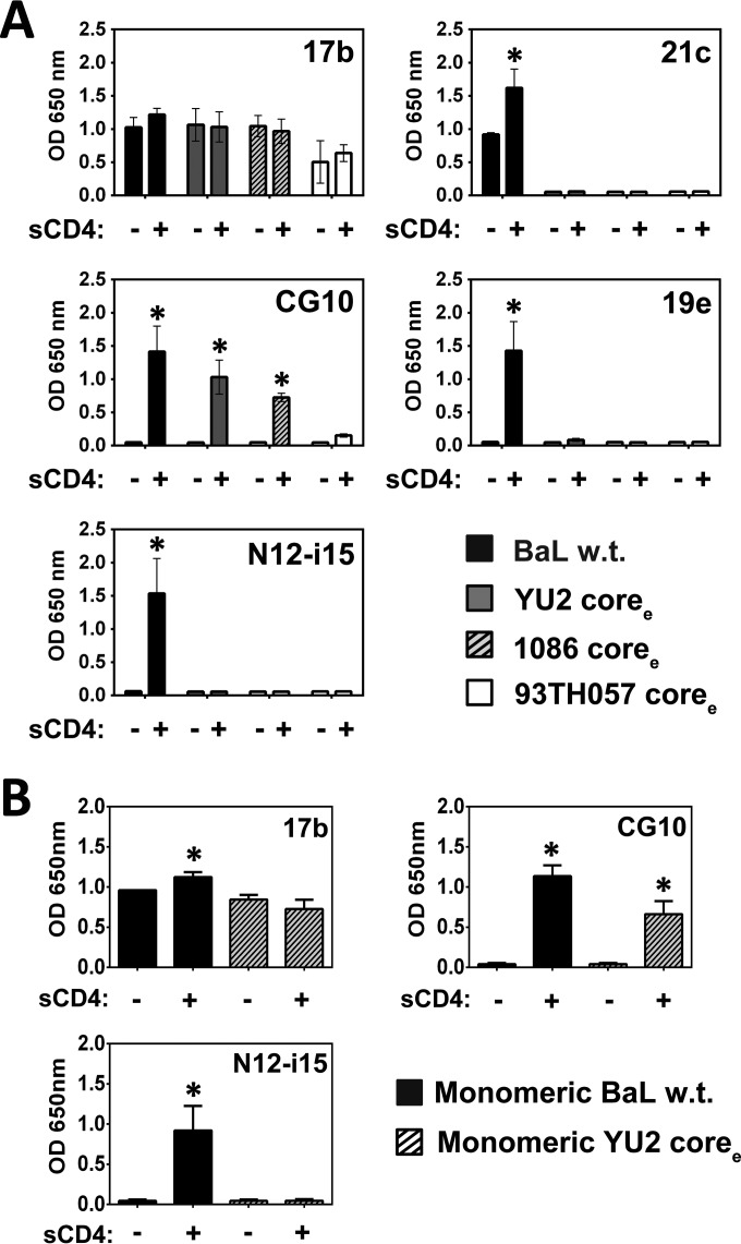
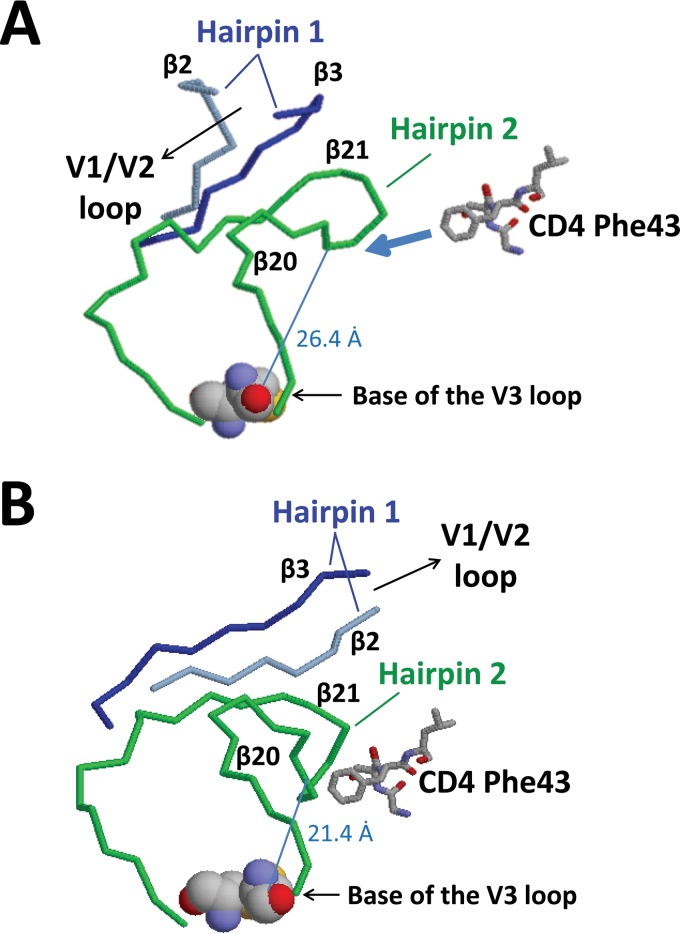
The HIV envelope binds cellular CD4 and undergoes a range of conformational changes that lead to membrane fusion and delivery of the viral nucleocapsid into the cellular cytoplasm. This binding to CD4 reveals cryptic and highly conserved epitopes, the molecular nature of which is still not fully understood. The atomic structures of CD4 complexed with gp120 core molecules (a form of gp120 in which the V1, V2, and V3 loops and N and C termini have been truncated) have indicated that a hallmark feature of the CD4-bound conformation is the bridging sheet minidomain. Variations in the orientation of the bridging sheet hairpins have been revealed when CD4-liganded gp120 was compared to CD4-unliganded trimeric envelope structures. Hence, there appears to be a number of conformational transitions possible in HIV-1 monomeric gp120 that are affected by CD4 binding. The spectrum of CD4-bound conformations has been interrogated in this study by using a well-characterized panel of conditional, CD4-induced (CD4i) monoclonal antibodies (MAbs) that bind HIV-1 gp120 and its mutations under various conditions. Two distinct CD4i epitopes of the outer domain were studied: the first comprises the bridging sheet, while the second contains elements of the V2 loop. Furthermore, we show that the unliganded extended monomeric core of gp120 (coree) assumes an intermediate CD4i conformation in solution that further undergoes detectable rearrangements upon association with CD4. These discoveries impact both accepted paradigms concerning gp120 structure and the field of HIV immunogen design.

Importance: Elucidation of the conformational transitions that the HIV-1 envelope protein undergoes during the course of entry into CD4(+)cells is fundamental to our understanding of HIV biology. The binding of CD4 triggers a range of gp120 structural rearrangements that could present targets for future drug design and development of preventive vaccines. Here we have systematically interrogated and scrutinized these conformational transitions using a panel of antibody probes that share a specific preference for the CD4i conformations. These have been employed to study a collection of gp120 mutations and truncations. Through these analyses, we propose 4 distinct sequential steps in CD4i transitions of gp120 conformations, each defined by antibody specificities and structural requirements of the HIV envelope monomer. As a result, we not only provide new insights into this dynamic process but also define probes to further investigate HIV infection.


Figures

FIG 1
FIG 1
Stringent CD4i MAbs do not bind gp120 in the absence of sCD4. An ELISA was used to analyze CD4i MAb binding to gp120s from different strains and to a gp140 trimer. MAbs 17b and 21c show binding to gp120 independent of sCD4 (relaxed binding). MAbs CG10, 19e, and N12-i15 show no binding to gp120 in the absence of sCD4 (stringent binding). Only MAb 17b was cross-reactive with the clade A BG505 SOSIP trimer and its monomer, showing relaxed binding to the monomer and stringent binding to the trimer. Statistically significant differences (P < 0.05) between the “−sCD4” and “+sCD4” columns are marked with an asterisk. OD, optical density.
FIG 2
FIG 2
MAb N12-i15 defines a CD4i region upon gp120 distinct from the bridging sheet. Competition ELISAs were carried out in order to discern whether all the detected CD4i epitopes cluster to one area on gp120 or not. Binding of CD4i MAbs to gp120BaL-sCD4 was measured in either the absence or the presence of prebound MAb CG10. MAbs 17b, 21c, and 19e compete strongly with MAb CG10. MAb N12-i15 defines a CD4i epitope sufficiently separated from the bridging sheet so as to show no competition with MAb CG10. Statistically significant differences (P < 0.05) between the “−CG10” and “+CG10” columns are marked with an asterisk.
FIG 3
FIG 3
Schematic of the wt and modified forms of the gp120 V1/V2 and V3 loops. The sequences of wt and modified V3 (A) and V1/V2 (B) loops are shown with disulfide bonds depicted as black lines. Amino acid numbering corresponds to BaL gp120. Landmark cysteine residues are numbered for convenience. wt sequences are shown in black, and altered sequences are shown in gray. (A) V3 loop mutants, including (i) the wt V3 loop, (ii) V3 base (gp120 with a partially truncated V3 loop), and (iii) ΔV3 (gp120 with a truncated V3 loop). (B) V1/V2 loop mutants, including (i) wt V1/V2 loops, (ii) ΔV1 (gp120 with a truncated V1 loop), (iii) ΔV2 (gp120 with a truncated V2 loop), (iv) ΔV1V2 (gp120 with truncated V1/V2 loops), and (v) ΔH1 (gp120 with truncated V1/V2 loops and stem).
FIG 4
FIG 4
Comparison of wt V1/V2 loops and the V2 core mutant. The crystal structure of the V1/V2 loops from gp120 of strain ZM109 reveals that the V2 loop assumes a Greek key fold (PDB accession number 3U2S) (78). The Greek key fold is comprised of four antiparallel β-strands (designated a to d) connected by the V1 loop (dashed) and two small loops (designated L1 and L2), which are part of the V2 loop (black). Based upon this crystal structure, the V2 core mutant was constructed by using gp120 from the BaL strain (the same strain upon which all of the envelope mutants in this study are based). The V2 core mutant was constructed by removing most of the V1 loop and replacing the parts of the V2 loop that do not participate in the Greek fold (loop L2) with a Ser-Gly linker. Numbering refers to adjacent cysteine residues and is based on the BaL strain, with disulfide bonds shown as black bars. Shown are the V1 loop (dashed), the V2 loop (black), and disulfide bonds (black bars between cysteine residues). (A) Linearized schematic of the wt V1/V2 loop fold. (B) Schematic of the fold of the wt V1/V2 loops. (C) Schematic of the fold of the V2 core mutant. (D) Sequence of the V2 core mutant.
FIG 5
FIG 5
Orientation of the bridging sheet hairpins in relation to the V3 loop. The hairpins that constitute the bridging sheet (hairpins 1 and 2, shown in blue and red, respectively) and the V3 loop (green) are shown in the CD4-liganded conformation (PDB accession number 2B4C). Note that hairpin 1 (β2-β3) is the base for the V1/V2 loops (the V1/V2 loops are missing in the crystal structure) and that the V3 loop protrudes between the proximal and distal aspects of hairpin 2 (β20-β21). The locations of the different V3 loop truncations (V3 base and ΔV3) are marked with dashed lines. For the sake of consistency with our mutants, the numbering is based on BaL gp120 (see Materials and Methods).
FIG 6
FIG 6
V3 loop modifications affect binding by relaxed CD4i MAbs. A binding ELISA was carried out to ascertain the effects of V3 loop truncations on binding by the CD4i MAb panel to gp120BaL. Full truncation of the V3 loop (ΔV3) results in stringent binding by MAbs 17b and 21c, an effect not seen for the V3 base mutant. Binding by the stringent CD4i MAbs CG10, 19e, and N12-i15 was not affected by V3 loop truncations. Statistically significant differences (P < 0.05) between the “−sCD4” and “+sCD4” columns are marked with an asterisk.
FIG 7
FIG 7
CD4i MAbs 21c and N12-i15 are sensitive to V1/V2 loop modifications. A binding ELISA was performed to test whether binding by the CD4i MAbs to gp120BaL is affected by the complete truncation of the V1/V2 loops (ΔV1V2) (A) or selective modification of the V1 or V2 loop (B). (A) MAb 21c becomes stringent and MAb N12-i15 loses all binding when the V1/V2 loops are fully truncated. (B) Selective modifications within the V1/V2 loops indicate that the V1 loop is not necessary for binding by MAbs 21c and N12-i15, but the core of the V2 loop (the V2 loop residues which fold into a Greek key conformation) (Fig. 4) is required. Statistically significant differences (P < 0.05) between the “−sCD4” and “+sCD4” columns are marked with an asterisk.
FIG 8
FIG 8
Sequence alignment of the gp120 V2 loop from different HIV strains. The gp120 V2 loop sequences from the different HIV strains tested against MAb N12-i15 for binding were aligned by using the Clustal Omega program (http://www.ebi.ac.uk/Tools/msa/clustalo/) and compared. The numbering system used is based on the BaL strain for consistency. Strand c of the Greek key fold based on the crystal structure of V1/V2 loops from the ZM109 strain (78) is shown. As MAb N12-i15 was shown to bind to elements within the Greek key fold of the V2 loop, the four residues different in the R2 V2 loop located within the Greek key fold are highlighted in dark gray. The V2 loop of the BG505 strain (both the monomer and the BG505 SOSIP trimer) is shown for comparison only, as MAb N12-i15 does not bind this strain (Fig. 1).
FIG 9
FIG 9
MAb 17b binds gp120 in the absence of bridging sheet hairpin 1 in a CD4-dependent manner. A binding ELISA was performed to test whether the CD4i MAbs and sCD4 bind wt gp120BaL or the following bridging sheet hairpin 1 gp120BaL mutants (truncation sequences shown in Fig. 3): ΔH1 (gp120 with truncated hairpin 1), ΔH1-V3 base (gp120 with truncated hairpin 1 and a partially truncated V3 loop), and ΔH1-ΔV3 (gp120 with truncated hairpin 1 and a fully truncated V3 loop). Only MAb 17b showed binding to the ΔH1 mutant and only in the presence of sCD4, provided that at least the base of the V3 loop was retained. Note that as sCD4 levels were detected by using a biotinylated anti-CD4 MAb and HRP-conjugated streptavidin (see Materials and Methods), a slightly different OD at 650 nm scale (y axis) is used. Although ELISAs are semiquantitative, the strict requirement for CD4 binding is considerable and statistically significant (P < 0.05), as indicated by an asterisk.
FIG 10
FIG 10
Binding of MAbs 17b, CG10, and N12-i15 to FPLC-purified monomeric gp120. The gp120 monomers of the truncations described in the legends of Fig. 3B, 7, and 9 were purified by FPLC and tested by an ELISA for MAb binding in the presence and absence of CD4, as indicated. Binding to purified monomeric full-length BaL gp120 is given for comparison. Patterns of binding to monomeric gp120 were identical to the patterns of binding to mixed monomeric and oligomeric preparations. Statistically significant differences (P < 0.05) between the “−sCD4” and “+sCD4” columns are marked with an asterisk.
FIG 11
FIG 11
MAb CG10 retains stringent binding to coree gp120s. A binding ELISA was performed to test whether the CD4i MAbs bind the different coree gp120s. (A) MAb 17b binds all of the coree gp120s with or without sCD4, while MAbs 21c and 19e do not bind coree gp120s at all. MAb CG10 retained stringent binding to the coree gp120s. (B) A binding ELISA was performed to test whether monomeric, FPLC-purified YU2 coree protein bound the CD4i MAbs similarly to the mixed monomeric and oligomeric preparations shown in panel A. Binding to purified monomeric full-length BaL gp120 is given for comparison. Patterns of binding to the CD4i MAbs tested persisted. Statistically significant differences (P < 0.05) between the “−sCD4” and “+sCD4” columns are marked with an asterisk.
FIG 12
FIG 12
Comparison of the gp120 bridging sheet in the CD4-unliganded and CD4-bound conformations. The structures of the bridging sheet elements in the crystal of CD4-unliganded trimer (PDB accession number 4NCO) (A) and the crystal of CD4-bound gp120 core (PDB accession number 1G9M) (B) were compared. Hairpin 1 (β2 [blue]-β3 [gray]), hairpin 2 (β20-β21 [green]), and the orientation of the V1/V2 loops are indicated (V1/V2 loops are not shown in the unliganded crystal and are truncated in the CD4-bound crystal). Residue Phe43 of CD4 is also shown (schematically in the unliganded crystal). The distance between carbon α of Trp422 in the tip of hairpin 2 and carbon α of Cys297 at the base of the V3 loop is also indicated (the cysteine residues at the base of the V3 loop are shown in space-fill for orientation). (A) In the unliganded crystal, β3 of hairpin 1 forms hydrogen bonds with β21 of hairpin 2, and the V1/V2 loops are oriented toward the trimer apex. (B) After binding to CD4, hairpin 1 twists and flips. Now, β2 of hairpin 1 forms hydrogen bonds with β21 of hairpin 2, and the V1/V2 loops are oriented away from the trimer apex. In addition, the distance between the tip of hairpin 2 and the base of the V3 loop is decreased by 5 Å.

Similar articles

Cited by

References

    1. Klatzmann D, Champagne E, Chamaret S, Gruest J, Guetard D, Hercend T, Gluckman JC, Montagnier L. 1984. T-lymphocyte T4 molecule behaves as the receptor for human retrovirus LAV. Nature 312:767–768. doi: 10.1038/312767a0. - DOI - PubMed
    1. McDougal JS, Mawle A, Cort SP, Nicholson JK, Cross GD, Scheppler-Campbell JA, Hicks D, Sligh J. 1985. Cellular tropism of the human retrovirus HTLV-III/LAV. I. Role of T cell activation and expression of the T4 antigen. J Immunol 135:3151–3162. - PubMed
    1. Dalgleish AG, Beverley PC, Clapham PR, Crawford DH, Greaves MF, Weiss RA. 1984. The CD4 (T4) antigen is an essential component of the receptor for the AIDS retrovirus. Nature 312:763–767. doi: 10.1038/312763a0. - DOI - PubMed
    1. Feng Y, Broder CC, Kennedy PE, Berger EA. 1996. HIV-1 entry cofactor: functional cDNA cloning of a seven-transmembrane, G protein-coupled receptor. Science 272:872–877. doi: 10.1126/science.272.5263.872. - DOI - PubMed
    1. Alkhatib G, Combadiere C, Broder CC, Feng Y, Kennedy PE, Murphy PM, Berger EA. 1996. CC CKR5: a RANTES, MIP-1alpha, MIP-1beta receptor as a fusion cofactor for macrophage-tropic HIV-1. Science 272:1955–1958. doi: 10.1126/science.272.5270.1955. - DOI - PubMed

Publication types

MeSH terms

LinkOut - more resources