. 2016 May;15(5):1572-83.
doi: 10.1074/mcp.M115.053603. Epub 2016 Feb 17.

Quantitative Proteomics Analysis Reveals the Min System of Escherichia coli Modulates Reversible Protein Association with the Inner Membrane

Affiliations

Quantitative Proteomics Analysis Reveals the Min System of Escherichia coli Modulates Reversible Protein Association with the Inner Membrane

Hsiao-Lin Lee et al. Mol Cell Proteomics. 2016 May.

Abstract

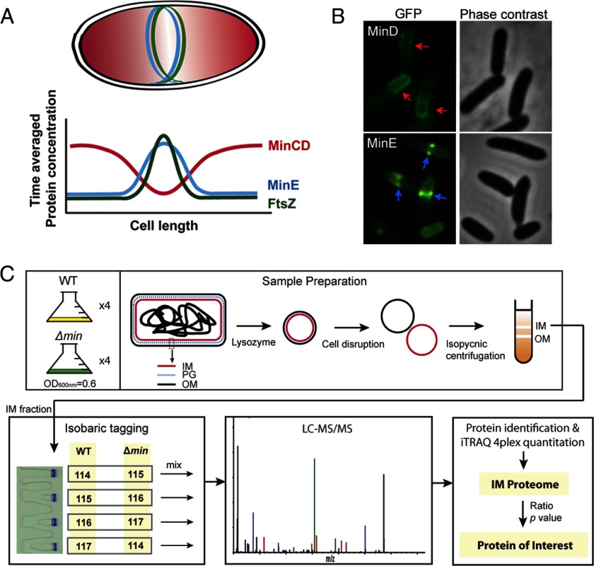
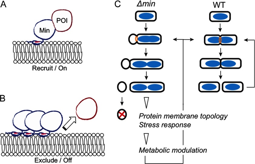
The Min system of Escherichia coli mediates placement of the division septum at the midcell. It oscillates from pole to pole to establish a concentration gradient of the division inhibition that is high at the poles but low at the midcell; the cell middle thereby becomes the most favorable site for division. Although Min oscillation is well studied from molecular and biophysical perspectives, it is still an enigma as to whether such a continuous, energy-consuming, and organized movement of the Min proteins would affect cellular processes other than the division site selection. To tackle this question, we compared the inner membrane proteome of the wild-type and Δmin strains using a quantitative approach. Forty proteins that showed differential abundance on the inner membrane of the mutant cells were identified and defined as proteins of interest (POIs). More than half of the POIs were peripheral membrane proteins, suggesting that the Min system affects mainly reversible protein association with the inner membrane. In addition, 6 out of 10 selected POIs directly interacted with at least one of the Min proteins, confirming the correlation between POIs and the Min system.Further analysis revealed a functional relationship between metabolism and the Min system. Metabolic enzymes accounted for 45% of the POIs, and there was a change of metabolites in the related reactions. We hypothesize that the Min system could alter the membrane location of proteins to modulate their enzymatic activity. Thus, the metabolic modulation in the Δmin mutant is likely an adaptive phenotype in cells of abnormal size and chromosome number due to an imbalanced abundance of proteins on the inner membrane. Taken together, the current work reports novel interactions of the Min system and reveals a global physiological impact of the Min system in addition to the division site placement.


Figures

Fig. 1.
Fig. 1.
Background and experimental procedures of the quantitative proteomic analysis in this work. (A) Pole-to-pole oscillation of the Min proteins establishes a concentration gradient of the division inhibition that is high at the poles (red shade), allowing formation of the FtsZ ring at the midcell. The cyan and green lines indicate the MinE and FtsZ rings, respectively. The chart shows the time-averaged protein concentration along the cell length. The red, cyan, and green lines represent the concentration of MinCD, MinE, and FtsZ, respectively. FtsZ is a tubulin homologue that assembles into a ring structure, the FtsZ ring, to recruit a series of septal proteins to the division site for the formation of the division septum. The FtsZ ring may also contribute constriction forces during cell division. (B) Micrographs showing GFP-MinD localization into a polar zone (red arrow) and MinE-GFP localization into a ring-like structure (blue arrow). (C) Experimental scheme. For clarity, only critical information is shown. The illustration of the intact cell is enlarged and is not in proportion to the size of the IM and OM vesicles.
Fig. 2.
Fig. 2.
Subcellular localization of the IM proteome proteins. (A) A Venn diagram showing coverage of the IM proteome proteins in comparison with the membrane proteins documented in STEPdb, PSORTdb, and Dynamic Localizome. (B) Enrichment of PIM proteins demonstrated by comparison of subcellular localization of membrane proteins in reference library, IM proteome, and POI. The percentage of PIM and IM proteins is labeled in the chart. The number listed below each category is the population size (n).
Fig. 3.
Fig. 3.
Functional enrichment analysis of POI. (A) A network view showing that 12 POIs are interconnected. The red shade (positive value) is applied to the nodes according to the ratio of protein abundance on the inner membrane. The thickness of the edges is drawn according to the mentha scores (34) that demonstrate evidence of PPI. (B) A table summarizing the node information in A.
Fig. 4.
Fig. 4.
Interaction between MinCDE and POIs. (A) Direct interaction examined by the B2H assay. Interaction of minC, minD, and minE with bfr, cyoA, dadA, and ftsY (upper panel), rpsA, ubiB, uspE, and pka (middle panel), and pfkA, pgk, and ygiC (lower panel). RpsA, the 30S ribosomal subunit protein S1 was included as a control. The color was developed at 30 °C for 18 h before taking photographs. Control reactions: c1, pT25-minD/pT18-minE; c2, pT18-minC/pT25; c3, pT25-minD/pT18; c4, pT18-minD/pT25-minD; c5, pT25-minE/pT18-minE; c6, pT18-minE/pT25. (B) Direct interaction examined by the pull-down assay. The His-tagged MinC (top), MinD (middle), and MinE (bottom) were used to pull down the GFP-tagged POIs in the cell lysate. Each set contains two blots. The upper blot was hybridized with the anti-GFP antibody, and the lower blot was hybridized with the anti-His6 antibody. The red arrow indicates the signal detected at the expected molecular weight. The black arrow indicates the signal that was not at the expected molecular weight. (C) A summary of the interaction between MinCDE and POIs. The images were converted to 8-bit and the intensity of the colony was measured using ImageJ. The measurements were subtracted by the intensity of the corresponding negative control in each set of measurements. The scores represent the strength of intensity that was determined by the ratio of intensity between the examined pair of interaction and the positive control. The scoring criteria are as follows: 5—positive control as 100%; 4—greater than 50%; 3—30 to 50%; 2—10 to 30%; 1—less than 10% and regarded as the background. The two numbers are the scores of minC, minD, or minE when cloned in pT18 and pT25 for cross-examination. The interactions detected in the pull-down assays were shown in italics, bold letters. The asterisks indicated the reproducible interactions in both assays.
Fig. 5.
Fig. 5.
Hypothetical models. (A) The POI is recruited to the membrane proximity by MinC, MinD, or MinE (Min). It may subsequently interact with the inner membrane directly. (B) Exclusion of POI from the inner membrane by the Min proteins. (C) The aberrant division in the Δmin mutant shows a metabolic modulation phenotype that could be due to an imbalance between membrane-bound and cytosolic pools of the metabolic enzymes. Alternatively, uncharacterized stress responses could be involved in modulating the metabolic flux. Blue ovoid, nucleoid; orange circle, FtsZ ring.

References

    1. Rowlett V. W., and Margolin W. (2015) The Min system and other nucleoid-independent regulators of Z ring positioning. Frontiers Microbiol. 6, 478 - PMC - PubMed
    1. de Boer P. A., Crossley R. E., and Rothfield L. I. (1989) A division inhibitor and a topological specificity factor coded for by the minicell locus determine proper placement of the division septum in E. coli. Cell 56, 641–649 - PubMed
    1. Hu Z., Mukherjee A., Pichoff S., and Lutkenhaus J. (1999) The MinC component of the division site selection system in Escherichia coli interacts with FtsZ to prevent polymerization. Proc. Natl. Acad. Sci. U.S.A. 96, 14819–14824 - PMC - PubMed
    1. Raskin D. M., and de Boer P. A. (1999) Rapid pole-to-pole oscillation of a protein required for directing division to the middle of Escherichia coli. Proc. Natl. Acad. Sci. U.S.A. 96, 4971–4976 - PMC - PubMed
    1. Fu X., Shih Y. L., Zhang Y., and Rothfield L. I. (2001) The MinE ring required for proper placement of the division site is a mobile structure that changes its cellular location during the Escherichia coli division cycle. Proc. Natl. Acad. Sci. U.S.A. 98, 980–985 - PMC - PubMed

Substances