Solamargine triggers cellular necrosis selectively in different types of human melanoma cancer cells through extrinsic lysosomal mitochondrial death pathway
- PMID: 26889092
- PMCID: PMC4756414
- DOI: 10.1186/s12935-016-0287-4
Solamargine triggers cellular necrosis selectively in different types of human melanoma cancer cells through extrinsic lysosomal mitochondrial death pathway
Abstract
Background: Previous reports showed that the Steroidal Glycoalkaloid Solamargine inhibited proliferation of non-melanoma skin cancer cells. However, Solamargine was not tested systematically on different types of melanoma cells and was not simultaneously tested on normal cells either. In this study we aimed to investigate the effect of Solamargine and the mechanism involved in inhibiting the growth of different types of melanoma cells.
Methods: Solamargine effect was tested on normal cells and on another three melanoma cell lines. Vertical growth phase metastatic and primary melanoma cell lines WM239 and WM115, respectively and the radial growth phase benign melanoma cells WM35 were used. The half inhibitory concentration IC50 of Solamargine was determined using Alamarblue assay. The cellular and subcellular changes were assessed using light and Transmission Electron Microscope, respectively. The percentage of cells undergoing apoptosis and necrosis were measured using Flow cytometry. The different protein expression was detected and measured using western blotting. The efficacy of Solamargine was determined by performing the clonogenic assay. The data collected was analyzed statistically on the means of the triplicate of at least three independent repeated experiments using one-way ANOVA test for parametric data and Kruskal-Wallis for non-parametric data. Differences were considered significant when the P values were less than 0.05.
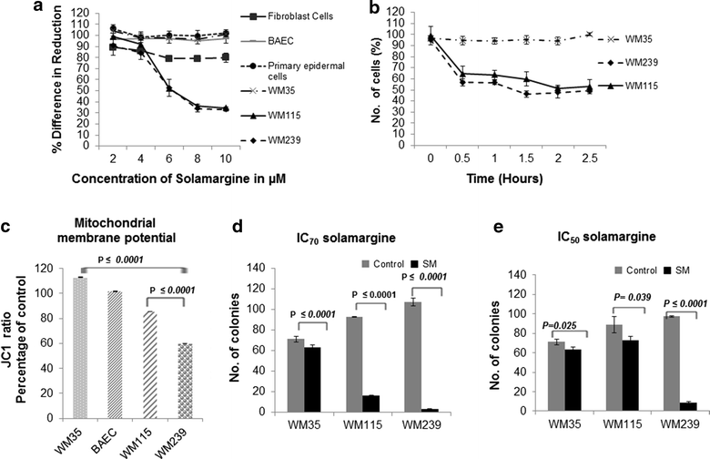
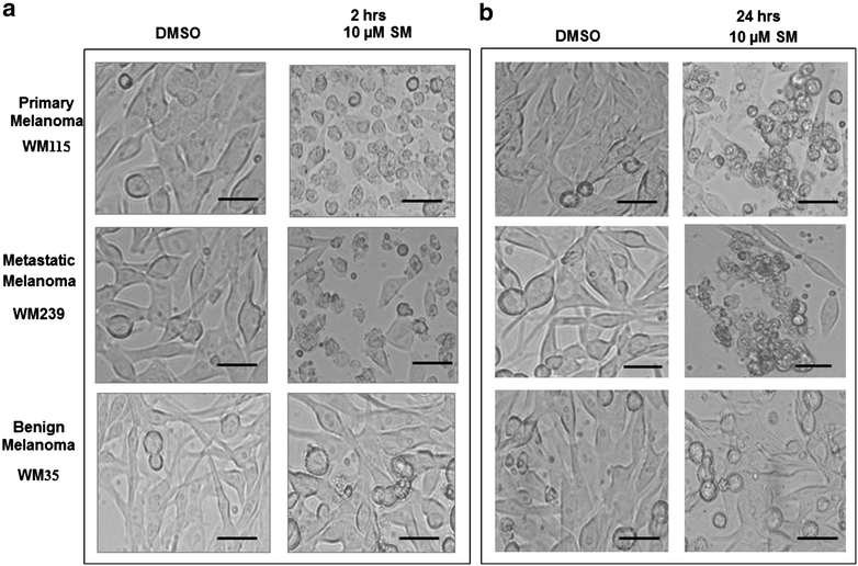
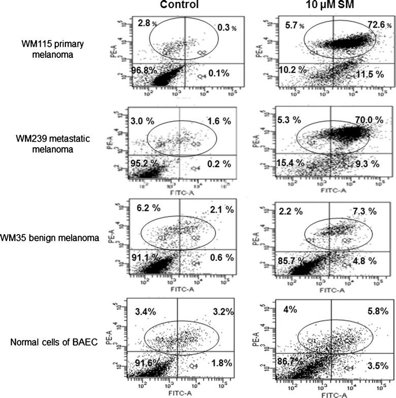
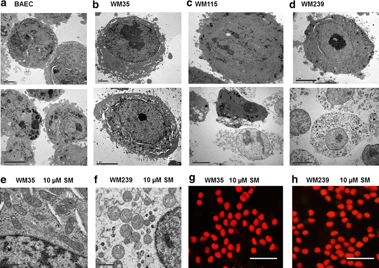
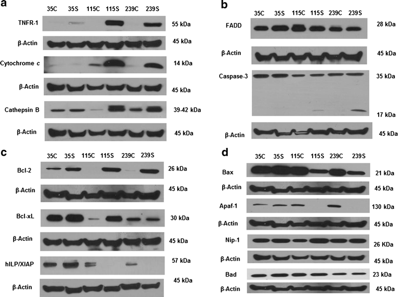
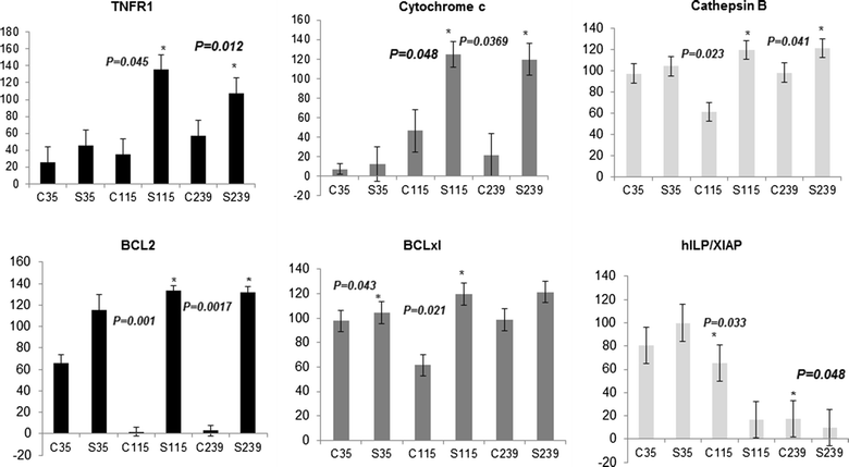
Results: Hereby, we demonstrate that Solamargine rapidly, selectively and effectively inhibited the growth of metastatic and primary melanoma cells WM239 and WM115 respectively, with minimum effect on normal and benign WM35 cells. Solamargine caused cellular necrosis to the two malignant melanoma cell lines (WM115, WM239), by rapid induction of lysosomal membrane permeabilization as confirmed by cathepsin B upregulation which triggered the extrinsic mitochondrial death pathway represented by the release of cytochrome c and upregulation of TNFR1. Solamargine disrupted the intrinsic apoptosis pathway as revealed by the down regulation of hILP/XIAP, resulting in caspase-3 cleavage, upregulation of Bcl-xL, and Bcl2, and down regulation of Apaf-1 and Bax in WM115 and WM239 cells only. Solamargine showed high efficacy in vitro particularly against the vertical growth phase melanoma cells.
Conclusion: Our findings suggest that Solamargine is a promising anti-malignant melanoma drug which warrants further attention.
Keywords: Cathepsin B; Cytochrome c; Melanoma; Solamargine; TNFR1.
Figures
Similar articles
-
Solamargine derived from Solanum nigrum induces apoptosis of human cholangiocarcinoma QBC939 cells.Oncol Lett. 2018 May;15(5):6329-6335. doi: 10.3892/ol.2018.8171. Epub 2018 Mar 5. Oncol Lett. 2018. PMID: 29731848 Free PMC article.
-
Solamargine induces apoptosis and sensitizes breast cancer cells to cisplatin.Food Chem Toxicol. 2007 Nov;45(11):2155-64. doi: 10.1016/j.fct.2007.05.009. Epub 2007 May 24. Food Chem Toxicol. 2007. PMID: 17619073
-
A lysosomal-mitochondrial death pathway is induced by solamargine in human K562 leukemia cells.Toxicol In Vitro. 2010 Sep;24(6):1504-11. doi: 10.1016/j.tiv.2010.07.013. Epub 2010 Jul 18. Toxicol In Vitro. 2010. PMID: 20647040
-
Casticin induces caspase-mediated apoptosis via activation of mitochondrial pathway and upregulation of DR5 in human lung cancer cells.Asian Pac J Trop Med. 2013 May 13;6(5):372-8. doi: 10.1016/S1995-7645(13)60041-3. Asian Pac J Trop Med. 2013. PMID: 23608376
-
Anticancer activity evaluation of the solanum glycoalkaloid solamargine. Triggering apoptosis in human hepatoma cells.Biochem Pharmacol. 2000 Dec 15;60(12):1865-73. doi: 10.1016/s0006-2952(00)00506-2. Biochem Pharmacol. 2000. PMID: 11108802
Cited by
-
An Updated Insight into Phytomolecules and Novel Approaches used in the Management of Breast Cancer.Curr Drug Targets. 2024;25(3):201-219. doi: 10.2174/0113894501277556231221072938. Curr Drug Targets. 2024. PMID: 38231060 Review.
-
Solamargine inhibits the growth of hepatocellular carcinoma and enhances the anticancer effect of sorafenib by regulating HOTTIP-TUG1/miR-4726-5p/MUC1 pathway.Mol Carcinog. 2022 Apr;61(4):417-432. doi: 10.1002/mc.23389. Epub 2022 Jan 17. Mol Carcinog. 2022. PMID: 35040191 Free PMC article.
-
Therapeutic Potential of Solanum Alkaloids with Special Emphasis on Cancer: A Comprehensive Review.Drug Des Devel Ther. 2024 Jul 16;18:3063-3074. doi: 10.2147/DDDT.S470925. eCollection 2024. Drug Des Devel Ther. 2024. PMID: 39050799 Free PMC article. Review.
-
Targeting EP4 downstream c-Jun through ERK1/2-mediated reduction of DNMT1 reveals novel mechanism of solamargine-inhibited growth of lung cancer cells.J Cell Mol Med. 2017 Feb;21(2):222-233. doi: 10.1111/jcmm.12958. Epub 2016 Sep 13. J Cell Mol Med. 2017. PMID: 27620163 Free PMC article.
-
Apoptotic Pathway as the Therapeutic Target for Anticancer Traditional Chinese Medicines.Front Pharmacol. 2019 Jul 12;10:758. doi: 10.3389/fphar.2019.00758. eCollection 2019. Front Pharmacol. 2019. PMID: 31354479 Free PMC article. Review.
References
-
- Leiter U, Eigentler T, Garbe C. Epidemiology of skin cancer. Adv Exp Med Biol. 2014;810:120–140. - PubMed
LinkOut - more resources
Full Text Sources
Other Literature Sources
Research Materials

