

Multimodal Imaging and Clinicopathologic Correlation in Primary Uveal Lymphoma
- PMID: 26889158
- PMCID: PMC4748778
- DOI: 10.1159/000442743
Multimodal Imaging and Clinicopathologic Correlation in Primary Uveal Lymphoma
Abstract
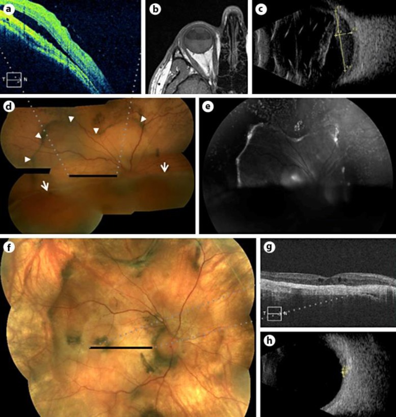
Purpose: We report a rare case of primary uveal lymphoma and characterize it using histopathology and multimodal imaging.
Patient and methods: A 41-year-old male presented with a 2-year history of increasingly blurry vision in his right eye and no systemic symptoms. Examination revealed a retinal detachment and mass lesion in the right eye. Radiologic and histologic testing was performed.
Results: Multimodal imaging localized the lesion to the choroid, and fine needle aspiration biopsy diagnosed the lesion as a low-grade B-cell lymphoma. The patient was treated with external beam radiation, resulting in regression of the mass and resolution of the retinal detachment.
Conclusions: Primary uveal lymphoma is a rare, usually indolent tumor that carries a good prognosis. In this case, we show that primary uveal lymphoma has distinct findings via histopathology and multimodal imaging, and that imaging after radiation treatment documents disease regression.
Keywords: Fluorescein angiography; Histopathology; Optical coherence tomography; Primary uveal lymphoma.
Figures

References
-
- Coupland SE, Foss H-D, Hidayat AA, Cockerham GC, Hummel M, Stein H. Extranodal marginal zone B cell lymphomas of the uvea: an analysis of 13 cases. J Pathol. 2002;197:333–340. - PubMed
-
- Crookes GP, Mullaney J. Lymphoid hyperplasia of the uveal tract simulating malignant lymphoma. Am J Ophthalmol. 1967;63:962–967. - PubMed
-
- Mauriello JA. A clinicopathologic and immunohistologic study of lymphoproliferative lesions of the uveal tract. ARVO Abstracts. Invest Ophthalmol Vis Sci. 1982;22:171.
Publication types
Grants and funding
LinkOut - more resources
Full Text Sources
Other Literature Sources
