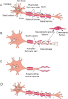
Dental pulp stem cells-derived schwann cells for peripheral nerve injury regeneration
- PMID: 26889180
- PMCID: PMC4730816
- DOI: 10.4103/1673-5374.172309
Dental pulp stem cells-derived schwann cells for peripheral nerve injury regeneration
Figures


Similar articles
-
Schwann-like cells differentiated from human dental pulp stem cells combined with a pulsed electromagnetic field can improve peripheral nerve regeneration.Bioelectromagnetics. 2016 Apr;37(3):163-174. doi: 10.1002/bem.21966. Epub 2016 Mar 15. Bioelectromagnetics. 2016. PMID: 26991921
-
Transplantation of Human Dental Pulp-Derived Stem Cells or Differentiated Neuronal Cells from Human Dental Pulp-Derived Stem Cells Identically Enhances Regeneration of the Injured Peripheral Nerve.Stem Cells Dev. 2017 Sep 1;26(17):1247-1257. doi: 10.1089/scd.2017.0068. Epub 2017 Jul 25. Stem Cells Dev. 2017. PMID: 28657463
-
Engineered neural tissue with Schwann cell differentiated human dental pulp stem cells: potential for peripheral nerve repair?J Tissue Eng Regen Med. 2017 Dec;11(12):3362-3372. doi: 10.1002/term.2249. Epub 2017 Jan 4. J Tissue Eng Regen Med. 2017. PMID: 28052540
-
Roles of neural stem cells in the repair of peripheral nerve injury.Neural Regen Res. 2017 Dec;12(12):2106-2112. doi: 10.4103/1673-5374.221171. Neural Regen Res. 2017. PMID: 29323053 Free PMC article. Review.
-
Practical considerations concerning the use of stem cells for peripheral nerve repair.Neurosurg Focus. 2009 Feb;26(2):E2. doi: 10.3171/FOC.2009.26.2.E2. Neurosurg Focus. 2009. PMID: 19435443 Review.
Cited by
-
Potential of dental pulp stem cells and their products in promoting peripheral nerve regeneration and their future applications.World J Stem Cells. 2023 Oct 26;15(10):960-978. doi: 10.4252/wjsc.v15.i10.960. World J Stem Cells. 2023. PMID: 37970238 Free PMC article. Review.
-
Equine Dental Pulp Connective Tissue Particles Reduced Lameness in Horses in a Controlled Clinical Trial.Front Vet Sci. 2017 Mar 10;4:31. doi: 10.3389/fvets.2017.00031. eCollection 2017. Front Vet Sci. 2017. PMID: 28344975 Free PMC article.
-
The application of collagen in the repair of peripheral nerve defect.Front Bioeng Biotechnol. 2022 Sep 23;10:973301. doi: 10.3389/fbioe.2022.973301. eCollection 2022. Front Bioeng Biotechnol. 2022. PMID: 36213073 Free PMC article. Review.
-
Age of the donor affects the nature of in vitro cultured human dental pulp stem cells.Saudi Dent J. 2021 Nov;33(7):524-532. doi: 10.1016/j.sdentj.2020.09.003. Epub 2020 Sep 24. Saudi Dent J. 2021. PMID: 34803296 Free PMC article.
-
Polycaprolactone/graphene oxide/acellular matrix nanofibrous scaffolds with antioxidant and promyelinating features for the treatment of peripheral demyelinating diseases.J Mater Sci Mater Med. 2023 Oct 5;34(10):49. doi: 10.1007/s10856-023-06750-2. J Mater Sci Mater Med. 2023. PMID: 37796399 Free PMC article.
References
-
- Al-Zer H, Apel C, Heiland M, Friedrich RE, Jung O, Kroeger N, Eichhorn W, Smeets R. Enrichment and schwann cell differentiation of neural crest-derived dental pulp stem cells. In Vivo. 2015;29:319–326. - PubMed
-
- Alluin O, Wittmann C, Marqueste T, Chabas JF, Garcia S, Lavaut MN, Guinard D, Feron F, Decherchi P. Functional recovery after peripheral nerve injury and implantation of a collagen guide. Biomaterials. 2009;30:363–373. - PubMed
-
- Arthur A, Shi S, Zannettino AC, Fujii N, Gronthos S, Koblar SA. Implanted adult human dental pulp stem cells induce endogenous axon guidance. Stem Cells. 2009;27:2229–2237. - PubMed
-
- Burnett MG, Zager EL. Pathophysiology of peripheral nerve injury: a brief review. Neurosurg Focus. 2004;16:E1. - PubMed
-
- Hall BK. Delamination, Migration and Potential. The Neural Crest and Neural Crest Cells in Vertebrate Development and Evolution. In: Hall BK, editor. New York: Springer; 2009.
LinkOut - more resources
Full Text Sources
Other Literature Sources
