

Prevalence of human papilloma virus (HPV) and its genotypes in cervical specimens of Egyptian women by linear array HPV genotyping test
- PMID: 26889206
- PMCID: PMC4756400
- DOI: 10.1186/s13027-016-0053-1
Prevalence of human papilloma virus (HPV) and its genotypes in cervical specimens of Egyptian women by linear array HPV genotyping test
Abstract
Background: The association of human papillomavirus (HPV) with cervical cancer is well established.
Aim: To investigate HPV genotype distribution and co-infection occurrence in cervical specimens from a group of Egyptian women.
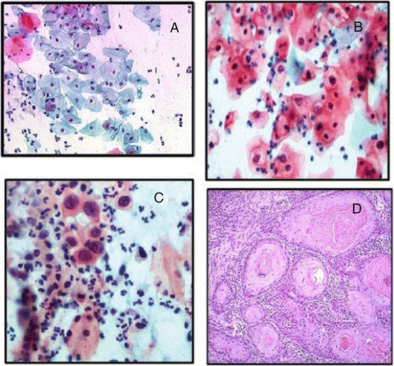
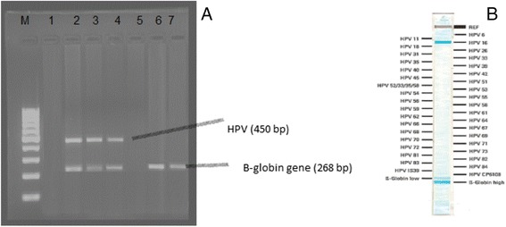
Methods: A group of 152 women with and without cervical lesions were studied. All women had cervical cytology and HPV testing. They were classified according to cytology into those with normal cytology, with squamous intraepithelial lesions (SIL) and invasive squamous cell carcinoma (SCC). Cervical samples were analyzed to identify the presence of HPV by PCR, and all positive HPV-DNA samples underwent viral genotype analysis by means of LINEAR ARRAY HPV Genotyping assay.
Results: A total of 26 HPV types with a prevalence of 40.8 % were detected. This prevalence was distributed as follows: 17.7 % among cytologically normal females, 56.5, 3.2, and 22.6 % among those with LSIL, HSIL and invasive SCC respectively. Low-risk HPV types were detected in 81.8 % of the cytologically-normal women, in 5.7 % of those in LSIL women, and in 14.3 % of infections with invasive SCC, while no low-risk types were detected in HSIL. High-risk HPV types were detected in 18.2 % of infections in the cytologically normal women, 14.3 % of infections in LSIL, and in 21.4 % of invasive lesions. The probable and possible carcinogenic HPV were not detected as single infections. Mixed infection was present in 80 % of women with LSIL, in 100 % of those with HSIL, and in 64.3 % of those with invasive SCC. This difference was statistically significant. HPV 16, 18 and 31 were the most prevalent HR HPV types, constituting 41.9, 29.03 and 12.9 % respectively, and HPV 6, 62 and CP6108 were the most prevalent LR HPV types constituting 11.3, 9.7 and 9.7 % respectively.
Conclusion: These data expand the knowledge concerning HPV prevalence and type distribution in Egypt which may help to create a national HPV prevention program. HPV testing using the LINEAR ARRAY HPV Genotyping assay is a useful tool when combined with cytology in the diagnosis of mixed and non-conventional HPV viral types.
Keywords: Cervical smear; Cervical squamous intraepithelial lesions; Human papillomavirus (HPV); Linear Array HPV genotyping.
Figures
Similar articles
-
HPV genotype distribution and anomalous association of HPV33 to cervical neoplastic lesions in San Luis Potosí, Mexico.Infect Agent Cancer. 2016 Mar 30;11:16. doi: 10.1186/s13027-016-0063-z. eCollection 2016. Infect Agent Cancer. 2016. PMID: 27030798 Free PMC article.
-
Prevalence of high-risk human papillomavirus (HR-HPV) infection among women with normal and abnormal cervical cytology in Myanmar.Acta Med Okayama. 2014;68(2):79-87. doi: 10.18926/AMO/52404. Acta Med Okayama. 2014. PMID: 24743783
-
Human papillomavirus infection and cervical cancer in Brazil: a retrospective study.Mem Inst Oswaldo Cruz. 1996 Jul-Aug;91(4):433-40. doi: 10.1590/s0074-02761996000400009. Mem Inst Oswaldo Cruz. 1996. PMID: 9070405
-
Evidence regarding human papillomavirus testing in secondary prevention of cervical cancer.Vaccine. 2012 Nov 20;30 Suppl 5:F88-99. doi: 10.1016/j.vaccine.2012.06.095. Vaccine. 2012. PMID: 23199969 Review.
-
Prevalence and distribution of human papillomavirus genotypes in women with abnormal cervical cytology in Ethiopia: a systematic review and meta-analysis.Front Oncol. 2024 Oct 15;14:1384994. doi: 10.3389/fonc.2024.1384994. eCollection 2024. Front Oncol. 2024. PMID: 39474105 Free PMC article.
Cited by
-
HPV prevalence and type distribution in Cypriot women with cervical cytological abnormalities.BMC Infect Dis. 2017 May 16;17(1):346. doi: 10.1186/s12879-017-2439-0. BMC Infect Dis. 2017. PMID: 28511636 Free PMC article.
-
Racial Disparities Associated with the Prevalence of Vaccine and Non-Vaccine HPV Types and Multiple HPV Infections between Asia and Africa: A Systematic Review and Meta-Analysis.Asian Pac J Cancer Prev. 2021 Sep 1;22(9):2729-2741. doi: 10.31557/APJCP.2021.22.9.2729. Asian Pac J Cancer Prev. 2021. PMID: 34582640 Free PMC article.
-
Human Papillomavirus Genotypes and Methylation of CADM1, PAX1, MAL and ADCYAP1 Genes in Epithelial Ovarian Cancer Patients.Asian Pac J Cancer Prev. 2017 Jan 1;18(1):169-176. doi: 10.22034/APJCP.2017.18.1.169. Asian Pac J Cancer Prev. 2017. PMID: 28240513 Free PMC article.
-
Human Papillomaviruses-Related Cancers: An Update on the Presence and Prevention Strategies in the Middle East and North African Regions.Pathogens. 2022 Nov 19;11(11):1380. doi: 10.3390/pathogens11111380. Pathogens. 2022. PMID: 36422631 Free PMC article. Review.
-
Correlation between vaginal flora and cervical immune function of human papilloma virus-infected patients with cervical cancer.Afr Health Sci. 2023 Jun;23(2):179-185. doi: 10.4314/ahs.v23i2.19. Afr Health Sci. 2023. PMID: 38223622 Free PMC article.
References
-
- Ozturk S, Kaleli I, Kaleli B, Bir F. Investigation of human papillomavirus DNA in cervical specimens by hybrid capture assay. Mikrobiyol Bul. 2004;38(3):223–32. - PubMed
-
- Ferlay J, Soerjomataram I, Ervik M, Dikshit R, Eser S, Mathers C, et al. GLOBOCAN 2012 v1.0, Cancer Incidence and Mortality Worldwide: IARC CancerBase No. 11 [Internet] Lyon, France: International Agency for Research on Cancer; 2013.
-
- Bruni L, Barrionuevo-Rosas L, Albero G, Aldea M, Serrano B, Valencia S et al. ICO Information Centre on HPV and Cancer (HPV Information Centre). Human Papillomavirus and Related Diseases in Egypt. Summary Report 2015-03-20. Available at: http://www.hpvcentre.net/statistics/reports/EGY.pdf.
-
- Giuliani L, Coletti A, Syrjanen K, Favalli C, Ciotti M. Comparison of DNA Sequencing and Roche Linear Array® in Human papillomavirus (HPV) Genotyping. Anticancer Research. 2006;26:3939–3942. - PubMed
LinkOut - more resources
Full Text Sources
Other Literature Sources
Research Materials
