Review
. 2016 Jun;34(6):1427-36.
doi: 10.1002/stem.2345. Epub 2016 Mar 16.

Concise Review: Control of Cell Fate Through Cell Cycle and Pluripotency Networks

Affiliations
Review

Concise Review: Control of Cell Fate Through Cell Cycle and Pluripotency Networks

Ben Boward et al. Stem Cells. 2016 Jun.

Abstract

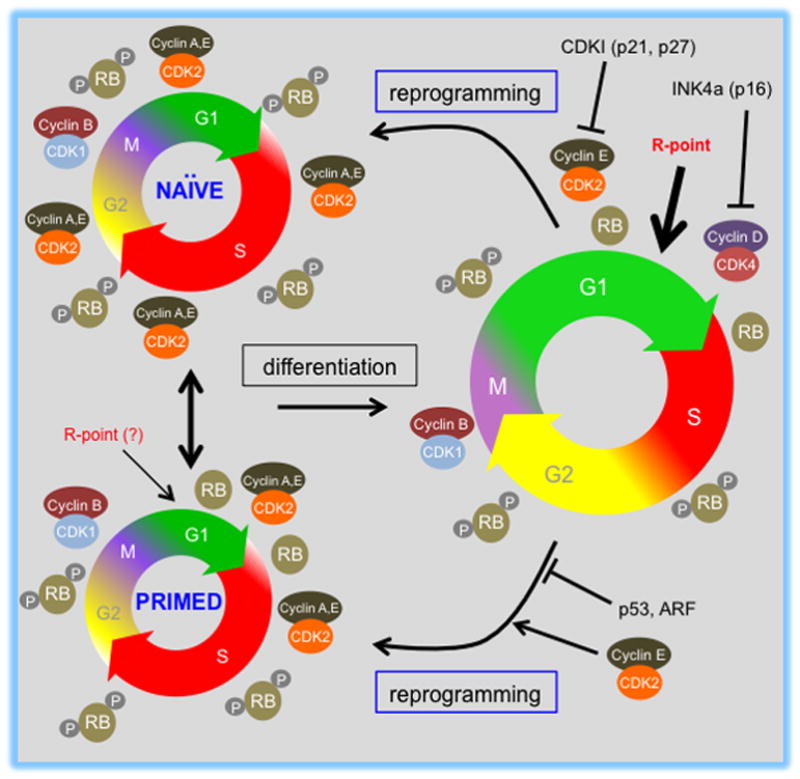
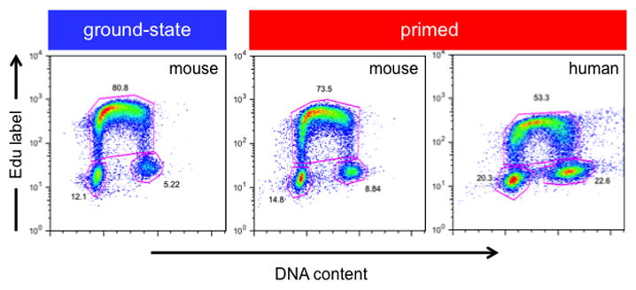
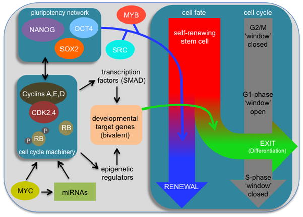
Pluripotent stem cells (PSCs) proliferate rapidly with a characteristic cell cycle structure consisting of short G1- and G2-gap phases. This applies broadly to PSCs of peri-implantation stage embryos, cultures of embryonic stem cells, induced pluripotent stem cells, and embryonal carcinoma cells. During the early stages of PSC differentiation however, cell division times increase as a consequence of cell cycle remodeling. Most notably, this is indicated by elongation of the G1-phase. Observations linking changes in the cell cycle with exit from pluripotency have raised questions about the role of cell cycle control in maintenance of the pluripotent state. Until recently however, this has been a difficult question to address because of limitations associated with experimental tools. Recent studies now show that pluripotency and cell cycle regulatory networks are intertwined and that cell cycle control mechanisms are an integral, mechanistic part of the PSC state. Studies in embryonal carcinoma, some 30 years ago, first suggested that pluripotent cells initiate differentiation when in the G1-phase. More recently, a molecular "priming" mechanism has been proposed to explain these observations in human embryonic stem cells. Complexity in this area has been increased by the realization that pluripotent cells exist in multiple developmental states and that in addition to each having their own characteristic gene expression and epigenetic signatures, they potentially have alternate modes of cell cycle regulation. This review will summarize current knowledge in these areas and will highlight important aspects of interconnections between the cell cycle, self-renewal, pluripotency, and cell fate decisions. Stem Cells 2016;34:1427-1436.

Keywords: Cell cycle; Differentiation; Embryonic stem cells; Pluripotent stem cells.


Figures

Figure 1
Figure 1. Kinetics and regulatory features of the cell cycle in ground-state, primed, and differentiated cells
Cell cycle structures of pluripotent stem cells (PSC) and differentiated derivatives are shown. Phases are shown as an approximation of their relative length. Naïve (mESCs) and primed (hESCs, EpiSCs) pluripotent cells (left) proliferate rapidly and are characterized by a short G1 phase. mESCs represent the naïve pluripotent state and its respective cell cycle characteristics, while hESCs and mEpiSCs represent the primed pluripotent state. The potential for conversion between the naïve and primed states of pluripotency is indicated by the double-sided arrow. At the right, a representation of the cell cycle of a differentiated PSC-derivative. Note the differences in the molecular regulation of cell cycle events such as periodicity of CDK-cyclin complex assembly/activity, RB phosphorylation status and the presence or absence of an intact R-point. The relative activity of CDK-cyclin complexes and RB phosphorylation status is shown in relation to cell cycle position for each cell type. During differentiation, G1 length is extended and overall cell cycle duration increases. This is associated with up-regulation of D-type cyclin complexes, down-regulation of global CDK activity, establishment of cell cycle dependent CDK2-cyclin A and E activities and activation of a functional R-point in G1 cells. As cells differentiate, CDKIs are expressed and can negatively control CDK activity (p21, p27 and p16, for example). The figure also indicates that pluripotent cell cycle controls are restored upon reprogramming. The effects of cell cycle complexes (CDK2-cyclin E) and growth regulators (p53, ARF) on reprogramming are indicated.
Figure 2
Figure 2. Cell cycle analysis of naïve and primed pluripotent cells
Cell cycle distribution of murine R1 naïve (2i) and primed (EpiSCs) cells and WA09 primed hPSCs. Cells were pulse-labeled with 5-ethynyl-2′-deoxyuridine (EdU, 10μM) to label actively replicating DNA shown on the Y-axis (S-phase cells), using the Click-iT Plus Edu Flow Cytometry Assay kit (Invitrogen). All cells were labeled for 15 mins. Cells were then fixed and stained with FxCycle Violet (Life Technologies; 30 mins, 1 μg/ml) to measure DNA content (X-axis). Cells were then analyzed using a Beckman Coulter CyAn ADP flow cytometer. Cells with low FxCycle Violet and low EdU fluorescence represent cells in G0/G1 phase. Cells with low to high FxCycle Violet and high EdU fluorescence represent cells in S-phase, while cells with high FxCycle Violet and low EdU fluorescence represent cells in G2/M-phase. The percentage of cells found in each respective fraction is indicated. Murine (naïve, primed) and human (primed) cells were cultured as described previously [15,43,62].
Figure 3
Figure 3. Regulatory networks impacting differentiation from G1-phase
The G1-phase represents a ‘window of opportunity’ when pluripotent cells decide to self-renew or exit the pluripotent state. This involves interplay between the pluripotency network (OCT4, NANOG, SOX2) and cell cycle machinery (CDK-cyclin complexes, RB) that are impacted by other factors such as MYC and miRNAs. The pluripotency network, under self-renewing conditions, ensures maintenance of the pluripotent state. In response to the differentiation signaling, signal-regulated transcription factors (such as SMAD2,3) and epigenetic remodeling enzymes prime and then activate developmental genes in G1-phase. Activation of bivalent genes from G1-phase then initiates the exit from pluripotency and the differentiation program. Other factors such as MYB and SRC regulate this network. Impact that the pluripotency network has on the decision to ‘renew’ is indicated. Effects of developmental regulators, MYB and SRC on pluripotency ‘exit’ are also indicated. The G1-phase is shown as the ‘window’ in which these signals are integrated and where cell fate decisions are initially made. This window closes as cells transit through S-phase and remains closed until the subsequent G1.

References

    1. Chambers I, Tomlinson SR. The transcriptional foundation of pluripotency. DEVELOPMENT. 2009;136(14):2311–2322. - PMC - PubMed
    1. Takahashi K, Yamanaka S. Induction of pluripotent stem cells from mouse embryonic and adult fibroblast cultures by defined factors. CELL. 2006;126(4):663–676. - PubMed
    1. Heasman J. Patterning the early Xenopus embryo. DEVELOPMENT. 2006;133(7):1205–1217. - PubMed
    1. Yarden A, Salomon D, Geiger B. Zebrafish cyclin D1 is differentially expressed during early embryogenesis. BIOCHIM BIOPHYS ACTA. 1995;1264(3):257–260. - PubMed
    1. Kane DA, Kimmel CB. The zebrafish midblastula transition. DEVELOPMENT. 1993;119(2):447–456. - PubMed

Publication types