Review
. 2016 Aug;34(8):1289-306.
doi: 10.1002/jor.23195. Epub 2016 Aug 12.

Molecular mechanisms of biological aging in intervertebral discs

Affiliations
Review

Molecular mechanisms of biological aging in intervertebral discs

Nam V Vo et al. J Orthop Res. 2016 Aug.

Abstract

Advanced age is the greatest risk factor for the majority of human ailments, including spine-related chronic disability and back pain, which stem from age-associated intervertebral disc degeneration (IDD). Given the rapid global rise in the aging population, understanding the biology of intervertebral disc aging in order to develop effective therapeutic interventions to combat the adverse effects of aging on disc health is now imperative. Fortunately, recent advances in aging research have begun to shed light on the basic biological process of aging. Here we review some of these insights and organize the complex process of disc aging into three different phases to guide research efforts to understand the biology of disc aging. The objective of this review is to provide an overview of the current knowledge and the recent progress made to elucidate specific molecular mechanisms underlying disc aging. In particular, studies over the last few years have uncovered cellular senescence and genomic instability as important drivers of disc aging. Supporting evidence comes from DNA repair-deficient animal models that show increased disc cellular senescence and accelerated disc aging. Additionally, stress-induced senescent cells have now been well documented to secrete catabolic factors, which can negatively impact the physiology of neighboring cells and ECM. These along with other molecular drivers of aging are reviewed in depth to shed crucial insights into the underlying mechanisms of age-related disc degeneration. We also highlight molecular targets for novel therapies and emerging candidate therapeutics that may mitigate age-associated IDD. © 2016 Orthopaedic Research Society. Published by Wiley Periodicals, Inc. J Orthop Res 34:1289-1306, 2016.

Keywords: aging; cellular senescence; inflammation; intervertebral disc; oxidative damage.


Figures

Figure 1
Figure 1. Gross features of the aged mammalian discs
Axial sections of young and aged lumbar discs of 3 month- vs. 24 month-old mice (A, B), 3 week- vs. 3 year-old pigs (C, D) , and 16 year- vs. 55 year-old humans (E,F, courtesy of Dr. Ian Stokes), are shown. Old discs exhibit an overall loss of hydration, loss of demarcation between the AF and NP boundary, and tissue discoloration (old disc more yellowish). Average lumbar disc cross-sectional diameters are approximately 2–3mm for mice, 24–30 mm for pigs, and 45–55mm for humans .
Figure 2
Figure 2. Gross features of the aging spine
Young and aged lumbar spines are visually compared to illustrate the wide range of tissues and processes involved in aging of the spine. Muscle atrophy and fatty infiltration is evident at L1-2 in the aged spine. 25Similarly, a window into L3 depicts reduced vascularity and fewer capillaries reaching the endplate. Foraminal stenosis is shown at L2-3 (arrow), L3-4, and L4-5, and facet hypertrophy is evident at L3-4 (arrow) and L4-5. An annular lesion is present in the posterior portion of the L3-4 disc. Disc degeneration with evident loss of disc height and prominent anterior osteophytes occur at L4-5. Ligamentous thickening is indicated in the interspinous and supraspinous ligament; thickening of the ligamentum flavum occurs with aging but is not observable in the sagittal view. Finally, facet cartilage arthritis is revealed on the inferior facet of L5.
Figure 3
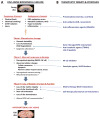
Figure 3. Proposed biochemical cascade in disc aging process and potential therapeutic targets
(A) With aging, there is time-dependent accumulation of biomolecular damage (Phase I), most importantly, DNA damage, in the disc due to exogenous and endogenous factors. Cellular responses to accumulated damage over time become dysregulated (Phase II) leading to more damage and eventual loss of disc biologic structure and function (Phase III). This results in degenerative changes observed in aged discs. * Observed in other tissues but not yet investigated in disc tissue. (B) Potential therapeutic targets to delay or ameliorate age-related degeneration. Oxidative and inflammatory stress can be reduced with anti-oxidants and anti-inflammatory drugs. Reducing chronic activation of NF-κB signaling by pharmacologic intervention may be efficacious in delaying age-related degeneration. Moreover, removal of senescent cells or blocking formation of SASP could potentially mitigate disc matrix catabolism. Finally, protein-, gene- and cell-based therapy could also conceivably delay or help restore age-related loss of disc matrix and functional cells.
Figure 4
Figure 4. Molecular and cellular features of the aging disc
Young and aged extracellular matrix (A) and cells (B) are schematically compared to summarize important changes that occur during disc aging. Panel A, young matrix is rich in elastin (green, coiled fiber), aggregated aggrecan (dark blue, bottle-brush aggregate), and collagen fibers (banded fibers). Aged matrix shows loss of elastin, increased collagen and collagen crosslinking, fragmented aggregan, diminished GAG quality, reduced aggregan aggregates, increased accumulation of advanced glycation end-products (AGEs) along with lower hydration. Panel B, young AF cells are elongated fibrochondrocytes and NP cells are a mixture of large, clustering, notochordal cells and smaller, chondrocyte-like cells. Aged cells show reduced cellularity, loss of notchordal cells, and incidence of senescence, apoptosis, and necrosis.

References

    1. World Population Prospects The 2015 Revision. at < http://esa.un.org/unpd/wpp/Publications/Files/Key_Findings_WPP_2015.pdf>.
    1. Christensen K, Doblhammer G, Rau R, Vaupel JW. Ageing populations: the challenges ahead. Lancet. 2009;374:1196–208. - PMC - PubMed
    1. The Burden of Musculoskeletal Diseases in the United States. American Academy of Orthopaedic Surgeons; 2008. at < http://www.boneandjointburden.org/docs/TheBurden of Musculoskeletal Dise...>.
    1. Derby R, Lee SH, Kim BJ. Discography. In: Slipman CW, Derby R, Simeone FA, Mayer TG , editors. Interventional Spine: an algorithmic approach. Elsevier; 2008. pp. 291–302.
    1. Prevalence of knee osteoarthritis in the United States: arthritis data from the Third National Health and Nutrition Examination Survey. 1991–1994 at < http://www.jrheum.org/content/33/11/2271.full.pdf>. - PubMed

Publication types

MeSH terms