Repression of Wnt/β-catenin response elements by p63 (TP63)
- PMID: 26890356
- PMCID: PMC4845946
- DOI: 10.1080/15384101.2016.1148837
Repression of Wnt/β-catenin response elements by p63 (TP63)
Abstract
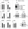
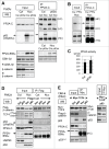
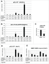
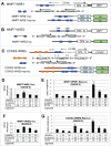
Submitted: TP63 (p63), a member of the tumor suppressor TP53 (p53) gene family, is expressed in keratinocyte stem cells and well-differentiated squamous cell carcinomas to maintain cellular potential for growth and differentiation. Controversially, activation of the Wnt/β-catenin signaling by p63 (Patturajan M. et al., 2002, Cancer Cells) and inhibition of the target gene expression (Drewelus I. et al., 2010, Cell Cycle) have been reported. Upon p63 RNA-silencing in squamous cell carcinoma (SCC) lines, a few Wnt target gene expression substantially increased, while several target genes moderately decreased. Although ΔNp63α, the most abundant isoform of p63, appeared to interact with protein phosphatase PP2A, neither GSK-3β phosphorylation nor β-catenin nuclear localization was altered by the loss of p63. As reported earlier, ΔNp63α enhanced β-catenin-dependent luc gene expression from pGL3-OT having 3 artificial Wnt response elements (WREs). However, this activation was detectable only in HEK293 cells examined so far, and involved a p53 family-related sequence 5' to the WREs. In Wnt3-expressing SAOS-2 cells, ΔNp63α rather strongly inhibited transcription of pGL3-OT. Importantly, ΔNp63α repressed WREs isolated from the regulatory regions of MMP7. ΔNp63α-TCF4 association occurred in their soluble forms in the nucleus. Furthermore, p63 and TCF4 coexisted at a WRE of MMP7 on the chromatin, where β-catenin recruitment was attenuated. The combined results indicate that ΔNp63α serves as a repressor that regulates β-catenin-mediated gene expression.
Keywords: TCF4; TP63; Wnt; p63; β-catenin.
Figures


Similar articles
-
C-terminal α Domain of p63 Binds to p300 to Coactivate β-Catenin.Neoplasia. 2019 May;21(5):494-503. doi: 10.1016/j.neo.2019.03.010. Epub 2019 Apr 12. Neoplasia. 2019. PMID: 30986748 Free PMC article.
-
A dynamic exchange of TCF3 and TCF4 transcription factors controls MYC expression in colorectal cancer cells.Cell Cycle. 2015;14(3):323-32. doi: 10.4161/15384101.2014.980643. Cell Cycle. 2015. PMID: 25659031 Free PMC article.
-
Human telomerase reverse transcriptase (hTERT) is a novel target of the Wnt/β-catenin pathway in human cancer.J Biol Chem. 2012 Sep 21;287(39):32494-511. doi: 10.1074/jbc.M112.368282. Epub 2012 Jul 31. J Biol Chem. 2012. PMID: 22854964 Free PMC article.
-
TP63 links chromatin remodeling and enhancer reprogramming to epidermal differentiation and squamous cell carcinoma development.Cell Mol Life Sci. 2020 Nov;77(21):4325-4346. doi: 10.1007/s00018-020-03539-2. Epub 2020 May 23. Cell Mol Life Sci. 2020. PMID: 32447427 Free PMC article. Review.
-
Discovery of small molecule inhibitors of the Wnt/β-catenin signaling pathway by targeting β-catenin/Tcf4 interactions.Exp Biol Med (Maywood). 2017 Jun;242(11):1185-1197. doi: 10.1177/1535370217708198. Epub 2017 May 5. Exp Biol Med (Maywood). 2017. PMID: 28474989 Free PMC article. Review.
Cited by
-
Novel insights into the pathways regulating the canine hair cycle and their deregulation in alopecia X.PLoS One. 2017 Oct 24;12(10):e0186469. doi: 10.1371/journal.pone.0186469. eCollection 2017. PLoS One. 2017. PMID: 29065140 Free PMC article.
-
C-terminal α Domain of p63 Binds to p300 to Coactivate β-Catenin.Neoplasia. 2019 May;21(5):494-503. doi: 10.1016/j.neo.2019.03.010. Epub 2019 Apr 12. Neoplasia. 2019. PMID: 30986748 Free PMC article.
-
Distinct interactors define the p63 transcriptional signature in epithelial development or cancer.Biochem J. 2022 Jun 30;479(12):1375-1392. doi: 10.1042/BCJ20210737. Biochem J. 2022. PMID: 35748701 Free PMC article.
-
Wnt signaling in orofacial clefts: crosstalk, pathogenesis and models.Dis Model Mech. 2019 Feb 4;12(2):dmm037051. doi: 10.1242/dmm.037051. Dis Model Mech. 2019. PMID: 30760477 Free PMC article. Review.
-
TRP63/TP63 loss accelerates skin tumorigenesis through activation of Wnt/β-catenin signaling.J Dermatol Sci. 2018 Sep;91(3):325-328. doi: 10.1016/j.jdermsci.2018.05.011. Epub 2018 Jun 1. J Dermatol Sci. 2018. PMID: 29885760 Free PMC article. No abstract available.
References
-
- Yang A, Kaghad M, Wang Y, Gillett E, Fleming MD, Dotsch V, Andrews NC, Caput D, McKeon F. p63, a p53 homolog at 3q27-29, encodes multiple products with transactivating, death-inducing, and dominant-negative activities. Mol Cell 1998; 2:305-16; PMID:9774969; http://dx.doi.org/10.1016/S1097-2765(00)80275-0 - DOI - PubMed
-
- Pellegrini G, Dellambra E, Golisano O, Martinelli E, Fantozzi I, Bondanza S, Ponzin D, McKeon F, De Luca M. p63 identifies keratinocyte stem cells. Proc Natl Acad Sci U S A 2001; 98:3156-61; PMID:11248048; http://dx.doi.org/10.1073/pnas.061032098 - DOI - PMC - PubMed
-
- Osada M, Ohba M, Kawahara C, Ishioka C, Kanamaru R, Katoh I, Ikawa Y, Nimura Y, Nakagawara A, Obinata M, et al.. Cloning and functional analysis of human p51, which structurally and functionally resembles p53. Nat Med 1998; 4:839-43; PMID:9662378; http://dx.doi.org/10.1038/nm0798-839 - DOI - PubMed
-
- Senoo M, Pinto F, Crum CP, McKeon F. p63 Is Essential for the Proliferative Potential of Stem Cells in Stratified Epithelia. Cell 2007; 129:523-36; PMID:17482546; http://dx.doi.org/10.1016/j.cell.2007.02.045 - DOI - PubMed
-
- Keyes WM, Pecoraro M, Aranda V, Vernersson-Lindahl E, Li W, Vogel H, Guo X, Garcia EL, Michurina TV, Enikolopov G, et al.. ΔNp63α is an oncogene that targets chromatin remodeler lsh to drive skin stem cell proliferation and tumorigenesis. Cell Stem Cell 2011; 8:164-76; PMID:21295273; http://dx.doi.org/10.1016/j.stem.2010.12.009 - DOI - PMC - PubMed
Publication types
MeSH terms
Substances
LinkOut - more resources
Full Text Sources
Other Literature Sources
Research Materials
Miscellaneous
