. 2016 Jun;35(6):736-40.
doi: 10.1097/ICO.0000000000000784.

Restoration of Fornix Tear Reservoir in Conjunctivochalasis With Fornix Reconstruction

Affiliations

Restoration of Fornix Tear Reservoir in Conjunctivochalasis With Fornix Reconstruction

Anny M S Cheng et al. Cornea. 2016 Jun.

Abstract

Purpose: To determine whether conjunctivochalasis (CCh) obliterates the fornix tear reservoir and to discern whether there is concomitant aqueous tear deficiency (ATD) dry eye.

Methods: Retrospective review of 18 eyes of 12 patients with CCh and ATD (CCh + ATD) and 18 eyes of 13 patients with CCh without ATD (CCh - ATD). Changes were compared before and after fornix reconstruction regarding symptoms, basal tear volumes, use of medications, conjunctival inflammation, and corneal staining.

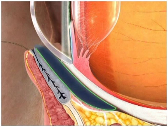
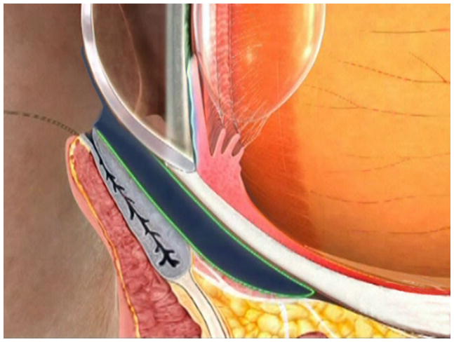
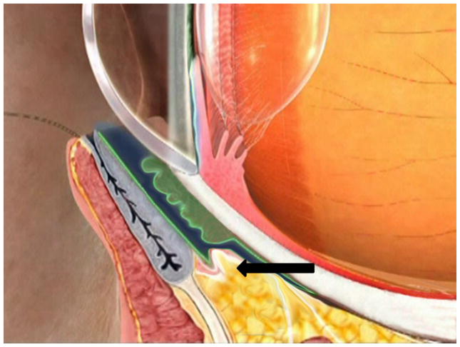
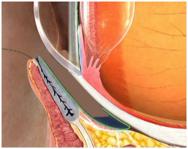
Results: Fornix reconstruction with conjunctival recession and amniotic membrane transplantation effectively restored the fornix tear reservoir as evidenced by a significant increase of the basal tear volume in both CCh - ATD and CCh + ATD groups. Multivariate regression analysis confirmed that such improvement was significantly correlated with symptomatic resolution (r = 1, P < 0.001), which was also accompanied by significant resolution of corneal staining, conjunctival inflammation, and reduction of topical medications. Intriguingly, the prior diagnosis of ATD was no longer existent in 10 of the 18 eyes (56%) with CCh + ATD suggesting that ATD could be secondary to obliteration of the fornix tear reservoir by CCh.

Conclusions: Obliteration of the fornix tear reservoir is a common pathogenic process regardless of whether CCh is associated with ATD dry eye. Restoration of the tear reservoir by fornix reconstruction with conjunctival recession and amniotic membrane transplantation results in significant resolution of symptoms and signs associated with ATD that is secondary to CCh and helps identify genuine ATD dry eye that is independent of CCh.


Figures

FIGURE 1
FIGURE 1. Schematic Demonstration of Fornix Tear Reservoir Obliterated by CCh and Restored by Fornix Reconstruction With Conjunctival Recession and AMT
Under the normal circumstance, the fornix tear reservoir depicted in dark blue is responsible for delivering tear fluids to the tear meniscus by blinking (A, B). In CCh, the fornix tear reservoir is obliterated by loose and wrinkled conjunctiva (C, green) and prolapse fat (C, arrow) due to degenerated Tenon’s capsule. This results in low basal wettings which mimick ATD dry eye. Restoration of the fornix tear reservoir is achieved by fornix deepening reconstruction, resulting in disappearance of ATD dry eye secondary to CCh (i.e., converted to a normal state). Consequently, fornix deepening reconstruction also helps identify ATD dry eye that is independent of CCh (D).

References

    1. Tseng SC, Tsubota K. Important concepts for treating ocular surface and tear disorders. Am J Ophthalmol. 1997;124(6):825–835. - PubMed
    1. Tseng SC. A practical treatment algorithm for managing ocular surface and tear disorders. Cornea. 2011;30(Suppl 1):S8–S14. - PubMed
    1. Mishima S, Gasset A, Klyce SD, et al. Determination of tear volume and tear flow. Invest Ophthalmol. 1966;5(3):264–276. - PubMed
    1. Meller D, Tseng SC. Conjunctivochalasis: literature review and possible pathophysiology. Surv Ophthalmol. 1998;43(3):225–232. - PubMed
    1. Di Pascuale MA, Espana EM, Kawakita T, et al. Clinical characteristics of conjunctivochalasis with or without aqueous tear deficiency. Br J Ophthalmol. 2004;88(3):388–392. - PMC - PubMed