. 2016 Mar;11(3):542-52.
doi: 10.1038/nprot.2016.031. Epub 2016 Feb 18.

A cryoinjury model in neonatal mice for cardiac translational and regeneration research

Affiliations

A cryoinjury model in neonatal mice for cardiac translational and regeneration research

Brian D Polizzotti et al. Nat Protoc. 2016 Mar.

Abstract

The introduction of injury models for neonatal mouse hearts has accelerated research on the mechanisms of cardiac regeneration in mammals. However, some existing models, such as apical resection and ligation of the left anterior descending artery, produce variable results, which may be due to technical difficulties associated with these methods. Here we present an alternative model for the study of cardiac regeneration in neonatal mice in which cryoinjury is used to induce heart injury. This model yields a reproducible injury size, does not induce known mechanisms of cardiac regeneration and leads to a sustained reduction of cardiac function. This protocol uses reusable cryoprobes that can be assembled in 5 min, with the entire procedure taking 15 min per pup. The subsequent heart collection and fixation takes 2 d to complete. Cryoinjury results in a myocardial scar, and the size of injury can be scaled by the use of different cryoprobes (0.5 and 1.5 mm). Cryoinjury models are medically relevant to diseases in human infants with heart disease. In summary, the myocardial cryoinjury model in neonatal mice described here is a useful tool for cardiac translational and regeneration research.


Conflict of interest statement

Competing financial interests

The authors declare that they have no competing financial interests.

Figures

Figure 1
Figure 1. Main steps of construction of cryoprobes and experimental setup
(A–C) Materials and assembly of cryoprobes. Materials needed for construction of cryoprobes (A): 1. Disposable 5 mL pipet, 2. 200 μL pipette tip, 3. 5 cm piece of tygon tubing, and 4. Vanadium or aluminum metal filament. Metal filament passed through the 200 uL pipette tip and attached to the disposable 5 mL pipette (B). Tygon tubing is used to secure the metal filament to the 5 mL pipette (C). (D) Photograph of the surgical setup used during the procedure (represented by arrows): 1. light source, 2. surgical stand, 3. sterile polydrape placed over the surgical area, and 4. ice cooled surgical bed.
Figure 2
Figure 2. Detailed description of cardiac cryoinjury in a neonatal mouse
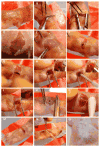
(A) Sterilization of the surgical site with Betadine swaps. (B) Transverse skin incision across the chest. (C) Separation of skin from underlying muscle using blunt dissection. (D–E) Lateral thoracotomy at the 4th intercostal space. (F–G) Separation of the intercostal muscles using blunt tipped forceps. (H) Exteriorization of the heart (inset illustrates the clear demarcation of right and left ventricles). (I) Application of the liquid nitrogen cooled cryoprobe to the surface of the left ventricle (inset, cryoprobe is placed on the left ventricular surface). (J) Visualization of the hematoma following cryoinjury. (K–M) The chest wall is closed with 8-0 nonabsorbable sutures. (N–O) The skin is closed with webglue. This protocol was approved by the Institutional Animal Care and Use Committee of Boston Children’s Hospital and University of Pittsburgh.
Figure 3
Figure 3. Resection of heart following cryoinjury at day of life 1 (P1)
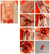
(A) Schematic representation of the main cuts required to expose the heart. 1. Abdominal transverse incision, 2. left, and 3. right lateral incisions extending from abdominal cavity to thoracic cavity. (B) Pups are injected with heparin (50 μL of heparin, 5000 USP units per mL) prior to euthanization to prevent clot formation in the heart. (C) Position of the first abdominal incision underneath the chest bone. D–E) Position of second and third incisions (D), which extend from the abdomen to the diaphram (E). (F) Removal of the diaphram to expose the heart. (G) Clamping of the major vessels prior to excision. (H) Excision of the heart using tapered microscissors. (I) Resected heart in cardioplegia solution. This protocol was approved by the Institutional Animal Care and Use Committee of Boston Children’s Hospital and University of Pittsburgh.
Figure 4
Figure 4. Cryoinjury in neonatal mice induces tissue damage
Mice underwent sham surgery or cryoinjury on day of life 1 (P1) with either a 0.5 mm or 1.5 mm probe and their hearts were resected the next day (P2). (A) Hematoma is seen at the site of injury, (B) Vital staining with 1% TTC (w/v; in phosphate buffer, pH 7.4) at 37°C for 20 min, and then fixed in 10% phosphate-buffered formaldehyde overnight shows injury zone indicated with white arrow in whole hearts. (C) Vital staining with TTC shows the degree of penetration of injury varies with probe size in sliced heart sections indicated by white dotted lines. Scale bars, 1 mm (A,B,C). Note the scalability of the injury size with different probes. Abbreviations: LV, Left ventricle, RV, Right ventricle, LA, Left atrium, RA, Right atrium. Figure 4A (first and third panel) was previously published.
Figure 5
Figure 5. Cryoinjury in neonatal mice induces apoptosis in myocardial cells
Cryoinjury was performed on day of life (DOL) 1 with either a 0.5 mm or a 1.5 mm probe and hearts were resected DOL 2. The scalability of injury is shown with myocardial cell death as visualized by TUNEL staining (green, ApopTag Red in Situ apoptosis detection kit, EMD Millipore Corporation, CA) and DNA staining with Hoechst (blue). Scale bars, 1 mm.
Figure 6
Figure 6. Cryoinjury induces scar formation in neonatal mice
Mice underwent sham surgery or cryoinjury on day of life 1 with either a 0.5 mm or a 1.5 mm probe. Time-series of AFOG-stained sections shows fibrin deposition at 1 days post injury (dpi, orange staining, black arrows). Scar (blue) is formed within 7 dpi (black arrow heads) and still present 30 days later. Note transmural scars were present only after 1.5 mm cryoinjury (lower right panel) but not after the 0.5 mm probe. Scale bar 1 mm. Photomicrographs of the lower right 2 panels (1.5 mm probe) have been published before.

References

    1. Naqvi N, et al. A proliferative burst during preadolescence establishes the final cardiomyocyte number. Cell. 2014;157:795–807. doi: 10.1016/j.cell.2014.03.035. - DOI - PMC - PubMed
    1. Wulfsohn D, Nyengaard JR, Tang Y. Postnatal growth of cardiomyocytes in the left ventricle of the rat. The anatomical record. Part A, Discoveries in molecular, cellular, and evolutionary biology. 2004;277:236–247. doi: 10.1002/ar.a.20009. - DOI - PubMed
    1. Li F, Wang X, Capasso JM, Gerdes AM. Rapid transition of cardiac myocytes from hyperplasia to hypertrophy during postnatal development. Journal of molecular and cellular cardiology. 1996;28:1737–1746. - PubMed
    1. Mollova M, et al. Cardiomyocyte proliferation contributes to heart growth in young humans. Proceedings of the National Academy of Sciences of the United States of America. 2013;110:1446–1451. doi: 10.1073/pnas.1214608110. - DOI - PMC - PubMed
    1. Bergmann O, et al. Evidence for cardiomyocyte renewal in humans. Science. 2009;324:98–102. - PMC - PubMed

Publication types