

Targeted Lesion Generation Through the Skull Without Aberration Correction Using Histotripsy
- PMID: 26890732
- PMCID: PMC7371448
- DOI: 10.1109/TUFFC.2016.2531504
Targeted Lesion Generation Through the Skull Without Aberration Correction Using Histotripsy
Abstract
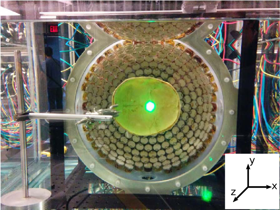
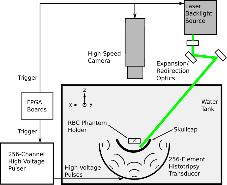
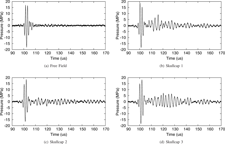
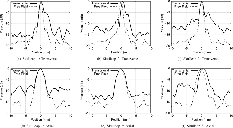
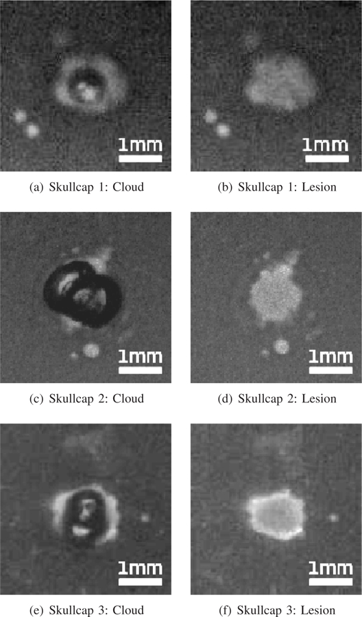
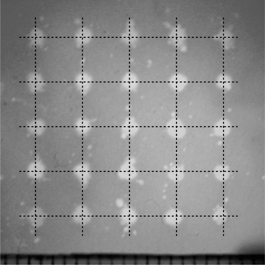
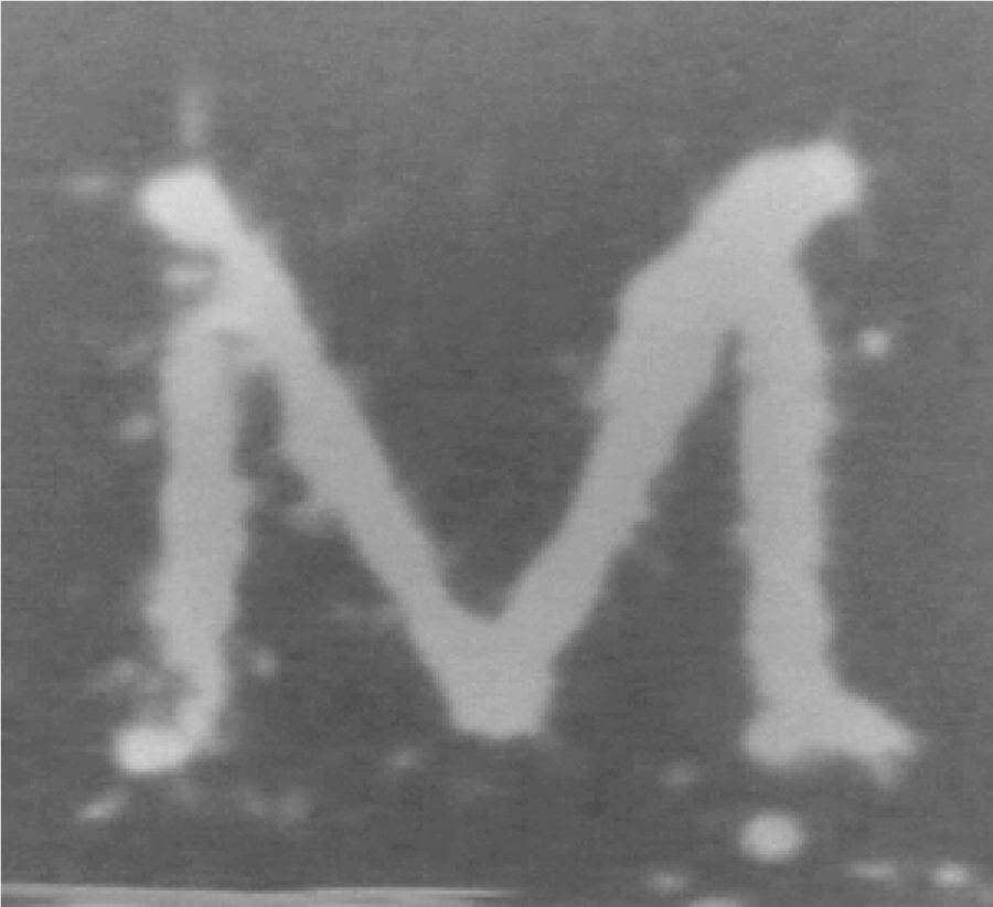
This study demonstrates the ability of histotripsy to generate targeted lesions through the skullcap without using aberration correction. Histotripsy therapy was delivered using a 500 kHz, 256-element hemispherical transducer with an aperture diameter of 30 cm and a focal distance of 15 cm fabricated in our lab. This transducer is theoretically capable of producing peak rarefactional pressures, based on linear estimation, (p-)LE, in the free field in excess of 200MPa with pulse durations 2 acoustic cycles. Three excised human skullcaps were used displaying attenuations of 73-81% of the acoustic pressure without aberration correction. Through all three skullcaps, compact lesions with radii less than 1mm were generated in red blood cell (RBC) agarose tissue phantoms without aberration correction, using estimated (p-)LE of 28-39MPa, a pulse repetition frequency of 1Hz, and a total number of 300 pulses. Lesion generation was consistently observed at the geometric focus of the transducer as the position of the skullcap with respect to the transducer was varied, and multiple patterned lesions were generated transcranially by mechanically adjusting the position of the skullcap with respect to the transducer to target different regions within. These results show that compact, targeted lesions with sharp boundaries can be generated through intact skullcaps using histotripsy with very short pulses without using aberration correction. Such capability has the potential to greatly simplify transcranial ultrasound therapy for non-invasive transcranial applications, as current ultrasound transcranial therapy techniques all require sophisticated aberration correction.
Figures

References
-
- Fry WJ, Mosberg W Jr, Barnard J, and Fry F, “Production of focal destructive lesions in the central nervous system with ultrasound*,” Journal of neurosurgery, vol. 11, no. 5, pp. 471–478, 1954. - PubMed
-
- Barnard J, Fry W, Fry F, and Brennan J, “Small localized ultrasonic lesions in the white and gray matter of the cat brain,” AMA Archives of Neurology & Psychiatry, vol. 75, no. 1, pp. 15–35, 1956. - PubMed
-
- Young G and Lele P, “Focal lesions in the brain of growing rabbits produced by focused ultrasound,” Experimental Neurology, vol. 9, no. 6, pp. 502–511, 1964. - PubMed
-
- Fry F, “Transkull transmission of an intense focused ultrasonic beam,” Ultrasound in medicine & biology, vol. 3, no. 2, pp. 179–184, 1977. - PubMed
Grants and funding
LinkOut - more resources
Full Text Sources
Other Literature Sources
Miscellaneous
