. 2016 May;79(5):726-738.
doi: 10.1002/ana.24620.

Dynamic Imaging of Individual Remyelination Profiles in Multiple Sclerosis

Affiliations

Dynamic Imaging of Individual Remyelination Profiles in Multiple Sclerosis

Benedetta Bodini et al. Ann Neurol. 2016 May.

Abstract

Background: Quantitative in vivo imaging of myelin loss and repair in patients with multiple sclerosis (MS) is essential to understand the pathogenesis of the disease and to evaluate promyelinating therapies. Selectively binding myelin in the central nervous system white matter, Pittsburgh compound B ([11 C]PiB) can be used as a positron emission tomography (PET) tracer to explore myelin dynamics in MS.

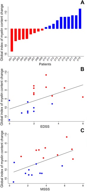
Methods: Patients with active relapsing-remitting MS (n = 20) and healthy controls (n = 8) were included in a longitudinal trial combining PET with [11 C]PiB and magnetic resonance imaging. Voxel-wise maps of [11 C]PiB distribution volume ratio, reflecting myelin content, were derived. Three dynamic indices were calculated for each patient: the global index of myelin content change; the index of demyelination; and the index of remyelination.

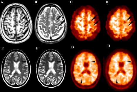
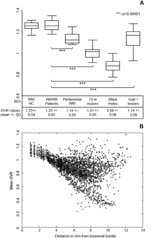
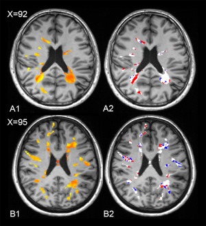
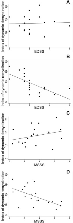
Results: At baseline, there was a progressive reduction in [11 C]PiB binding from the normal-appearing white matter to MS lesions, reflecting a decline in myelin content. White matter lesions were characterized by a centripetal decrease in the tracer binding at the voxel level. During follow-up, high between-patient variability was found for all indices of myelin content change. Dynamic remyelination was inversely correlated with clinical disability (p = 0.006 and beta-coefficient = -0.67 with the Expanded Disability Status Scale; p = 0.003 and beta-coefficient = -0.68 with the MS Severity Scale), whereas no significant clinical correlation was found for the demyelination index.

Interpretation: [11 C]PiB PET allows quantification of myelin dynamics in MS and enables stratification of patients depending on their individual remyelination potential, which significantly correlates with clinical disability. This technique should be considered to assess novel promyelinating drugs. Ann Neurol 2016;79:726-738.


Figures

Figure 1
Figure 1
Representative magnetic resonance imaging (MRI) and positron emission tomography (PET) images from MS patients. (A–D) T1‐weighted MRI (A), T2‐weighted MRI (B), Pittsburgh compound B ([11C]PiB) standard uptake value (SUV) map (C), and [11C]PiB distribution volume ratio (DVR) parametric map (D) of a single patient at study entry. SUV maps are semi‐quantitative measures of the tracer's uptake obtained by averaging the PET frames acquired between the minutes 30 and 70 of the examination and correcting the values for the tracer's injected dose and the patient's weight. DVR maps are quantitative parametric maps obtained with the automatic extraction of a reference region and the subsequent application of the Logan graphical method. Arrows indicate two typical multiple sclerosis white matter lesions appearing as areas of decreased uptake both on SUV and DVR images. T2‐weighed MRI at study entry (E) and after 3 months (F) and [11C]PiB DVR parametric map at baseline (G) and at follow‐up (H) of a single patient. Arrowheads (G and H) indicate two lesions visible on MRI scans that appear as regions of decreased DVR values on PET images and point to parts of the lesions where a subtle local increase in DVR value between the first and second PET scan is visible, suggesting local myelin regeneration developing during follow‐up. Note that the same lesion appears unchanged on T2‐weighed images.
Figure 2
Figure 2
Gradient in Pittsburgh compound B ([11C]PiB) binding from normal‐appearing WM to the center of lesions. (A) Box plot diagrams showing the median DVR (middle line) and range for each ROI at baseline in healthy controls and patients (from left to right: WM in healthy controls, normal‐appearing WM in patients, perilesional WM, T2‐weighted lesions, black holes, and gadolinium‐enhancing lesions). These box plots show that the lowest myelin content was detected in the “black holes”, the hypointense lesions on T1 spin‐echo scans that are known to represent the most severely demyelinated lesions in MS brains. A paired t test was used in a within‐patient analysis to test for between‐region differences in myelin content. (B) [11C]PiB binding values are negatively correlated with the distance from the lesional border. Each point in this scatter plot diagram represents the mean DVR value (y axis) of all the voxels localized at any given distance in millimeters from the lesional border (x axis) in any given patient. Although voxels closer to the lesional border, on average, present higher myelin content values, those located far from the lesional border tend to present lower myelin content values. The correlation between each voxel's distance in millimeters from the lesional border and its corresponding DVR value, which was tested using a mixed‐effect linear model in which the subject was included as random effect and age and gender were covariates, was highly significant (p = 0.00001). DVR = distribution volume ratio; MS = multiple sclerosis; NAWM = normal‐appearing white matter; ROI = region of interest; WM = white matter. SD = standard deviation; T2‐w = T2‐weighted.
Figure 3
Figure 3
Between‐patient heterogeneity in the global index of myelin content change values. (A) Bar chart diagram displaying the global index of myelin content change value for each patient, which is defined as the difference in demyelinated voxels between the second time point and baseline. This index reflects the individual balance between dynamic demyelination and dynamic remyelination. Patients with positive values on the global index of myelin content change, which indicate a predominant dynamic demyelinating process, are displayed in red. Patients with negative values, characterized by a prevalent dynamic process of remyelination, are indicated in blue. (B and C) Scatter plot diagrams and fitting lines representing the correlations between the global index of myelin content change and clinical scores. Although only a trend toward a significant correlation was found between the global index of myelin content change and EDSS (B), a significant correlation was found between this index and MSSS (C). EDSS = Expanded Disability Status Scale; MSSS = Multiple Sclerosis Severity Scale.
Figure 4
Figure 4
Dynamic myelin loss and regeneration: images from two patients. In A1 and B1, the myelin content of lesional voxels in 2 patients at baseline (patient A: male, 33 years old, disease duration 4 years, EDSS 3; patient B: female, 32 years old, disease duration 3 years, EDSS 0), as measured by Pittsburgh compound B ([11C]PiB) binding (voxels in red correspond to the values in the lower range, reflecting more severely demyelinated areas), is represented in red and yellow. In A2 and B2, the longitudinal follow‐up of the same patients is displayed, with the demyelinating voxels over time reported in red and the remyelinating voxels reported in blue. The dynamically demyelinating voxels (in red) were defined as normally myelinated voxels at baseline that were classified as demyelinated at the second time point. Dynamically remyelinating voxels (in blue) were those demyelinated voxels at baseline that reached a myelin level within normal limits at follow‐up. EDSS = Expanded Disability Status Scale.
Figure 5
Figure 5
Clinical relevance of remyelination. Scatter plot diagrams and fitting lines representing the correlations between EDSS individual scores and the indices of dynamic demyelination (A) and dynamic remyelination (B) are reported. Although no significant correlation was found between the index of dynamic demyelination and EDSS, a strong inverse correlation was found between the index of dynamic remyelination and EDSS. Patients with lower disability were those presenting higher proportions of remyelinating voxels over total lesion load. Scatter plot diagrams and fitting lines representing the correlations between MSSS individual scores and the indices of dynamic demyelination (C) and dynamic remyelination (D) are also reported. EDSS = Expanded Disability Status Scale; MSSS = Multiple Sclerosis Severity Scale.

Comment in

References

    1. Ramagopalan SV, Dobson R, Meier UC, Giovannoni G. Multiple sclerosis: risk factors, prodromes, and potential causal pathways. Lancet Neurol 2010;9:727–739. - PubMed
    1. Kieseier BC, Wiendl H. Multiple sclerosis: advances, excitements, disenchantments. Lancet Neurol 2006;5:2–3. - PubMed
    1. Crawford AH, Chambers C, Franklin RJ. Remyelination: the true regeneration of the central nervous system. J Comp Pathol 2013;149:242–254. - PubMed
    1. Franklin RJ, Gallo V. The translational biology of remyelination: past, present, and future. Glia 2014;62:1905–1915. - PubMed
    1. Filippi M, Rocca MA, Barkhof F, et al. Association between pathological and MRI findings in multiple sclerosis. Lancet Neurol 2012;11:349–360. - PubMed