Review
. 2016 Mar 15:594:8-17.
doi: 10.1016/j.abb.2016.02.016. Epub 2016 Feb 15.

Structural determinants of muscle thin filament cooperativity

Affiliations
Review

Structural determinants of muscle thin filament cooperativity

Jeffrey R Moore et al. Arch Biochem Biophys. .

Abstract

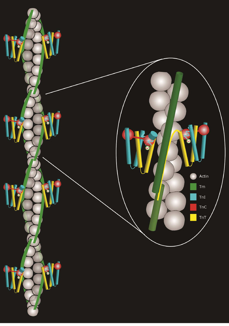
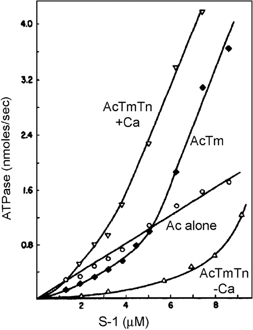
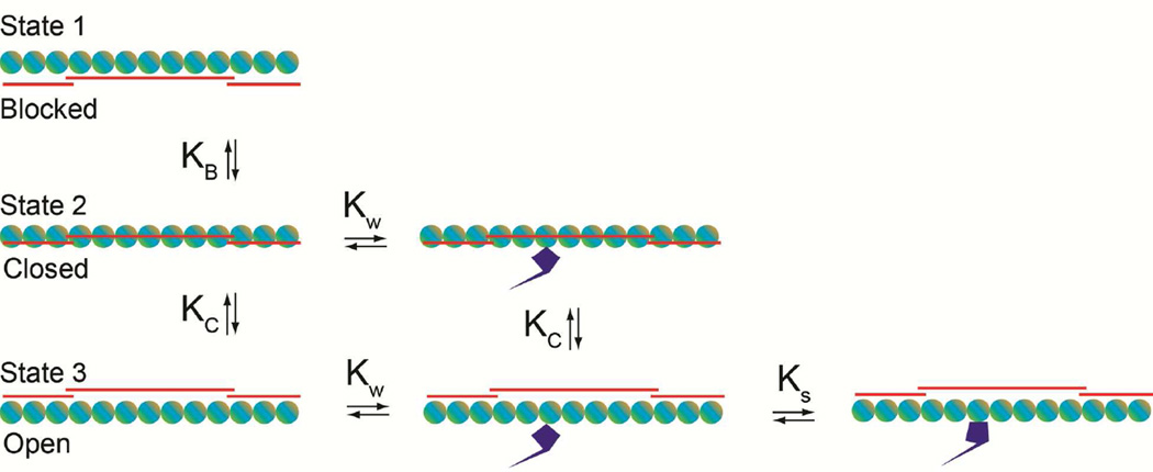
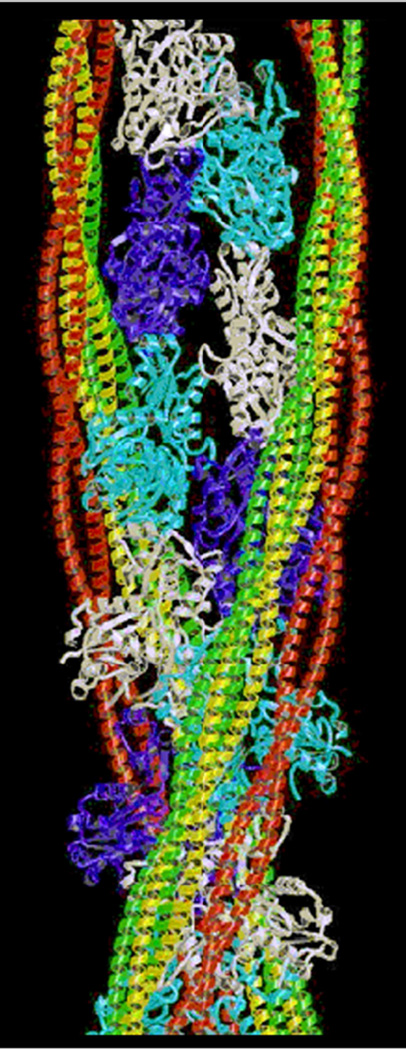
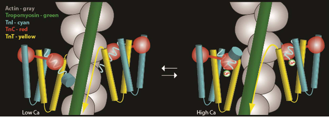
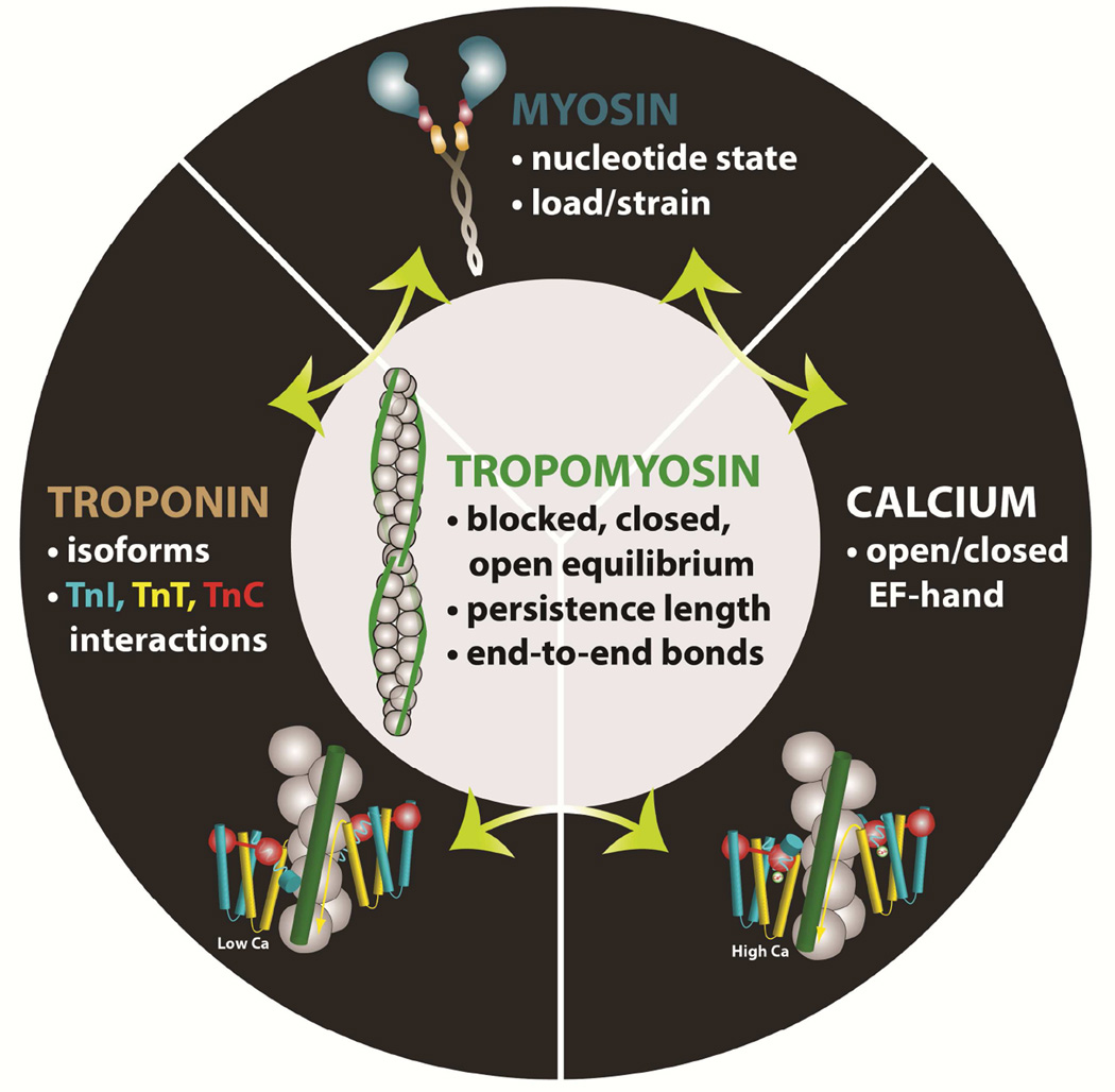
End-to-end connections between adjacent tropomyosin molecules along the muscle thin filament allow long-range conformational rearrangement of the multicomponent filament structure. This process is influenced by Ca(2+) and the troponin regulatory complexes, as well as by myosin crossbridge heads that bind to and activate the filament. Access of myosin crossbridges onto actin is gated by tropomyosin, and in the case of striated muscle filaments, troponin acts as a gatekeeper. The resulting tropomyosin-troponin-myosin on-off switching mechanism that controls muscle contractility is a complex cooperative and dynamic system with highly nonlinear behavior. Here, we review key information that leads us to view tropomyosin as central to the communication pathway that coordinates the multifaceted effectors that modulate and tune striated muscle contraction. We posit that an understanding of this communication pathway provides a framework for more in-depth mechanistic characterization of myopathy-associated mutational perturbations currently under investigation by many research groups.

Keywords: Actin; Cooperativity; Myosin; Thin filament; Tropomyosin; Troponin.


Figures

Figure 1
Figure 1
Cartoon representation of the thin filament showing the double stranded helical array of actin subunits (gray), end-to-end bonded tropomyosin molecules (green) and the troponin complex (TnI, cyan; TnC, red; TnT, yellow). Left, four successive troponin-tropomyosin regulatory units on each actin helical strand; Right, enlargement of a portion of the cartoon.
Figure 2
Figure 2
Dual effect of tropomyosin and troponin-tropomyosin on actomyosin subfragment 1 ATPase. Acto-S1 ATPase assays indicate that the troponin-tropomyosin regulated thin filament activation of myosin ATPase is Ca2+-dependent, while troponin-free F-actin or F-actin-tropomyosin activate the ATPase but confer no Ca2+-dependence. The activation of S1 ATPase by “unregulated” tropomyosin-free F-actin is linear as a function of added S1, yet sigmoidal when either tropomyosin or troponin-tropomyosin is present (saturation by very high S1 levels not shown). Thus, relative to control F-actin values, tropomyosin, in the presence and absence of troponin and Ca2+, inhibits actomyosin ATPase at low S1:F-actin ratios, and stimulates ATPase at high S1:F-actin ratios. Inhibition and activation by tropomyosin are greatest when troponin is present. Figure adapted, with permission, from reference [31]. This figure was originally published in The Journal of Biological Chemistry by S.S. Lehrer and E.P. Morris in an article “Dual effects of tropomyosin and tropomyosin-troponin on skeletal muscle subfragment 1 ATPase” J. Biol. Chem. 1982; 257:8073–8080 © the American Society for Biochemistry and Molecular Biology.
Figure 3
Figure 3
Schematic diagram of the McKillop-Geeves three-state regulatory model of the thin filament. (A) Thin filament regulatory units consist of single lines depicting tropomyosin (red) over seven circles representing actin (aquamarine/tan); myosin is drawn as blue S1-like objects. Blocked, closed, open states (a.k.a. B-, C-, M-states [41]) are indicated. The dynamic equilibrium between states is illustrated as an azimuthal translocation of tropomyosin on actin and an isomerization of myosin into a strongly attached configuration. KB and KC identify equilibrium transitions between blocked and closed states, while KW and KS represent equilibria for initial weak and then strong myosin binding steps on actin.
Figure 4
Figure 4
Atomic coordinates of actin, tropomyosin and myosin based on single particle reconstructions of thin filaments [99]. Tropomyosin in its three average regulatory positions superposed on actin for comparison; tropomyosin shown in the blocking, closed and open states (red, yellow, green, respectively). Figure adapted, with permission, from [6].
Figure 5
Figure 5
Cartoon representations of the movement of tropomyosin under the influence of conformational changes in troponin dependent on Ca2+. Low-Ca2+ – left, High-Ca2+ – right. Figure from references [12] and [67] with permission. Note the movement of tropomyosin and the changing association of the TnI C-terminus (depicted as a disc), at low-Ca2+ associating with TnC and at high-Ca2+ with actin-tropomyosin.
Figure 6
Figure 6
The interconnected effects of thin filament cooperative activation operate through tropomyosin. Several effectors, acting through tropomyosin, contribute to cooperative activation of the striated muscle thin filament. Tropomyosin exists in three equilibrium states: blocked, closed, and open that control access of myosin to its binding site on actin. Calcium-binding to TnC, which is itself moderately cooperative in skeletal muscle, shifts the equilibrium via the multicomponent troponin complex allowing myosin to bind weakly to actin. Myosin-binding shifts tropomyosin further over actin exposing neighboring binding sites on actin, which can slightly increase Ca2+-binding by the thin filament. The predominant player in the final step of cooperative activation appears to be strongly attached myosin molecules on actin that perturb tropomyosin-defined regulatory units locally, thereby increasing myosin-attachment in neighboring units by shifting tropomyosin. The efficacy of this cooperative switching mechanism of tropomyosin depends on the degree and duration of strongly bound actin-myosin attachments, the tropomyosin flexibility and the tropomyosin-tropomyosin end-to-end bond strength along the end-to-end linked tropomyosin cable on the thin filament.

References

    1. Haselgrove JC. X-ray evidence for a conformational change in actin-containing filaments of vertebrate striated muscle. Cold Spring Harbor Symp. Quant. Biol. 1972;37:341–352.
    1. Huxley HE. Structural changes in actin and myosin-containing filaments during contraction. Cold Spring Harbor Symp. Quant. Biol. 1972;37:361–376.
    1. Parry DAD, Squire JM. Structural role of tropomyosin in muscle regulation: Analysis of the X-ray diffraction patterns from relaxed and contracting muscles. J. Mol. Biol. 1973;75:33–55. - PubMed
    1. Lehman W, Craig R, Vibert P. Ca2+-induced tropomyosin movement in Limulus thin filaments revealed by three-dimensional reconstruction. Nature. 1994;368:65–67. - PubMed
    1. Vibert P, Craig R, Lehman W. Steric-model for activation of muscle thin filaments. J. Mol. Biol. 1997;266:8–14. - PubMed

Publication types

LinkOut - more resources