. 2016 Feb 19:6:21094.
doi: 10.1038/srep21094.

Differential effects of gram-positive and gram-negative bacterial products on morphine induced inhibition of phagocytosis

Affiliations

Differential effects of gram-positive and gram-negative bacterial products on morphine induced inhibition of phagocytosis

Jana Ninkovic et al. Sci Rep. .

Erratum in

Abstract

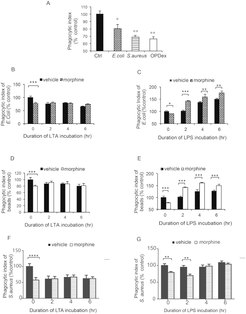
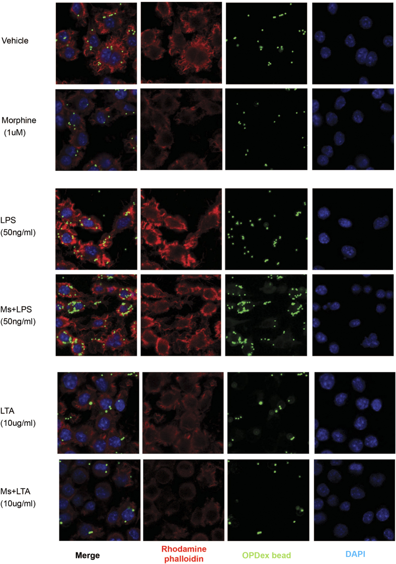
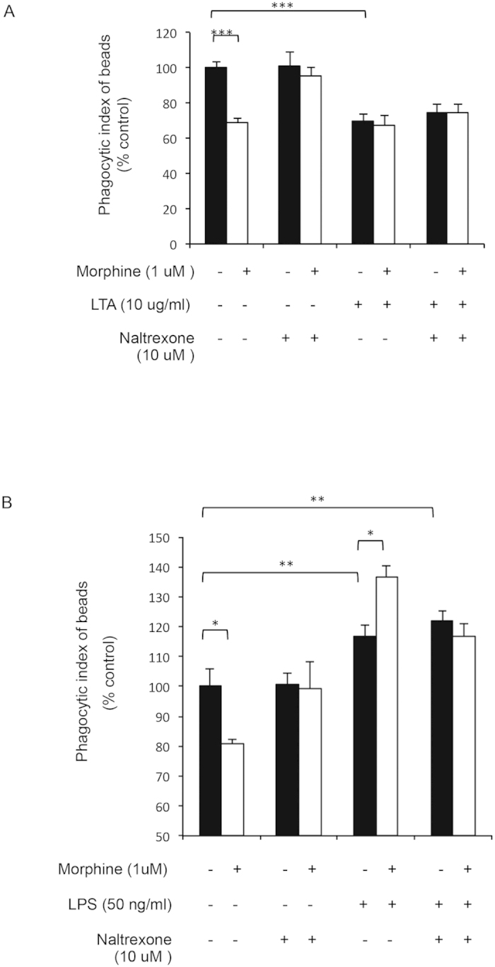
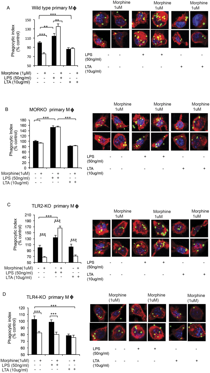
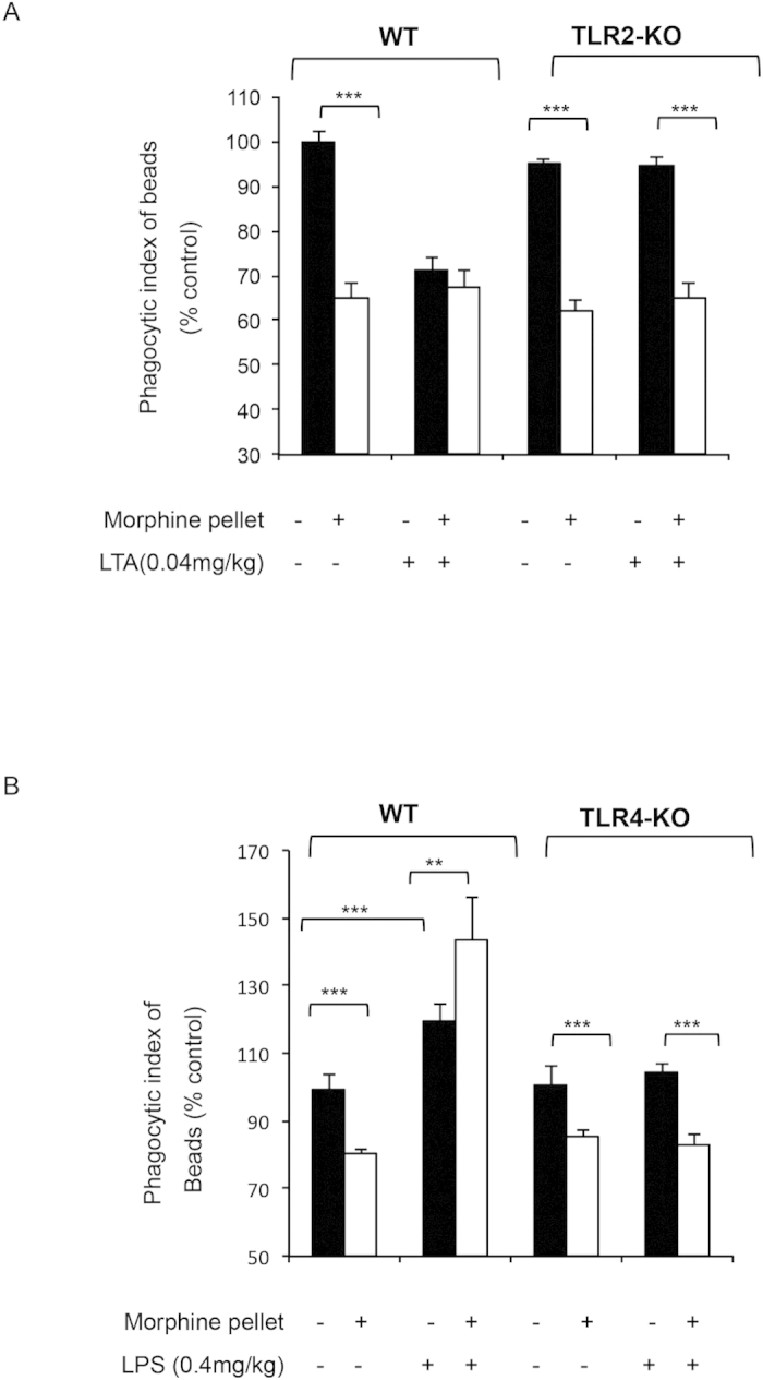
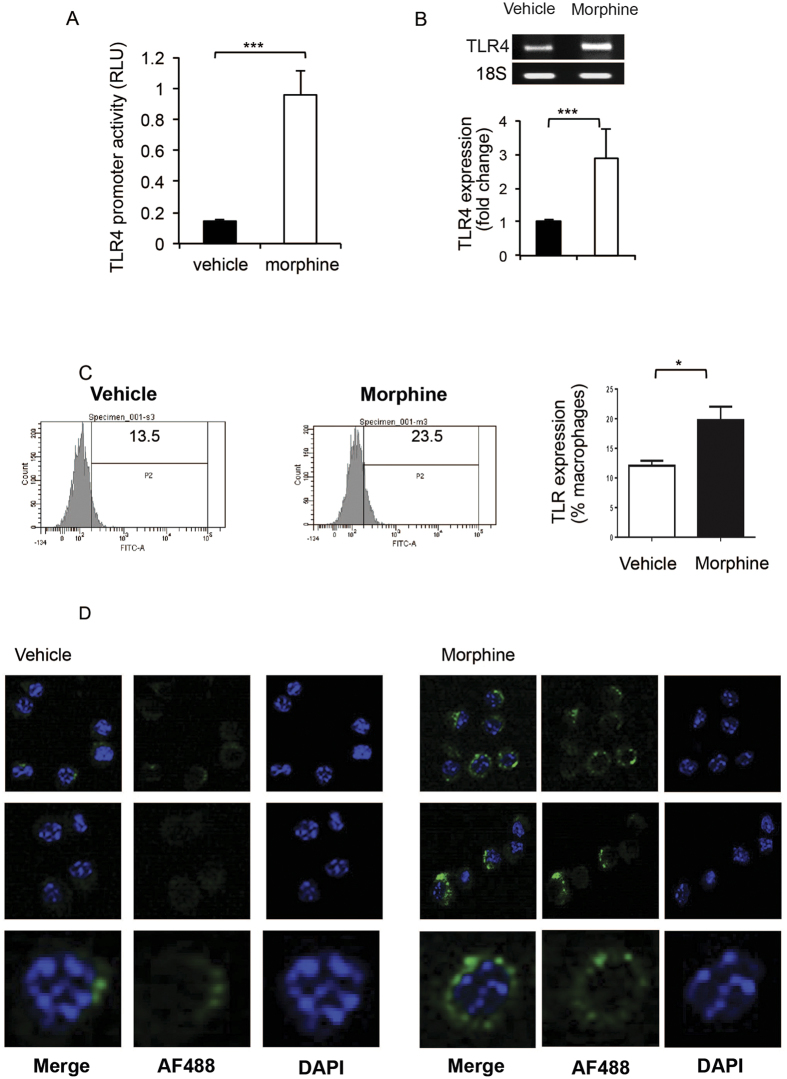
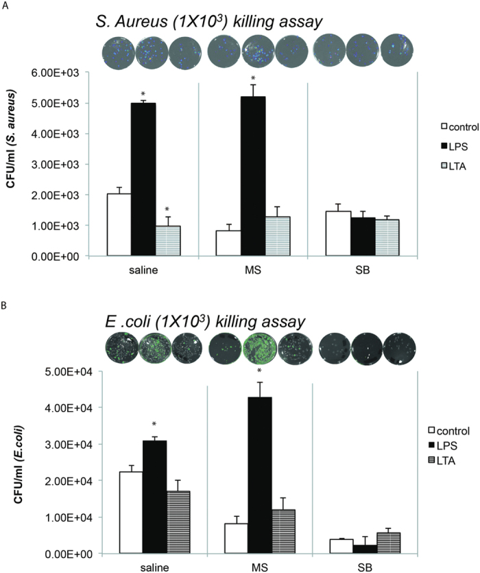
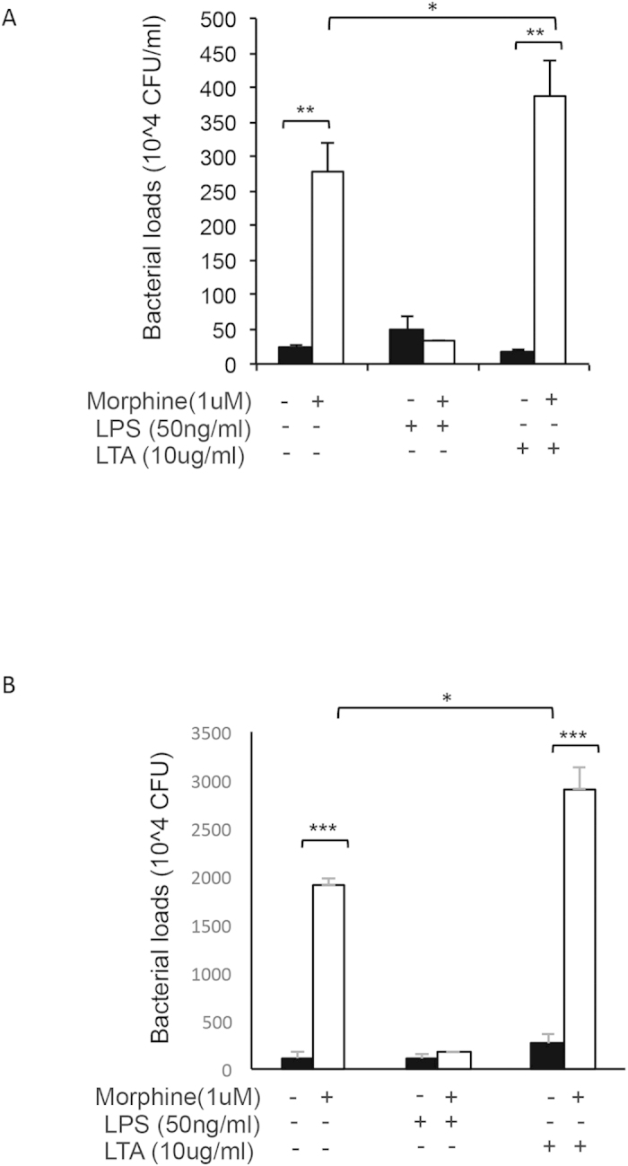
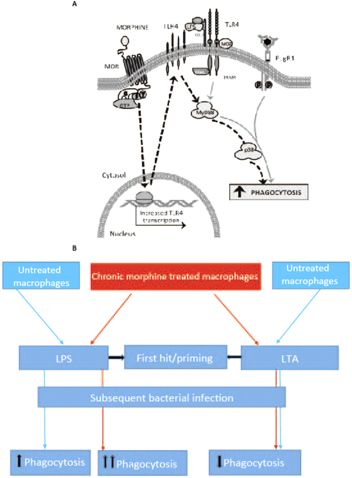
Opioid drug abusers have a greater susceptibility to gram positive (Gram (+)) bacterial infections. However, the mechanism underlying opioid modulation of Gram (+) versus Gram (-) bacterial clearance has not been investigated. In this study, we show that opioid treatment resulted in reduced phagocytosis of Gram (+), when compared to Gram (-) bacteria. We further established that LPS priming of chronic morphine treated macrophages leads to potentiated phagocytosis and killing of both Gram (+) and Gram (-) bacteria in a P-38 MAP kinase dependent signaling pathway. In contrast, LTA priming lead to inhibition of both phagocytosis and bacterial killing. This study demonstrates for the first time the differential effects of TLR4 and TLR2 agonists on morphine induced inhibition of phagocytosis. Our results suggest that the incidence and severity of secondary infections with Gram (+) bacteria would be higher in opioid abusers.


Figures

Figure 1
Figure 1. Differential effects of TLR4 (LPS) and TLR2 (LTA) ligands on phagocytosis of chronic morphine treated macrophages.
(A) Fluorometric analysis of phagocytosis of HKOE. coli (30 min), HKOS. aureus (30 min) and OPDex bead (60 min) by J774 following chronic morphine treatment (1 μM overnight). Phagocytosis of opsonized HKO E. coli (B,C) HKOE. coli and (D,E) OPDex bead (F,G) HKOS. aureus by morphine/vehicle treated J774 macrophages following LTA (10 μg/ml) (B,D,F) or LPS (50 ng/ml) (C,E,G) treatment for 2,4 and 6 hours. Phagocytic index expressed as ratio of relative fluorescence units (FITC/DAPI) expressed as % from vehicle control. Bars illustrate mean of three independent experiments ± SE (student’s t-test ***p < 0.001, **p < 0.01, *p < 0.05-significance to respective vehicle control).
Figure 2
Figure 2. Microscopic analysis of morphine modulation of phagocytosis following TLR4 and TLR2 activation.
Cells undergoing chronic morphine treatment (1 μM overnight) were exposed to (A) vehicle, (B) LTA (10 μg/ml) or (C) LPS (50 ng/ml) 4 hours prior to phagocytosis. Following internalization of FITC conjugated OPDex bead (1:20, cell:bead ratio) for 60 min, cells were washed with trypan and PBS, fixed with paraformaldehyde, permeabilized with acetone and stained for f-actin using rhodamine phalloidin (red), and DAPI (blue). Images illustrate confocal microscopic analysis at 60× with additional digital magnification.
Figure 3
Figure 3. Modulation of phagocytosis following activation of TLR 4 and TLR 2 in morphine treated primary macrophages is naltrexone reversible.
Fluorometric quantification of phagocytosis by J774 macrophages. Naltrexone (10 μM) was added 2 hr prior to overnight morphine (1 μM) treatment of primary macrophages from WT mice. (A) LTA (10 μg/ml) and (B) LPS (50 ng/ml) were added 4hr before addition of OPDex beads. Phagocytosis was allowed for 60 min and cells were analyzed via fluorometric analysis and expressed as % control (Phagocytic index = FITC (RFU)/DAPI(RFU)). Data quantified as % from vehicle control, mean ± standard error. Significance was determined using student’s t-test (***p < 0.0001, **p< 0.001, *p < 0.05).
Figure 4
Figure 4. Effects of chronic morphine and TLR ligands in knock-out mice.
Macrophages extracted from (A) wild-type, (B) MORKO, (C) TLR2-KO and (D) TLR4-KO were treated ex vivo with morphine, LPS and LTA as previously described for 4 hours, and analyzed using fluorometric assay and confocal microscopy (single cells) following 60 min of phagocytosis with OPDex bead (***p < 0.0001).
Figure 5
Figure 5. Effects of chronic morphine and TLR ligands (LPS and LTA) in knock-out mice.
Fluorometric analysis of ex vivo phagocytosis by primary macrophages from WT, TLR2-KO or TLR4-KO mice treated in vivo with morphine (75 mg slow releasing pellet for 3 days), and injected IP with LTA (A) or LPS (B) for 4 hours prior to sacrifice and cell harvesting. Phagocytosis was assessed ex vivo following 60 min of OPDex incubation, using fluorometric analysis and expressed as % control (Phagocytic index = FITC (RFU)/DAPI(RFU). Data quantified as % from vehicle control, mean ± standard error. Significance was determined using student’s t-test (**p < 0.001, ***p < 0.0001).
Figure 6
Figure 6. Chronic morphine treatment increases TLR-4 expression.
(A) TLR4 promoter activity as measured by luciferase. J774 cells were transfected using Fugene HD transfection reagent (roche) with a plasmid containing firefly luciferase, driven by the TLR4 full-length (~2.2 kb) promoter. Chronic morphine treatment was started 24 hours post-transfection. Promoter activity was measured by luciferase activity normalized to renilla luciferase values and represented as Relative Luciferase Units (RLU). (B) QT-PCR analysis of TLR4 expression following overnight 1 uM morphine treatment. (C) After undergoing chronic morphine (1 μM) treatment overnight J774 cells were collected, fixed and stained with Anti-Mouse CD284 (TLR4) Alexa Flour 488 antibody (eBioscience; cat# 53-9041-80) for 30 min at 4 °C and analyzed using Diva software (BD Bioscience). Data quantified as % positive cells, mean ± standard error. Significance was determined using student’s t-test (*p < 0.05, **p < 0.001, ***p < 0.0001). (D) Confocal microscopy analysis of the cytospin from the cells collected as in (C). Images are 4.2 times zoomed after collecting at 40×.
Figure 7
Figure 7
Modulation of phagocytosis of live bacteria by chronic morphine treatment following LPS and LTA priming: J774 macrophage cells were pretreated with morphine (1 um) overnight and than exposed to either vehicle, LPS (50 ng/ml) or LTA (10 ug/ml) for 4 hours and then incubated with either live GFP-tagged E. coli (1 × 103) (A) or live luciferase tagged S. aureus (1 × 103) , bioluminescent strain 100) (B). Cell to bacteria ratio was maintained at 1:20. To determine the mechanism underlying increase in phagocytosis by morphine treated LPS, J774 macrophages were treated with p38 MAP kinase inhibitor (SB 202190-10 uM) for 4 hours and then treated with morphine overnight and then incubated with live bacteria as described above. Representative bioluminescence photographs (upper insert) of bacterial culture plates after overnight culture and histograms represents mean±SE of colony-forming units (CFUs) of S. aureus/E. coli (lower panel) recovered after lysing of J774 macrophage cell line (0.2 × 106/200 μl). *p< 0.05 (student T test).
Figure 8
Figure 8. LPS and LTA in presence of morphine differentially modulates bacterial killing.
J774 cells undergoing morphine treatment (1 μM overnight), were treated with LPS (50 ng/ml) or LTA (10 μg/ml) for 4 hours (in antibiotic free media) prior to addition of GFP E. coli (A) and S. pneumoniae (B). Following 30 min of phagocytosis cells were washed four times with PBS containing 1% penicillin/streptomycin and then washed with antibiotic free medium and incubated overnight in antibiotic free media containing morphine, LPS or LTA (where appropriate). Next day cells were lysed and plated on LB plates (Ampicillin 100 μg/ml) for GFP E. coli and Kanamycin (50 ug/ml) blood agar plates for S. pneumoniae, and evaluated for bacterial colony formation (CFU) the next day. Significance was determined using student’s t-test (*p < 0.05, **p < 0.001,***p < 0.0001).
Figure 9
Figure 9
(A) Role of MOR and TLR in modulation of FcγR mediated phagocytosis. Morphine via activation of MOR increases TLR4 expression. In presence of LPS, TLR4 is activated to turn on MyD88 and p38 MAPK. By p38 MAPK phosphorylation, actin polymerization in increased leading to increased phagocytosis. (B) Schematic showing differential effects of LPS and LTA priming on morphine induced inhibition of phagocytosis. Chronic morphine treatment decreases phagocytosis in macrophages. Priming with LPS, increases phagocytosis in morphine treated macrophages which is greater than the effects of LPS alone. This potentiation is mediated by increase in TLR4 message and surface expression by morphine. LTA priming is associated with inhibition of phagocytosis which is independent to morphine’s effects and is neither additive nor synergistic.

References

    1. Roy S. et al. Opioid drug abuse and modulation of immune function: consequences in the susceptibility to opportunistic infections. Journal of neuroimmune pharmacology: the official journal of the Society on NeuroImmune Pharmacology 6, 442–465 (2011). - PMC - PubMed
    1. Peterson P. K., Sharp B., Gekker G., Brummitt C. & Keane W. F. Opioid-mediated suppression of interferon-gamma production by cultured peripheral blood mononuclear cells. J Clin Invest 80, 824–831 (1987). - PMC - PubMed
    1. Aderem A. & Underhill D. M. Mechanisms of phagocytosis in macrophages. Annu Rev Immunol 17, 593–623 (1999). - PubMed
    1. Hind C. R. Pulmonary complications of intravenous drug misuse. 1. Epidemiology and non-infective complications. Thorax 45, 891–898 (1990). - PMC - PubMed
    1. Palepu A. et al. Hospital utilization and costs in a cohort of injection drug users. Cmaj 165, 415–420 (2001). - PMC - PubMed

Publication types

MeSH terms