. 2017 Sep;25(9):2904-2913.
doi: 10.1007/s00167-016-4045-x. Epub 2016 Feb 18.

Gait analysis before and after corrective osteotomy in patients with knee osteoarthritis and a valgus deformity

Affiliations

Gait analysis before and after corrective osteotomy in patients with knee osteoarthritis and a valgus deformity

N van Egmond et al. Knee Surg Sports Traumatol Arthrosc. 2017 Sep.

Abstract

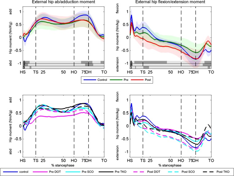
Purpose: In this prospective study, the changes in kinetics and kinematics of gait and clinical outcomes after a varus osteotomy (tibial, femoral or double osteotomy) in patients with osteoarthritis (OA) of the knee and a valgus leg alignment were analysed and compared to healthy subjects.

Methods: Twelve patients and ten healthy controls were included. Both kinetics and kinematics of gait and clinical and radiographic outcomes were evaluated.

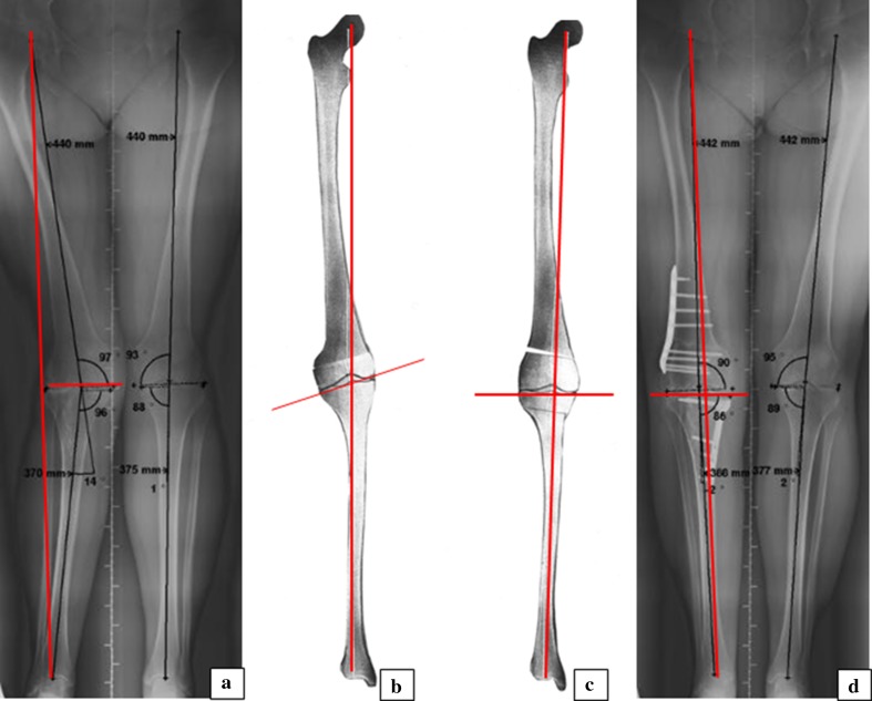
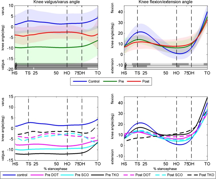
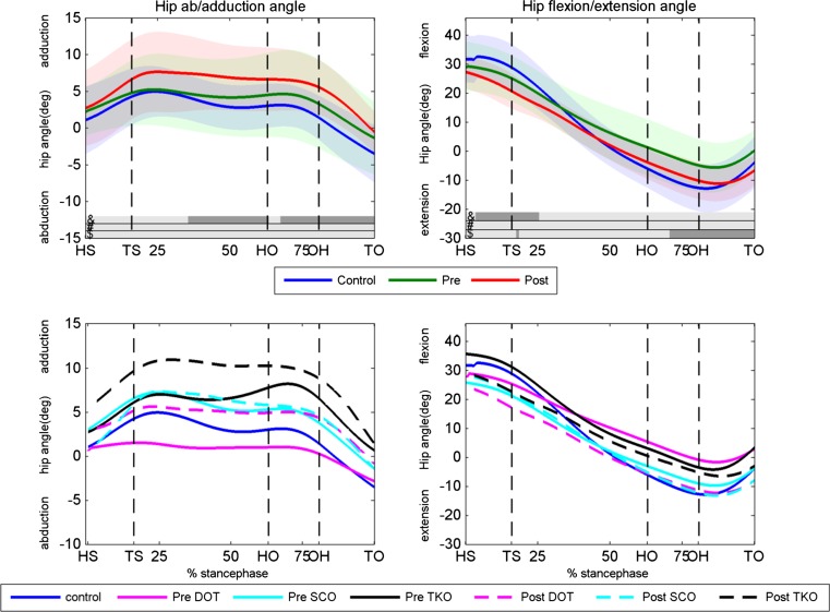
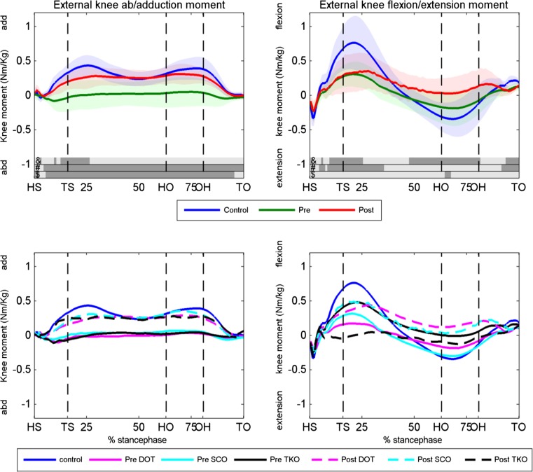
Results: The knee adduction moment increased significantly postoperatively (p < 0.05) and almost similar to the control group. Patients showed less knee and hip flexion/extension motion and moment during gait pre- and postoperatively compared to the controls. A significant improvement was found in WOMAC [80.8 (SD 16.1), p = 0.000], KOS [74.9 (SD 14.7), p = 0.018], OKS [21.2 (SD 7.5), p = 0.000] and VAS-pain [32.9 (SD 20.9), p = 0.003] in all patients irrespective of the osteotomy technique used. The radiographic measurements showed a mean hip knee ankle (HKA) angle correction of 10.4° (95 % CI 6.4°-14.4°).

Conclusion: In patients with knee OA combined with a valgus leg alignment, the varus-producing osteotomy is a successful treatment. Postoperatively, the patients showed kinetics and kinematics of gait similar as that of a healthy control group. A significant increase in the knee adduction moment during stance phase was found, which was related to the degree of correction. The HKA angle towards zero degrees caused a medial shift in the dynamic knee loading. The medial shift will optimally restore cartilage loading forces and knee ligament balance and reduces progression of OA or the risk of OA. A significant improvement in all clinical outcomes was also found.

Level of evidence: III.

Keywords: Closed wedge medial high tibial osteotomy; Double osteotomy; Gait analysis; Supracondylar femoral osteotomy; Valgus alignment.


Conflict of interest statement

The authors declare that they have no conflict of interest.

Figures

Fig. 1
Fig. 1
Rationale of double osteotomy in valgus corrective surgery. Weight-bearing long-leg radiographs and planning drawings including weight-bearing lines (WBL) and knee joint orientation lines (KJOL) of one of the study patients. a Preoperative valgus leg alignment caused by femoral and tibial bone deformity, WBL lateral and KJOL neutral. b Planning drawing of medial closing wedge distal femur osteotomy resulting in neutral WBL and severe valgus KJOL. c Planning drawing of double osteotomy, i.e. lateral open wedge distal femur and medial closing proximal tibial osteotomy, resulting in neutral WBL and neutral KJOL. d Postoperative leg alignment after double osteotomy
Fig. 2
Fig. 2
Knee valgus/varus and flexion/extension angles. Upper panels the knee angles of the controls, preoperative condition and postoperative condition for the valgus/varus (left panel) and flexion/extension (right panel) angle. Dark areas in the bars right above the x-axis indicate significant differences (p < 0.05) between: &, postoperative and controls; #, preoperative and controls; $, pre- and postoperative. Lower panels knee varus/valgus and flexion/extension angles for the DOT, SCO and TKO group. Pre- and postoperative as well as the control data are displayed. HS heel strike, TS toe strike, HO heel off, OH opposite heel strike, TO toe-off, Deg degrees, Pre preoperative, Post postoperative, DOT double osteotomy, SCO supracondylar osteotomy, TKO high tibial osteotomy
Fig. 3
Fig. 3
Hip abduction/adduction and flexion/extension angles. Upper panels the hip angles of the controls, preoperative condition and postoperative condition for the abduction/adduction (left panel) and flexion/extension (right panel) angle. Dark areas in the bars right above the x-axis indicate significant differences (p < 0.05) between: &, postoperative and controls, #, preoperative and controls, $, pre- and postoperative. Lower panels hip abduction/adduction and flexion/extension angles for the DOT, SCO and TKO group. Pre- and postoperative as well as the control data are displayed. HS heel strike, TS toe strike, HO heel off, OH opposite heel strike, TO toe-off, Deg degrees, Pre preoperative, Post postoperative, DOT double osteotomy, SCO supracondylar osteotomy, TKO high tibial osteotomy
Fig. 4
Fig. 4
Knee external knee abduction/adduction and flexion/extension moments. Upper panels the knee moments of the controls, preoperative condition and postoperative condition for the abduction/adduction (left panel) and flexion/extension (right panel) moment. Dark areas in the bars right above the x-axis indicate significant differences (p < 0.05) between: &, postoperative and controls, #, preoperative and controls, $, pre- and postoperative. Lower panels knee abduction/adduction and flexion/extension moments for the DOT, SCO and TKO group and the controls. HS heel strike, TS toe strike, HO heel off, OH opposite heel strike, TO toe-off, Deg degrees, Pre preoperative, Post postoperative, DOT double osteotomy, SCO supracondylar osteotomy, TKO high tibial osteotomy
Fig. 5
Fig. 5
Hip abduction/adduction and flexion/extension moments. Upper panels the hip moments of the controls, preoperative condition and postoperative condition for the abduction/adduction (left panel) and flexion/extension (right panel) moment. Dark areas in the bars right above the x-axis indicate significant differences (p < 0.05) between: &, postoperative and controls, #, preoperative and controls, $, pre,- and postoperative. Lower panels hip abduction/adduction and flexion/extension moments for the DOT, SCO and TKO group and controls. HS heel strike, TS toe strike, HO heel off, OH opposite heel strike, TO toe-off, Deg degrees, Pre preoperative, Post postoperative, DOT double osteotomy, SCO supracondylar osteotomy, TKO high tibial osteotomy

References

    1. Aglietti P, Menchetti PP. Distal femoral varus osteotomy in the valgus osteoarthritic knee. Am J Knee Surg. 2000;13(2):89–95. - PubMed
    1. Babis GC, An KN, Chao EY, et al. Double level osteotomy of the knee: a method to retain joint-line obliquity. J Bone Joint Surg Am. 2002;84-A:1380–1388. doi: 10.2106/00004623-200208000-00013. - DOI - PubMed
    1. Brinkmann JR, Perry J. Rate and range of knee motion during ambulation in healthy and arthritic subjects. Phys Ther. 1985;65:1055–1060. doi: 10.1093/ptj/65.7.1055. - DOI - PubMed
    1. Brouwer RW, Huizinga MR, Duivenvoorden T, van Raaij TM, Verhagen AP, Bierma-Zeinstra SMA, Verhaar JAN. Osteotomy for treating knee osteoarthritis (Review) Cochrane Database Syst Rev. 2014;13(12):CD004019. - PMC - PubMed
    1. Bus SA, de Lange A. 1,2,3 step protocol: a comparison of the 1-step, 2-step, and 3-step protocols for obtaining barefoot plantar pressure data in the diabetic neuropathic foot. Clin Biomech. 2005;20(9):892–899. doi: 10.1016/j.clinbiomech.2005.05.004. - DOI - PubMed