

Detection of metastatic tumors after γ-irradiation using longitudinal molecular imaging and gene expression profiling of metastatic tumor nodules
- PMID: 26892334
- PMCID: PMC4777593
- DOI: 10.3892/ijo.2016.3384
Detection of metastatic tumors after γ-irradiation using longitudinal molecular imaging and gene expression profiling of metastatic tumor nodules
Abstract
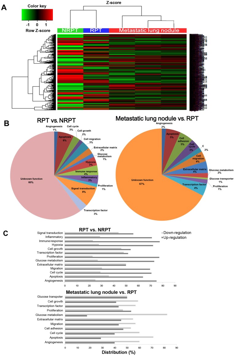
A few recent reports have indicated that metastatic growth of several human cancer cells could be promoted by radiotherapy. C6-L cells expressing the firefly luciferase (fLuc) gene were implanted subcutaneously into the right thigh of BALB/c nu/nu mice. C6-L xenograft mice were treated locally with 50-Gy γ-irradiation (γ-IR) in five 10-Gy fractions. Metastatic tumors were evaluated after γ-IR by imaging techniques. Total RNA from non-irradiated primary tumor (NRPT), γ-irradiated primary tumor (RPT), and three metastatic lung nodule was isolated and analyzed by microarray. Metastatic lung nodules were detected by BLI and PET/CT after 6-9 weeks of γ-IR in 6 (17.1%) of the 35 mice. The images clearly demonstrated high [18F]FLT and [18F]FDG uptake into metastatic lung nodules. Whole mRNA expression patterns were analyzed by microarray to elucidate the changes among NRPT, RPT and metastatic lung nodules after γ-IR. In particular, expression changes in the cancer stem cell markers were highly significant in RPT. We observed the metastatic tumors after γ-IR in a tumor-bearing animal model using molecular imaging methods and analyzed the gene expression profile to elucidate genetic changes after γ-IR.
Figures


Similar articles
-
Establishment of animal model for the analysis of cancer cell metastasis during radiotherapy.Radiat Oncol. 2012 Sep 11;7:153. doi: 10.1186/1748-717X-7-153. Radiat Oncol. 2012. PMID: 22963683 Free PMC article.
-
Using dual-tracer PET to predict the biologic behavior of human colorectal cancer.J Nucl Med. 2009 Nov;50(11):1857-64. doi: 10.2967/jnumed.109.064238. Epub 2009 Oct 16. J Nucl Med. 2009. PMID: 19837754
-
Application of carbon-ion beams or gamma-rays on primary tumors does not change the expression profiles of metastatic tumors in an in vivo murine model.Int J Radiat Oncol Biol Phys. 2009 May 1;74(1):210-8. doi: 10.1016/j.ijrobp.2008.12.078. Int J Radiat Oncol Biol Phys. 2009. PMID: 19362239
-
[Correlation of 3'-deoxy-3'-18F-fluorothymidine uptake to cell proliferation in lung carcinoma xenografts].Ai Zheng. 2006 Dec;25(12):1512-6. Ai Zheng. 2006. PMID: 17166377 Chinese.
-
p53 deficiency linked to B cell translocation gene 2 (BTG2) loss enhances metastatic potential by promoting tumor growth in primary and metastatic sites in patient-derived xenograft (PDX) models of triple-negative breast cancer.Breast Cancer Res. 2016 Jan 27;18(1):13. doi: 10.1186/s13058-016-0673-9. Breast Cancer Res. 2016. PMID: 26818199 Free PMC article.
Cited by
-
The increased adhesion of tumor cells to endothelial cells after irradiation can be reduced by FAK-inhibition.Radiat Oncol. 2019 Feb 4;14(1):25. doi: 10.1186/s13014-019-1230-3. Radiat Oncol. 2019. PMID: 30717801 Free PMC article.
References
Publication types
MeSH terms
LinkOut - more resources
Full Text Sources
Other Literature Sources
Research Materials
