

Parotid gland lesions: separate and combined diagnostic value of conventional MRI, diffusion-weighted imaging and dynamic contrast-enhanced MRI
- PMID: 26892378
- PMCID: PMC4846216
- DOI: 10.1259/bjr.20150912
Parotid gland lesions: separate and combined diagnostic value of conventional MRI, diffusion-weighted imaging and dynamic contrast-enhanced MRI
Abstract
Objective: To evaluate the ability of conventional MRI, diffusion-weighted imaging (DWI) and dynamic contrast-enhanced (DCE) MRI to differentiate malignant and benign parotid lesions.
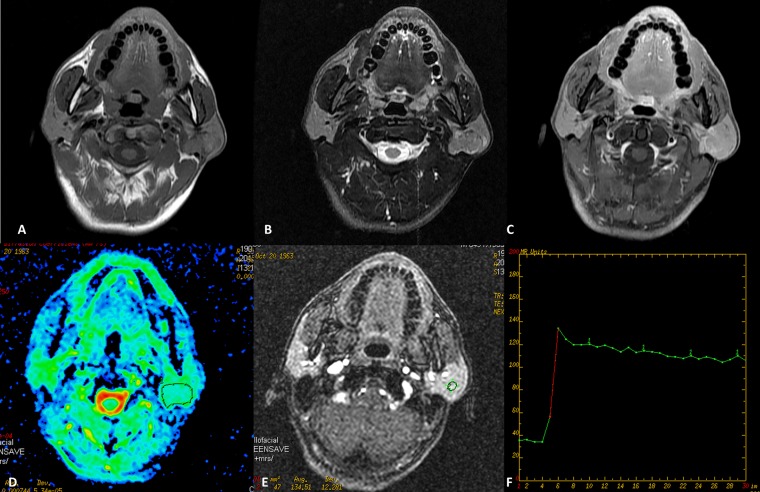
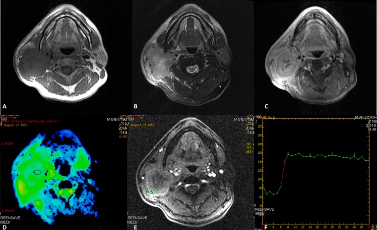
Methods: A retrospective review of MRI findings was performed in patients with pathologically confirmed parotid lesions between January 2010 and December 2014. Morphological MRI characteristics and functional characteristics such as apparent diffusion coefficient (ADC) and pattern of time-signal intensity curve (TIC) were recorded and compared. For each lesion, summed scores were respectively calculated for conventional MRI alone, conventional MRI with DWI and/or with DCE-MRI. Statistical analyses were performed to assess the association of these characteristics and summed scores with malignancy.
Results: A total of 207 patients (111 males and 96 females; age: 48.4 ± 17.0 years) were included, consisting of 156 benign and 51 malignant tumours. After adjusting for age, sex, smoking status, alcohol use and tumour size, the lesions with ill-defined margin, adjacent tissue infiltration, cervical lymphadenopathy, ADC ≤1.01 × 10(-3) mm(2) s(-1) and plateau TIC pattern are more likely to be malignant than those without these findings. Significant difference in receiver operator characteristic was detected after adding DWI to conventional MRI (p = 0.003), generating a sensitivity and specificity of 54.05% and 91.30%, respectively. Compared with lesions score <3, lesions with score ≥5 in the combination of conventional MRI and DWI were approximately 90 times more likely to be malignant parotid tumour. Additional DCE-MRI did not improve differential ability of conventional MRI.
Conclusion: Morphological and functional MRI characteristics are associated with malignancy in parotid gland. The diagnostic value of MRI would increase when DWI is applied in combination with conventional MRI.
Advances in knowledge: The parotid lesions with ill-defined margin, adjacent tissue infiltration, cervical lymphadenopathy, ADC ≤1.01 × 10(-3) mm(2) s(-1) and plateau TIC pattern are more likely to be malignant. The diagnostic value of conventional MRI would be increased when DWI is applied in combination, whereas additional DCE-MRI did not improve differential ability of conventional MRI.
Figures
References
Publication types
MeSH terms
Substances
LinkOut - more resources
Full Text Sources
Other Literature Sources
Medical
