Review
. 2016 Feb 19;118(4):653-67.
doi: 10.1161/CIRCRESAHA.115.306256.

Macrophage Phenotype and Function in Different Stages of Atherosclerosis

Affiliations
Review

Macrophage Phenotype and Function in Different Stages of Atherosclerosis

Ira Tabas et al. Circ Res. .

Abstract

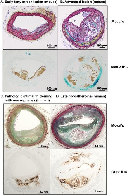
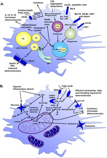
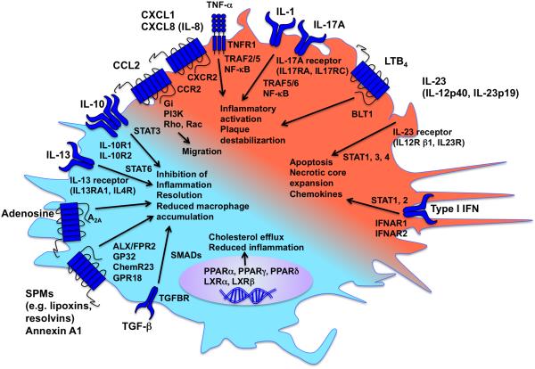
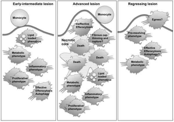
The remarkable plasticity and plethora of biological functions performed by macrophages have enticed scientists to study these cells in relation to atherosclerosis for >50 years, and major discoveries continue to be made today. It is now understood that macrophages play important roles in all stages of atherosclerosis, from initiation of lesions and lesion expansion, to necrosis leading to rupture and the clinical manifestations of atherosclerosis, to resolution and regression of atherosclerotic lesions. Lesional macrophages are derived primarily from blood monocytes, although recent research has shown that lesional macrophage-like cells can also be derived from smooth muscle cells. Lesional macrophages take on different phenotypes depending on their environment and which intracellular signaling pathways are activated. Rather than a few distinct populations of macrophages, the phenotype of the lesional macrophage is more complex and likely changes during the different phases of atherosclerosis and with the extent of lipid and cholesterol loading, activation by a plethora of receptors, and metabolic state of the cells. These different phenotypes allow the macrophage to engulf lipids, dead cells, and other substances perceived as danger signals; efflux cholesterol to high-density lipoprotein; proliferate and migrate; undergo apoptosis and death; and secrete a large number of inflammatory and proresolving molecules. This review article, part of the Compendium on Atherosclerosis, discusses recent advances in our understanding of lesional macrophage phenotype and function in different stages of atherosclerosis. With the increasing understanding of the roles of lesional macrophages, new research areas and treatment strategies are beginning to emerge.

Keywords: atherosclerosis; cholesterol; foam cells; macrophage; phenotype.


Figures

Figure 1
Figure 1. Cross-sections of different stages of lesions
A-B. Cross-sections of lesions in the brachiocephalic artery of Ldlr−/− mice fed a high-fat diet. A. Early fatty streak lesion with some smooth muscle cell involvement. B. Advanced lesion containing large necrotic cores in macrophage-rich areas. Macrophages are visualized by anti-Mac-2 immunohistochemistry (brown reaction product) on adjacent sections. C-D. Cross-sections of lesions of human coronary arteries. C. Pathologic intimal thickening with macrophages. D. Human advanced lesion (late fibroatheroma with macrophages and necrotic core). Macrophages are visualized by anti-CD68 immunohistochemistry (brown reaction product) on adjacent sections. Panels C-D are from Atherosclerosis, 241, Otsuka et al. Natural progression of atherosclerosis from pathologic intimal thickening to late fibroatheroma in human coronary arteries: A pathology study, pages 772-82, Copyright (2015), with permission from Elsevier. Sections were stained by Movat’s pentachrome. Scale bar, 100 μm (A-B); 1 mm (C-D).
Figure 2
Figure 2. Lipids and metabolism determine lesion macrophage phenotype and function
A. Lipoproteins impact macrophage phenotype. Lipoproteins are taken up by macrophages through macro-pinocytosis, phagocytosis and scavenger receptors, including SR-A, CD36, SR-BI, and LOX1. The scavenger receptors also have signaling capacities. As the lipoproteins are degraded in endosomes-lysosomes, CE is converted to free cholesterol (FC) by lysosomal acid lipase (LAL) and then redistributed to other cellular compartments through NPC (Niemann-Pick disease, type C) proteins. FC is converted back to CE by acyl-CoA cholesterol acyltransferase (ACAT) in the ER and then again to FC before it can be effluxed from the cell through the cholesterol exporters ABCA1 and ABCG1 to apolipoprotein A-I (ApoA-I) or HDL. FC can exert detrimental effects if it is not re-esterified to CE in lipid droplets or effluxed through ABCA1 and ABCG1. For example, cholesterol crystal formation leads to inflammasome activation, and FC accumulation causes ER stress and cell death. These processes promote progression of lesions of atherosclerosis and necrotic core expansion. B. The metabolic state of the macrophage influences its phenotype. Increased glycolysis initiated by glucose entry into the cell through glucose transporters, such as GLUT1, is required for the inflammatory phenotype. Hypoxia and inflammatory stimuli increase glycolysis in part to allow the cell to generate sufficient energy under anaerobic conditions, but also to fight pathogens and infection. TCA cycle intermediates also play important roles in inflammatory activation of macrophages. For example, the amino acid glutamine replenishes the TCA cycle to produce succinate, which in turn stabilizes the hypoxia-inducible factor 1 (HIF1) complex. Whereas a relative increase in fatty acid oxidation is associated with a more resolving macrophage phenotype, fatty acids and their metabolites exert a number of important effects in macrophages. Fatty acids are taken up by the cell through CD36 and fatty acid transport proteins (FATP) or by transport across the plasma membrane, and are readily converted to acyl-CoAs by a group of enzymes with acyl-CoA synthetase activity, including long-chain acyl-CoA synthetases (ACSLs). Acyl-CoAs are channeled to different fates in the cell, including oxidative phosphorylation (OxPhos), neutral lipids, phospholipids (PL), sphingolipids, or are used for protein modification or signaling. Sphingolipids are found in lipid rafts and are believed to promote inflammatory activation by TLRs present in these rafts. Reduced levels of intracellular fatty acids or acyl-CoAs or reduced OxPhos lead to reduced inflammatory activation and atherosclerosis.
Figure 3
Figure 3. Pro-inflammatory and pro-resolving mediators balance macrophage phenotype
A large number of pro-inflammatory and pro-resolving mediators act on macrophages through binding to specific receptors and activation of intracellular signaling pathways. The balance between pro-inflammatory mediators (indicated by red) and pro-resolving mediators (indicated by turquoise) determines the macrophage phenotype. Effects on atherosclerosis are indicated by arrows.
Figure 4
Figure 4. Roles of macrophages in different stages of lesions
In early fatty streak lesions, monocytes and macrophages gather lipoproteins and become lipid-loaded foam cells. Inflammatory mediators and changes in macrophage metabolism govern lesional macrophage phenotype. We define “metabolic phenotype” as a phenotype induced by substrates and intermediates used in energy metabolism, i.e. generation of ATP from nutrients. Some macrophages become apoptotic, but are effectively cleared by efferocytosis. Hyperlipidemia also promotes proliferation of macrophages, which may contribute to lesion growth. In advanced lesions, the ability of lesional macrophages to effectively efferocytose dying macrophages is defective, and inflammatory processes and macrophage death are rampant, contributing to necrotic core expansion and plaque rupture. Macrophages also secrete proteases, which might promote plaque rupture. In regressing lesions, lesional macrophages are reduced, perhaps due to egress or reduced monocyte infiltration, and perhaps due to improved efferocytosis and a pro-resolving macrophage phenotype. The remaining macrophages adopt a phenotype characterized by altered gene expression, and lipid loaded macrophages are reduced due to reduced lipid uptake and/or increased efflux.

References

    1. Ross R. Atherosclerosis--an inflammatory disease. N Engl J Med. 1999;340:115–126. - PubMed
    1. Libby P, Ridker PM, Maseri A. Inflammation and atherosclerosis. Circulation. 2002;105:1135–1143. - PubMed
    1. Hansson GK. Inflammation, atherosclerosis, and coronary artery disease. N Engl J Med. 2005;352:1685–1695. - PubMed
    1. Williams KJ, Tabas I. The response-to-retention hypothesis of early atherogenesis. Arterioscler Thromb Vasc Biol. 1995;15:551–561. - PMC - PubMed
    1. Libby P. Inflammation in atherosclerosis. Arterioscler Thromb Vasc Biol. 2012;32:2045–2051. - PMC - PubMed

Publication types