. 2016 Mar 15;143(6):1041-54.
doi: 10.1242/dev.130575. Epub 2016 Feb 18.

Wnt/β-catenin signaling enables developmental transitions during valvulogenesis

Affiliations

Wnt/β-catenin signaling enables developmental transitions during valvulogenesis

Fernanda M Bosada et al. Development. .

Abstract

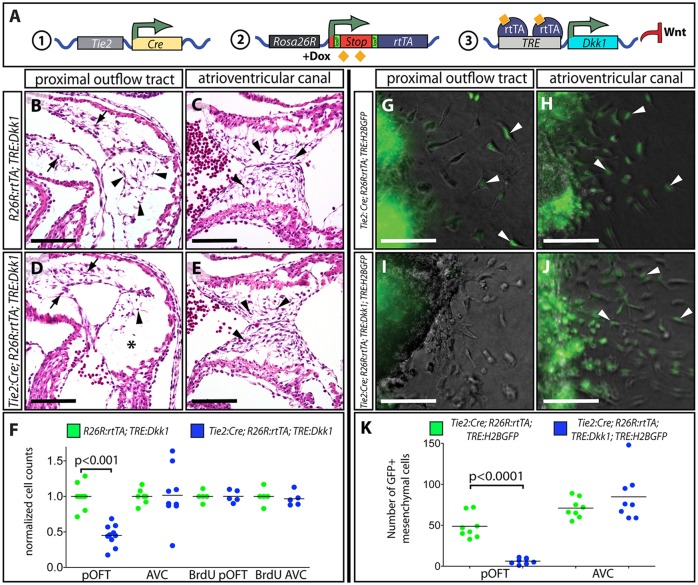
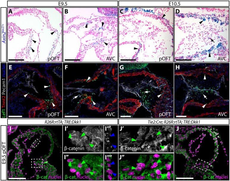
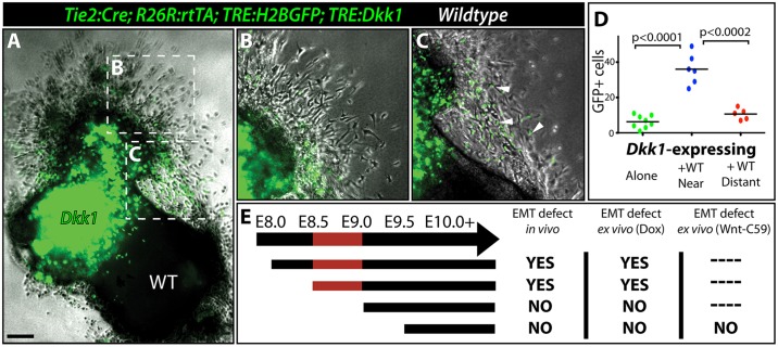
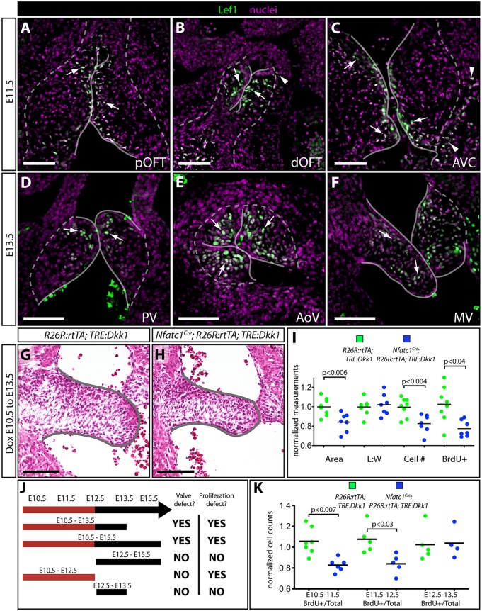
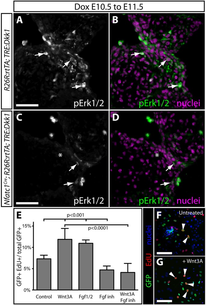
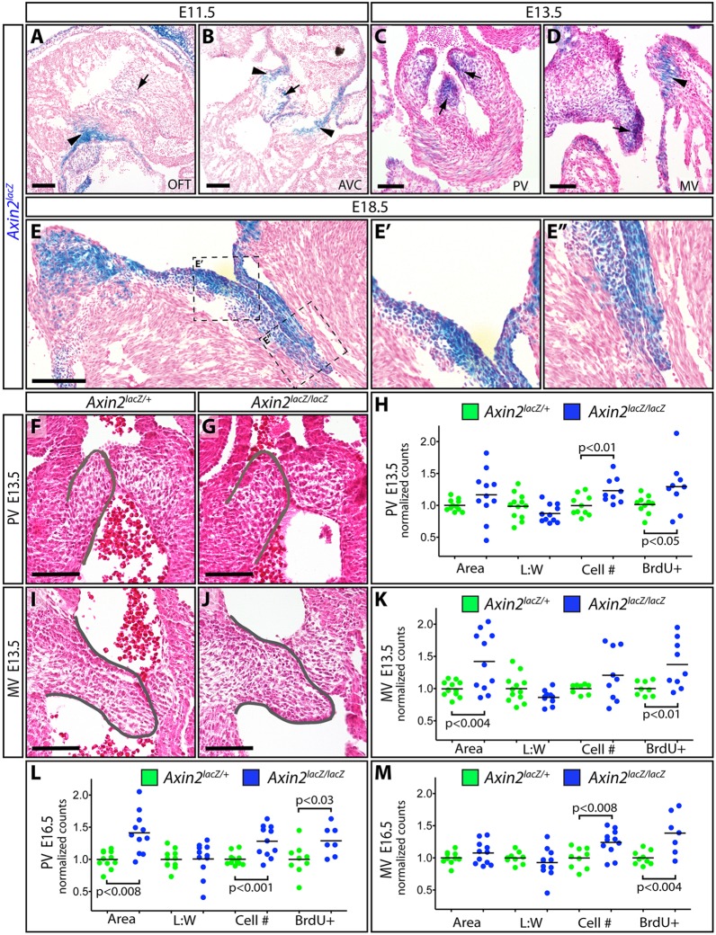
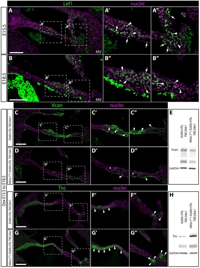
Heart valve development proceeds through coordinated steps by which endocardial cushions (ECs) form thin, elongated and stratified valves. Wnt signaling and its canonical effector β-catenin are proposed to contribute to endocardial-to-mesenchymal transformation (EMT) through postnatal steps of valvulogenesis. However, genetic redundancy and lethality have made it challenging to define specific roles of the canonical Wnt pathway at different stages of valve formation. We developed a transgenic mouse system that provides spatiotemporal inhibition of Wnt/β-catenin signaling by chemically inducible overexpression of Dkk1. Unexpectedly, this approach indicates canonical Wnt signaling is required for EMT in the proximal outflow tract (pOFT) but not atrioventricular canal (AVC) cushions. Furthermore, Wnt indirectly promotes pOFT EMT through its earlier activity in neighboring myocardial cells or their progenitors. Subsequently, Wnt/β-catenin signaling is activated in cushion mesenchymal cells where it supports FGF-driven expansion of ECs and then AVC valve extracellular matrix patterning. Mice lacking Axin2, a negative Wnt regulator, have larger valves, suggesting that accumulating Axin2 in maturing valves represents negative feedback that restrains tissue overgrowth rather than simply reporting Wnt activity. Disruption of these Wnt/β-catenin signaling roles that enable developmental transitions during valvulogenesis could account for common congenital valve defects.

Keywords: Atrioventricular canal; Axin2; Chordae tendineae; Cushion mesenchyme; EMT; Heart valves; Lef1; Mitral valve; Outflow tract; Spongiosa; Tenascin; Versican; Wnt/β-catenin signaling.


Conflict of interest statement

Competing interests

The authors declare no competing or financial interests.

Figures

Fig. 1.
Fig. 1.
Canonical Wnt signaling is required for proximal outflow tract (pOFT) but not atrioventricular canal (AVC) cushion EMT. (A) Schematic illustrating the transgenic mouse approach that uses doxycycline (Dox)-inducible expression of Dkk1 to provide spatiotemporal control over canonical Wnt signaling. (B-E) Hematoxylin and Eosin (H&E) stained sections of control (R26R:rtTA;TRE:Dkk1) (B,C) and Tie2:Cre;R26R:rtTA;TRE:Dkk1 (D,E) embryos treated with Dox from E8.5 to E10.5. Arrowheads show mesenchymal cells and the asterisk marks the lack of mesenchymal cells in the Dkk1-expressing pOFT cushion. (F) A quantitative comparison of the amount and proliferative rate (BrdU incorporation) of pOFT and AVC cushion mesenchyme between Dox-treated R26R:rtTA;TRE:Dkk1 and Tie2:Cre;R26R:rtTA;TRE:Dkk1 embryos. Numbers are normalized to the mean of the control samples in each litter. (G-J) Overlaid bright-field and fluorescence images of collagen gel cushion explants from Dox-treated E9.5 Tie2:Cre;R26R:rtTA;TRE:H2BGFP (G,H) and Tie2:Cre;R26R:rtTA;TRE:Dkk1;TRE:H2BGFP (I,J) embryos. EMT-derived GFP+ cells (green) are highlighted with arrowheads. (K) Scatterplot graph of the number of collagen-invading GFP+ mesenchymal cells 24 h after explanting Dox-treated E9.5 Tie2:Cre;R26R:rtTA;TRE:H2BGFP and Tie2:Cre;R26R:rtTA;TRE:Dkk1;TRE:H2BGFP pOFT and AVC cushions. P-values indicate a significant difference determined by Student's t-tests. Scale bars: 100 µm.
Fig. 2.
Fig. 2.
Expression of canonical Wnt signaling target genes and pathway components suggests Wnt activity is predominantly myocardial at the onset of EMT. (A-D) Cryo sections showing the pOFT and AVC of X-gal stained (blue) Axin2lacZ heterozygous embryos at E9.5 (A,B) and E10.5 (C,D). Sections are counterstained with Nuclear Fast Red. Black arrows and arrowheads indicate Axin2-expressing myocardium and mesenchyme, respectively. (E-H) Anti-Lef1 (green), troponin T (Tnnt2, myocardium, red) and Pecam1 (endocardium, gray) immunofluorescent stained paraffin sections of the pOFT and AVC of E9.5 (E,F) and E10.5 (G,H) wild-type embryos. Nuclei (stained with Hoechst) are in blue. White arrows and arrowheads indicate strongly expressing Lef1+ myocardial and mesenchymal cells, respectively. (I,J) Anti-β-catenin stained single optical sections of pOFT myocardium of R26R:rtTA;TRE:Dkk1 (I) and Tie2:Cre;R26R:rtTA;TRE:Dkk1 (J) embryos treated with Dox from E8.5 to E9.5. β-catenin staining is gray/green and nuclei are purple (Hoechst). Dashed boxes outline the zoomed regions shown in the central panels. Green and blue arrowheads mark myocardial and endocardial cell nuclei, respectively. Scale bars: 100 µm.
Fig. 3.
Fig. 3.
Canonical Wnt signaling enables OFT myocardium to produce an EMT-inducing factor(s). (A-C) Overlaid bright-field and fluorescence images of an explanted OFT from a Dox-exposed E9.5 Tie2:Cre;R26R:rtTA;TRE:Dkk1;TRE:H2BGFP embryo cultured adjacent to the OFT from an unlabeled wild-type embryo (A). Magnified regions distant (B) and near (C) to the wild-type OFT cushion are shown. Arrowheads indicate GFP-expressing EMT-derived mesenchymal cells (green). (D) Quantification of EMT-derived mesenchymal cells from co-culture experiments. (E) Chart showing results of in vivo and ex vivo explant Dox exposure timing experiments using Tie2:Cre;R26R:rtTA;TRE:Dkk1 embryos and a Wnt secretion inhibitor (Wnt-C59) to determine when pOFT EMT is sensitive to canonical Wnt signaling inhibition. The red-shaded region denotes the period of sensitivity to Wnt inhibition. P-values indicate a significant difference determined by Student's t-tests. Scale bar: 100 µm.
Fig. 4.
Fig. 4.
Canonical Wnt signaling supports AVC cushion expansion following EMT. (A-F) Wide-field fluorescent images of paraffin sections showing pOFT, dOFT and AVC from E11.5 (A-C) and pulmonic (PV), aortic (AoV) and mitral (MV) valves from E13.5 (D-F) wild-type embryos stained with anti-Lef1 antibody (green). Nuclei (stained with Hoechst) are purple. White arrowheads and arrows indicate strongly expressing Lef1+ mesenchymal and myocardial cells, respectively. (G,H) Hematoxylin and Eosin (H&E) stained paraffin sections showing the mitral valve (MV) of control R26R:rtTA;TRE:Dkk1 (E) and littermate Nfatc1Cre;R26R:rtTA;TRE:Dkk1 (F) embryos exposed to Dox from E10.5-E13.5. (I) Scatterplot graph showing MV sectional area, leaflet length to width ratio, number of mesenchymal cells, and fraction of BrdU+ proliferating cells in control and Dkk1-expressing E12.5 embryos (Dox exposed beginning at E10.5). Values are normalized to the mean of control samples within each litter. (J) Timeline showing outcomes of Dox dose timing experiments in terms of gross morphology and proliferation defects. (K) Quantification of the fraction of BrdU+ mesenchymal cells within the MV leaflets of R26R:rtTA;TRE:Dkk1 (control) and Nfatc1Cre;R26R:rtTA;TRE:Dkk1 embryos treated with Dox at E10.5-E11.5, E11.5-E12.5 or E12.5-E13.5. Solid lines outline cushion endocardium; dashed lines mark the extent of cushion myocardium. Scale bars: 100 µm.
Fig. 5.
Fig. 5.
Wnt/β-catenin signaling is a competence factor for FGF-driven AVC cushion expansion. (A-D) Exposure-matched immunofluorescent staining for pErk1/2 on AVC cushion sections of TRE:Dkk1;R26R:rtTA (A,B) and Nfatc1Cre;R26R:rtTA;TRE:Dkk1 (C,D) embryos exposed to Dox at E10.5-E11.5. pErk1/2 is in green/gray, and nuclei are purple (Hoechst). Arrows denote pErk1/2+ mesenchymal cells. (E) Graph showing extent of EdU incorporation by GFP+ primary cultured cushion mesenchymal cells left untreated (F) or treated with Wnt3A (G), Fgf1/2, FGF inhibitor (PD173074), Wnt-C59, or a combination of Wnt3A and FGF inhibitor. Bars show mean percentage of EdU+ cells across three independent experiments. Error bars are one s.d. Fisher's exact tests using pooled data were used to determine significance. Arrowheads mark GFP+/EdU+ mesenchymal cells. Scale bars: 50 µm.
Fig. 6.
Fig. 6.
Axin2 constrains Wnt-promoted growth of atrioventricular canal and semilunar valve mesenchyme. (A-E) X-gal stained cryo-sections from E11.5 (A,B), E13.5 (C,D) and E18.5 (E) Axin2lacZ heterozygous embryos. Sections are counterstained with Nuclear Fast Red. Arrowheads and arrows indicate Axin2-expressing myocardium and mesenchyme, respectively. (F,G,I,J) H&E stained sections from E13.5 control (Axin2lacZ/+; F,I) or Axin2-null (Axin2lacZ/lacZ; G,J) embryos. Pulmonic (PV) and mitral valves (MV) are shown. (H,K) Graphs comparing sectional area, length: width ratio, cell counting and BrdU incorporation rates between PVs and MVs of E13.5 control and Axin2-null embryos. Data are normalized to the mean of the control embryos within each litter. (L,M) Graphs show quantitative measurements as above for E16.5 PVs (L) and MVs (M). P-values indicate a significant difference determined by Student's t-tests. Scale bars: 100 µm.
Fig. 7.
Fig. 7.
Spatially restricted canonical Wnt signaling supports ECM remodeling of the mitral valve during late embryonic development. (A,B) Wide-field immunofluorescence images of paraffin sections showing the mitral valves of E15.5 (A) and E18.5 (B) wild-type embryos. Anti-Lef1 immunoreactivity is green and nuclei are purple (Hoechst). Arrows and arrowheads indicate Lef1+ endocardial and mesenchymal cells, respectively. (C-H) Wide-field fluorescent antibody stained MV sections from R26R:rtTA;TRE:Dkk1 (C,F) and Nfatc1Cre;R26R:rtTA;TRE:Dkk1 (D,G) embryos exposed to Dox from E13.5-E18.5. Anti-Vcan and anti-Tnc staining is in green, as indicated. Nuclei are purple (stained with Hoechst). Arrowheads indicate robustly expressing cells for the indicated ECM component. Asterisks indicate low-expressing regions. Western blots showing Vcan (E) or Tnc (H) expression levels in protein lysates prepared from hearts of R26R:rtTA;TRE:Dkk1 and Nfatc1Cre;R26R:rtTA;TRE:Dkk1 embryos that had been exposed to Dox at E13.5-E18.5. GAPDH serves as a loading control. Scale bars: 100 µm.

References

    1. Ai D., Fu X., Wang J., Lu M.-F., Chen L., Baldini A., Klein W. H. and Martin J. F. (2007). Canonical Wnt signaling functions in second heart field to promote right ventricular growth. Proc. Natl. Acad. Sci. USA 104, 9319-9324. 10.1073/pnas.0701212104 - DOI - PMC - PubMed
    1. Akerberg B. N., Sarangam M. L. and Stankunas K. (2015). Endocardial Brg1 disruption illustrates the developmental origins of semilunar valve disease. Dev. Biol. 407, 158-172. 10.1016/j.ydbio.2015.06.015 - DOI - PMC - PubMed
    1. Al Alam D., Green M., Tabatabai Irani R., Parsa S., Danopoulos S., Sala F. G., Branch J., El Agha E., Tiozzo C., Voswinckel R. et al. (2011). Contrasting expression of canonical Wnt signaling reporters TOPGAL, BATGAL and Axin2LacZ during murine lung development and repair. PLoS ONE 6, e23139 10.1371/journal.pone.0023139 - DOI - PMC - PubMed
    1. Alfieri C. M., Cheek J., Chakraborty S. and Yutzey K. E. (2010). Wnt signaling in heart valve development and osteogenic gene induction. Dev. Biol. 338, 127-135. 10.1016/j.ydbio.2009.11.030 - DOI - PMC - PubMed
    1. Bafico A., Liu G., Yaniv A., Gazit A. and Aaronson S. A. (2001). Novel mechanism of Wnt signalling inhibition mediated by Dickkopf-1 interaction with LRP6/Arrow. Nat. Cell Biol. 3, 683-686. 10.1038/35083081 - DOI - PubMed

Publication types

MeSH terms

LinkOut - more resources