. 2016 Feb;11(2):945-952.
doi: 10.3892/ol.2015.4000. Epub 2015 Dec 3.

miR-96 induces cisplatin chemoresistance in non-small cell lung cancer cells by downregulating SAMD9

Affiliations

miR-96 induces cisplatin chemoresistance in non-small cell lung cancer cells by downregulating SAMD9

Lin Wu et al. Oncol Lett. 2016 Feb.

Abstract

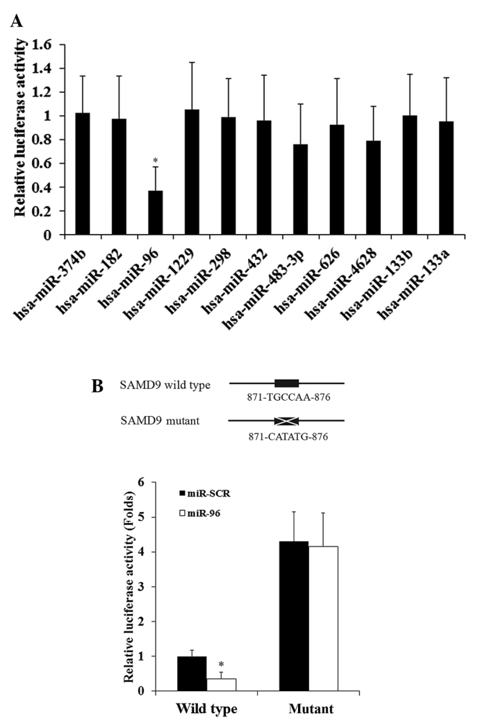
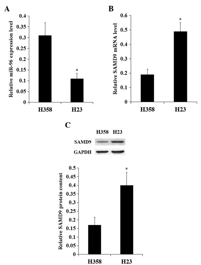
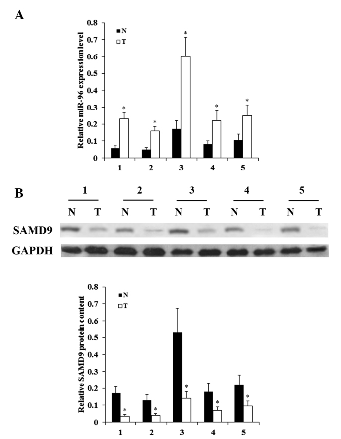
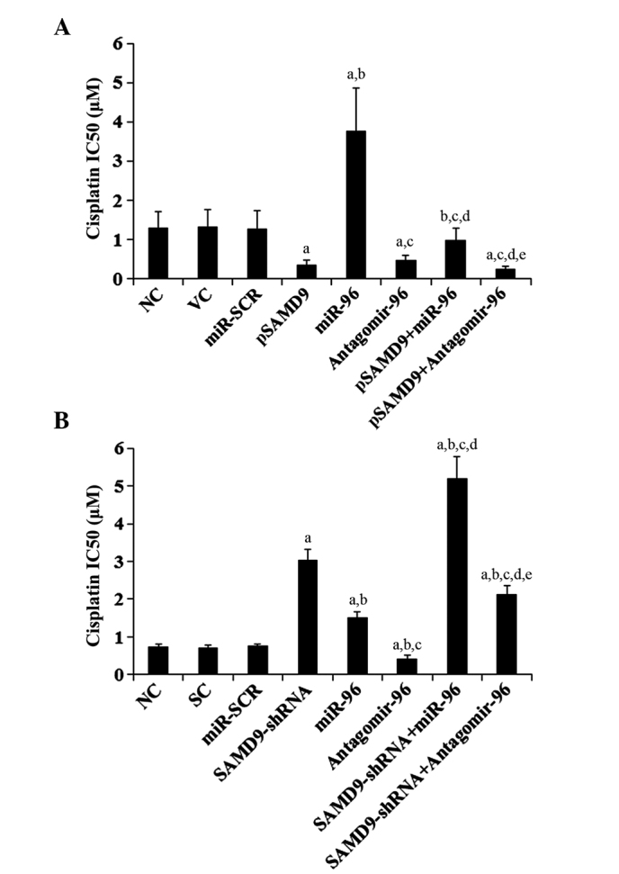
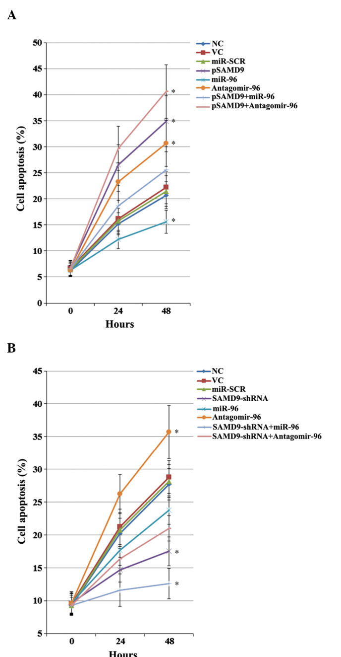
Cisplatin is effective as a single agent or in combination with other drugs for the treatment of non-small cell lung cancer (NSCLC). A concerning clinical challenge with cisplatin-based NSCLC chemotherapy is the intrinsic and acquired chemoresistance to cisplatin. The sterile α motif domain-containing (SAMD9) gene has been reported as a potent tumor suppressor gene that inhibits tumorigenesis and progression of NSCLC. microRNAs (miRNA) have been revealed to play important roles in the regulation of cancer chemoresistance. To the best of our knowledge the present study explored the role of miRNA/SAMD9 signaling in regulating cisplatin chemoresistance in NSCLC for the first time. Out of the several candidate miRNAs predicted to bind the 3'-untranslated region (UTR) of the SAMD9 gene, miRNA-96 (miR-96) demonstrated significant target-sequence-specific inhibition of the SAMD9 3'-UTR luciferase reporter activity in NSCLC cells. In addition, while NSCLC tumor samples exhibited significantly higher expression levels of miR-96 compared with adjacent normal tissues, the expression levels of SAMD9 were significantly lower than those in adjacent normal tissues. miR-96 and SAMD9 were overexpressed and knocked down in the human NSCLC H358 and H23 cell lines and the half maximal inhibitory concentration (IC50) of cisplatin and cell apoptosis rate under cisplatin treatment were used as measures of cisplatin chemoresistance. The present results identified that overexpression of miR-96 in NSCLC cells markedly decreased SAMD9 expression and cisplatin-induced apoptosis, and increased the cisplatin IC50, which could be eliminated by overexpression of SAMD9. By contrast, knocking down miR-96 in NSCLC cells using antagomir-96 significantly increased SAMD9 expression and the cisplatin-induced apoptosis and decreased cisplatin IC50, which could be completely reversed by a knockdown of SAMD9. In conclusion, the current study demonstrates that miR-96 targets and downregulates SAMD9 in NSCLC, which decreases cisplatin-induced apoptosis and induces cisplatin chemoresistance in NSCLC cells. The findings of the present study add novel insights into the function of miR-96 and SAMD9 in cancer, as well as into the molecular mechanisms underlying NSCLC chemoresistance.

Keywords: SAMD9; apoptosis; chemoresistance; cisplatin; miR-96; non-small cell lung cancer.


Figures

Figure 1.
Figure 1.
Effect of selected miRNAs on the SAMD9 3′-UTR luciferase reporter in NSCLC cells. (A) In total, 11 miRNAs potentially able to regulate SAMD9 were selected based on TargetScan prediction software. The miRNA mimics were co-transfected with a SAMD9 3′-UTR luciferase reporter in human NSCLC H358 cells. In total, 30 h subsequent to transfection, the luciferase activity was measured. *P<0.05 vs. 1.0 (cut off value). (B) Luciferase activities were measured in H358 cells co-transfected with miR-96 mimics and the SAMD9 3′-UTR luciferase reporter with wild-type or mutant miR-96-binding sequence. Cells co-transfected with miR-SCR instead of miR-96 mimics was used as a negative control. The luciferase activity was expressed as fold changes compared to cells co-transfected with miR-SCR and the wild-type SAMD9 3′-UTR luciferase reporter (designated as 1). *P<0.05 vs. miR-SCR. UTR, untranslated region; NSCLC, non-small cell lung cancer; miR, microRNA; SAMD9, sterile α motif domain-containing 9; miR-SCR, scramble miR.
Figure 2.
Figure 2.
Expression of miR-96 and SAMD9 in NSCLC cells. In H358 and H23 NSCLC cells, the expression levels of (A) miR-96 and (B) SAMD9 mRNA were determined with reverse transcription-quantitative polymerase chain reaction. (C) SAMD9 protein levels were determined by western blot analysis. Density of the SAMD9 blots was normalized against that of GAPDH to obtain a relative blot density to indicate the relative SAMD9 protein content. *P<0.05 vs. H358. NSCLC, non-small cell lung cancer; miR, microRNA; SAMD9, sterile α motif domain-containing 9.
Figure 3.
Figure 3.
Expression of miR-96 and SAMD9 in NSCLC and adjacent normal lung tissues. The expression levels of (A) miR-96 and (B) SAMD9 protein in T and N lung tissues from 5 consecutive patients were determined with reverse transcription-quantitative polymerase chain reaction and western blot analysis, respectively. In western blot analysis, the density of the SAMD9 blots was normalized against that of GAPDH to obtain a relative blot density to indicate the relative SAMD9 protein content. *P<0.05 vs. N. NSCLC, non-small cell lung cancer; miR microRNA; SAMD9, sterile α motif domain-containing 9; T, NSCLC tumor; N, adjacent normal tissue.
Figure 4.
Figure 4.
Effect of miR-96 on SAMD9 mRNA levels in non-small cell lung cancer cells (A) In H358 cells, the SAMD9 mRNA level was determined with RT-qPCR in NC, cells stably transfected with VC, transfected with miR-SCR, stably transfected with pSAMD9, transfected with miR-96 mimics, transfected with antagomir-96, stably transfected with pSAMD9 + miR-96, and stably transfected with pSAMD9 + antagomir-96. (B) In H23 cells, the SAMD9 mRNA level was determined with RT-qPCR in NC cells and cells stably transduced with SC, transfected with miR-SCR, stably transduced with SAMD9-shRNA, transfected with miR-96 mimics, transfected with antagomir-96, stably transduced with SAMD9-shRNA and transiently transfected with miR-96 mimics, and stably transduced with SAMD9-shRNA and transiently transfected with antagomir-96. In (A) H358 cells, aP<0.05 vs. NC, VC and miR-SCR; bP<0.05 vs. pSAMD9; cP<0.05 vs. miR-96; dP<0.05 vs. antagomir-96; eP<0.05 vs. pSAMD9 + miR-96. In (B) H23 cells, aP<0.05 vs. NC, SC and miR-SCR; bP<0.05 vs. SAMD9-shRNA; cP<0.05 vs. miR-96; dP<0.05 vs. antagomir-96; eP<0.05 vs. SAMD9-shRNA + miR-96. miR, microRNA; SAMD9, sterile α motif domain-containing 9; RT-qPCR, reverse transcription-quantitative polymerase chain reaction; NC, normal control cells; VC, pcDNA3.1 plasmid; miR-SCR, scramble miR; UTR, untranslated region; pSAMD9, pcDNA3.1-(SAMD9 cDNA plus UTR) plasmid; pSAMD9 + miR-96, pcDNA3.1-(SAMD9 cDNA plus UTR) plasmid and transiently transfected with miR-96 mimics; pSAMD9 + antagomir-96, pcDNA3.1-(SAMD9 cDNA plus UTR) plasmid and transiently transfected with antagomir-96; SC, scramble control short hairpin RNA.
Figure 5.
Figure 5.
Effect of miR-96 on SAMD9 protein levels in non-small cell lung cancer cells. (A) In H358 cells, the SAMD9 protein level was determined with western blot analysis in NC (lane 1), cells stably transfected with VC (lane 2), transfected with miR-SCR (lane 3), stably transfected with pSAMD9 (lane 4), transfected with miR-96 mimics (lane 5), transfected with antagomir-96 (lane 6), stably transfected with pSAMD9 + miR-96 (lane 7), and stably transfected with pSAMD9 + antagomir-96 (lane 8). (B) In H23 cells, the SAMD9 protein level was determined with western blot analysis in NC cells (lane 1), cells stably transduced with SC (lane 2), transfected with miR-SCR (lane 3), stably transduced with SAMD9-shRNA (lane 4), transfected with miR-96 mimics (lane 5), transfected with antagomir-96 (lane 6), stably transduced with SAMD9-shRNA and transiently transfected with miR-96 mimics (lane 7), and stably transduced with SAMD9-shRNA and transiently transfected with antagomir-96 (lane 8). Density of the SAMD9 blot was normalized against that of GAPDH to obtain a relative blot density to represent relative SAMD9 protein content. In (A) H358 cells, aP<0.05 vs. NC, VC and miR-SCR; bP<0.05 vs. pSAMD9; cP<0.05 vs. miR-96; dP<0.05 vs. antagomir-96; eP<0.05 vs. pSAMD9 + miR-96. In (B) H23 cells, aP<0.05 vs. NC, SC and miR-SCR; bP<0.05 vs. SAMD9-shRNA; cP<0.05 vs. miR-96; dP<0.05 vs. antagomir-96; eP<0.05 vs. SAMD9-shRNA + miR-96. miR, microRNA; SAMD9, sterile α motif domain-containing 9; NC, normal control cells; VC, pcDNA3.1 plasmid; miR-SCR, scramble miR; UTR, untranslated region; pSAMD9, pcDNA3.1-(SAMD9 cDNA plus UTR) plasmid; pSAMD9 + miR-96, pcDNA3.1-(SAMD9 cDNA plus UTR) plasmid and transiently transfected with miR-96 mimics; pSAMD9 + antagomir-96, pcDNA3.1-(SAMD9 cDNA plus UTR) plasmid and transiently transfected with antagomir-96; SC, scramble control short hairpin RNA.
Figure 6.
Figure 6.
Effect of miR-96/SAMD9 signaling on cisplatin chemoresistance in non-small cell lung cancer cells. H358 and H23 cells were treated with or without various concentrations of cisplatin (0.1, 0.25, 0.5, 1.0, 1.5, 3.0, 6.0, 15.0, 30.0 and 55.0 mM) for 96 h. (A) In H358 cells, IC50 was determined in NC, cells stably transfected with VC, transfected with miR-SCR, stably transfected with pSAMD9, transfected with miR-96 mimics, transfected with antagomir-96, stably transfected with pSAMD9 + miR-96, and stably transfected with pSAMD9 + antagomir-96. (B) In H23 cells, IC50 was determined in NC, cells stably transduced with SC, transfected with miR-SCR, stably transduced with SAMD9-shRNA, transfected with miR-96 mimics, transfected with antagomir-96, stably transduced with SAMD9-shRNA and transiently transfected with miR-96 mimics, and stably transduced with SAMD9-shRNA and transiently transfected with antagomir-96. In (A) H358 cells, aP<0.05 vs. NC, VC and miR-SCR; bP<0.05 vs. pSAMD9; cP<0.05 vs. miR-96; dP<0.05 vs. antagomir-96; eP<0.05 vs. pSAMD9 + miR-96. In (B) H23 cells, aP<0.05 vs. NC, SC and miR-SCR; bP<0.05 vs. SAMD9-shRNA; cP<0.05 vs. miR-96; dP<0.05 vs. antagomir-96; eP<0.05 vs. SAMD9-shRNA + miR-96. miR, microRNA; SAMD9, sterile α motif domain-containing 9; IC50, the half maximal inhibitory concentration; NC, normal control cells; VC, pcDNA3.1 plasmid; miR-SCR, scramble miR; UTR, untranslated region; pSAMD9, pcDNA3.1-(SAMD9 cDNA plus UTR) plasmid; pSAMD9 + miR-96, pcDNA3.1-(SAMD9 cDNA plus UTR) plasmid and transiently transfected with miR-96 mimics; pSAMD9 + antagomir-96, pcDNA3.1-(SAMD9 cDNA plus UTR) plasmid and transiently transfected with antagomir-96; SC, scramble control short hairpin RNA.
Figure 7.
Figure 7.
Effect of miR-96/SAMD9 signaling on cisplatin-induced apoptosis in non-small cell lung cancer (NSCLC) cells. H358 and H23 cells were treated with cisplatin (1 µM) for 24 h and 48 h. Apoptosis was measured with a microplate reader-based TiterTACS in situ apoptosis detection kit. (A) In H358 cells, apoptosis was determined in NC, cells stably transfected with VC, transfected with miR-SCR, stably transfected with pSAMD9, transfected with miR-96 mimics, transfected with antagomir-96, stably transfected with pSAMD9 + miR-96, and stably transfected with pSAMD9 + antagomir-96. (B) In H23 cells, apoptosis was determined in NC, cells stably transduced with SC, transfected with miR-SCR, stably transduced with SAMD9-shRNA, transfected with miR-96 mimics, transfected with antagomir-96, stably transduced with SAMD9-shRNA and transiently transfected with miR-96 mimics, and stably transduced with SAMD9-shRNA and transiently transfected with antagomir-96. The cell apoptosis rates at 24 h and 48 h were shown as the percentage of apoptotic cells (as compared to 100% cell apoptosis induced by nuclease treatment). In (A) H358 cells, *P<0.05 vs. NC, VC and miR-SCR. In (B) H23 cells, *P<0.05 vs. NC, SC and miR-SCR. miR microRNA; SAMD9, sterile α motif domain-containing 9; IC50, the half maximal inhibitory concentration; NC, normal control cells; VC, pcDNA3.1 plasmid; miR-SCR, scramble miR; UTR, untranslated region; pSAMD9, pcDNA3.1-(SAMD9 cDNA plus UTR) plasmid; pSAMD9 + miR-96, pcDNA3.1-(SAMD9 cDNA plus UTR) plasmid and transiently transfected with miR-96 mimics; pSAMD9 + antagomir-96, pcDNA3.1-(SAMD9 cDNA plus UTR) plasmid and transiently transfected with antagomir-96; SC, scramble control shRNA.

References

    1. Ma L, Huang Y, Zhu W, et al. An integrated analysis of miRNA and mRNA expressions in non-small cell lung cancers. PLoS One. 2011;6:e26502. doi: 10.1371/journal.pone.0026502. - DOI - PMC - PubMed
    1. Merk J, Rolff J, Dorn C, Leschber G, Fichtner I. Chemoresistance in non-small-cell lung cancer, Can multidrug resistance markers predict the response of xenograft lung cancer models to chemotherapy? Eur J Cardiothorac Surg. 2011;40:e29–e33. doi: 10.1016/j.ejcts.2011.02.010. - DOI - PubMed
    1. Chang A. Chemotherapy chemoresistance and the changing treatment landscape for NSCLC. Lung Cancer. 2011;71:3–10. doi: 10.1016/j.lungcan.2010.08.022. - DOI - PubMed
    1. Asou H, Matsui H, Ozaki Y, et al. Identification of a common microdeletion cluster in 7q21.3 subband among patients with myeloid leukemia and myelodysplastic syndrome. Biochem Biophys Res Commun. 2009;383:245–251. doi: 10.1016/j.bbrc.2009.04.004. - DOI - PubMed
    1. Li CF, MacDonald JR, Wei RY, et al. Human sterile alpha motif domain 9, a novel gene identified as down-regulated in aggressive fibromatosis, is absent in the mouse. BMC Genomics. 2007;8:92. doi: 10.1186/1471-2164-8-92. - DOI - PMC - PubMed

LinkOut - more resources