

Case Reports
doi: 10.1097/GOX.0000000000000567.
eCollection 2015 Dec.
Primary Squamous Cell Carcinoma Arising from a Breast Implant Capsule
Affiliations
- PMID: 26894011
- PMCID: PMC4727695
- DOI: 10.1097/GOX.0000000000000567
Item in Clipboard
Case Reports
Primary Squamous Cell Carcinoma Arising from a Breast Implant Capsule
Plast Reconstr Surg Glob Open.
.
Abstract
Primary squamous cell carcinoma (SCC) of the breast comprises less than 0.1% of all breast cancers. Literature review reveals only 1 reported case of an SCC arising from the capsule of a breast implant. The authors describe, herein, a primary SCC arising from the capsule of a long-standing silicone breast implant.
Conflict of interest statement
Figures
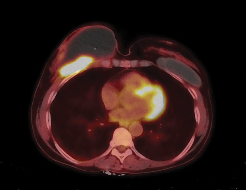
F-18 fluorodeoxyglucose (FDG) positron emission tomography-computed tomographic study demonstrates an ill-defined hypermetabolic soft tissue lesion located deep to the right breast and along the right anterior chest wall, concerning for neoplastic process. There is a large non-FDG avid fluid density collection in the right breast and overlying the hypermetabolic lesion, likely representing a seroma. Left breast implant is noted. No other suspicious FDG avid lesion or lymph node to suggest systemic involvement of malignancy was identified.
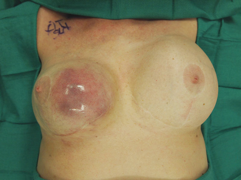
Intraoperative view of right breast (which has previously been explanted) demonstrating dramatic enlargement with obvious thinning and attenuation of the soft tissue envelope.
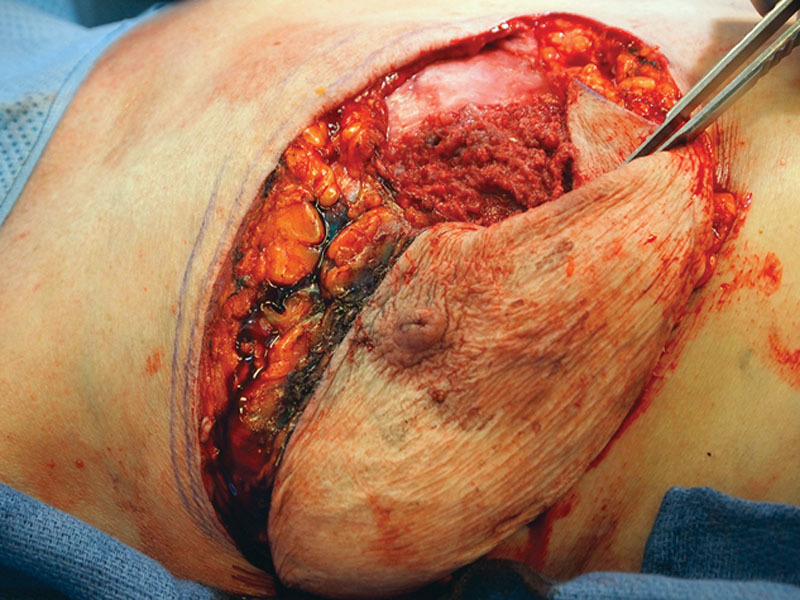
Intraoperative appearance of in situ posterior capsule.
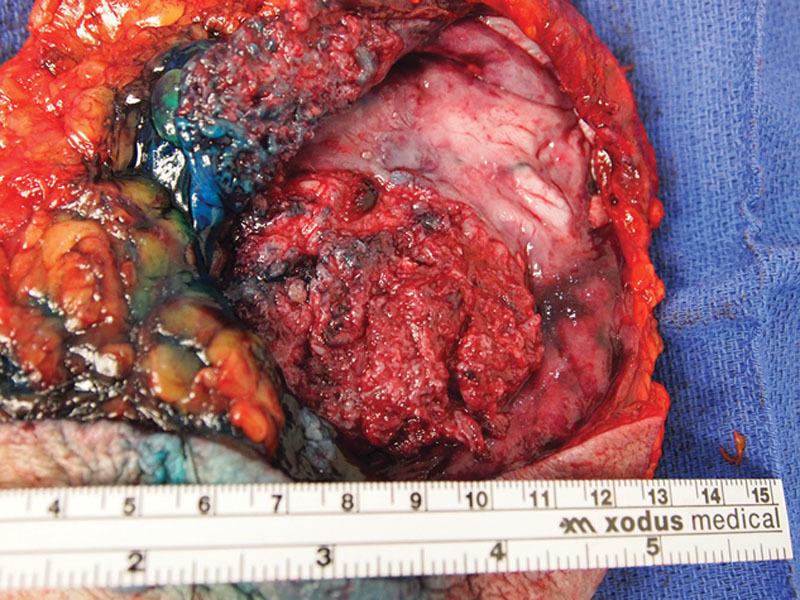
Mastectomy specimen with visible fungating mass on posterior capsule. Isosulfan blue dye present from aborted sentinel lymph node biopsy.
References
-
- Menes T, Schachter J, Morgenstern S, et al. Primary squamous cell carcinoma (SqCC) of the breast. Am J Clin Oncol. 2003;26:571–573. - PubMed
-
- Macia M, Ces JA, Becerra E, et al. Pure squamous carcinoma of the breast. Report of a case diagnosed by aspiration cytology. Acta Cytol. 1989;33:201–204. - PubMed
-
- Hennessy BT, Krishnamurthy S, Giordano S, et al. Squamous cell carcinoma of the breast. J Clin Oncol. 2005;23:7827–7835. - PubMed
-
- Grabowski J, Saltzstein SL, Sadler G, et al. Squamous cell carcinoma of the breast: a review of 177 cases. Am Surg. 2009;75:914–917. - PubMed
-
- Behranwala KA, Nasiri N, Abdullah N, et al. Squamous cell carcinoma of the breast: clinico-pathologic implications and outcome. Eur J Surg Oncol. 2003;29:386–389. - PubMed
Publication types
LinkOut - more resources
Full Text Sources
Research Materials