. 2016 Feb 2;3(2):e204.
doi: 10.1212/NXI.0000000000000204. eCollection 2016 Apr.

Serum peptide reactivities may distinguish neuromyelitis optica subgroups and multiple sclerosis

Affiliations

Serum peptide reactivities may distinguish neuromyelitis optica subgroups and multiple sclerosis

Imke Metz et al. Neurol Neuroimmunol Neuroinflamm. .

Abstract

Objective: To assess in an observational study whether serum peptide antibody reactivities may distinguish aquaporin-4 (AQP4) antibody (Ab)-positive and -negative neuromyelitis optica spectrum disorders (NMOSD) and relapsing-remitting multiple sclerosis (RRMS).

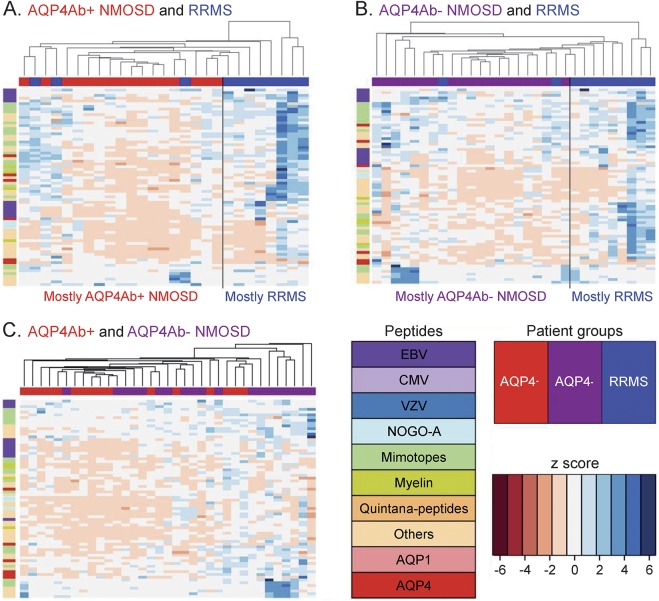
Methods: We screened 8,700 peptides that included human and viral antigens of potential relevance for inflammatory demyelinating diseases and random peptides with pooled sera from different patient groups and healthy controls to set up a customized microarray with 700 peptides. With this microarray, we tested sera from 66 patients with AQP4-Ab-positive (n = 16) and AQP4-Ab-negative (n = 19) NMOSD, RRMS (n = 11), and healthy controls (n = 20).

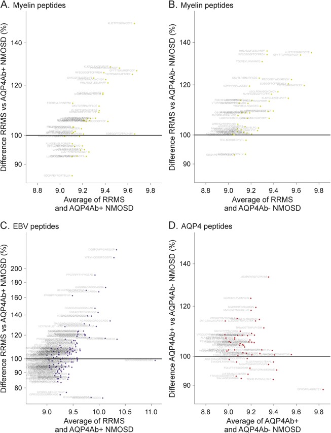
Results: Differential peptide reactivities distinguished NMOSD subgroups from RRMS in 80% of patients. However, the 2 NMOSD subgroups were not well-discriminated, although those patients are clearly separated by their antibody reactivities against AQP4 in cell-based assays. Elevated reactivities to myelin and Epstein-Barr virus peptides were present in RRMS and to AQP4 and AQP1 peptides in AQP4-Ab-positive NMOSD.

Conclusions: While AQP4-Ab-positive and -negative NMOSD subgroups are not well-discriminated by peptide antibody reactivities, our findings suggest that peptide antibody reactivities may have the potential to distinguish between both NMOSD subgroups and MS. Future studies should thus concentrate on evaluating peptide antibody reactivities for the differentiation of AQP4-Ab-negative NMOSD and MS.


Figures

Figure 1
Figure 1. Neuromyelitis optica spectrum disorder (NMOSD) subgroups and patients with relapsing-remitting multiple sclerosis (RRMS) show different antibody signatures
Heat maps (A: RRMS [blue] vs aquaporin-4 antibody [AQP4Ab]+ NMOSD [red]; B: RRMS [blue] vs AQP4Ab− NMOSD [purple]; C: AQP4Ab+ NMOSD [red] vs AQP4Ab− NMOSD [purple]). The top 20 peptides from all pairwise subgroup comparisons as well as all AQP4 peptides are shown as rows (see key for peptides). Patients from the different subgroups are shown as columns (see key for patient groups). The color (see key for z score) indicates the intensity of the peptides standardized to the healthy controls, with blue indicating an upregulation and red a downregulation of peptide reactivities, given in standard deviations. Heat maps show a clustering of RRMS and AQP4Ab+ NMOSD (A) as well as RRMS and AQP4Ab− NMOSD (B), suggesting different antibody signatures in these patient groups. In contrast, such a clustering is not evident comparing the 2 NMOSD subgroups (C). CMV = cytomegalovirus; EBV = Epstein-Barr virus; VZV = varicella-zoster virus.
Figure 2
Figure 2. Elevated antibody reactivities against myelin and Epstein-Barr virus (EBV) peptides in relapsing-remitting multiple sclerosis (RRMS) and higher anti–aquaporin-4 (AQP4) peptide reactivities in neuromyelitis optica spectrum disorders positive for AQP4−Abs (AQP4Ab+ NMOSD)
Comparison of selected peptide groups in different patient subsets shown with MA plots. (A) Myelin peptides RRMS vs AQP4Ab+ NMOSD. (B) Myelin peptides RRMS vs AQP4Ab− NMOSD. (C) EBV peptides RRMS vs AQP4Ab+ NMOSD. (D) AQP4 peptides AQP4Ab+ vs AQP4Ab− NMOSD. Each dot represents one peptide. The x-axis displays the average normalized signal to show the general level of peptide binding. The y-axis indicates the difference between the patient groups given as a percentage. Greater reactivities in the first mentioned group are found above the 100% horizontal line, and lower reactivities below this line. Patients with RRMS show higher antibody reactivities against myelin peptides and EBV-EBNA 1 peptides than do patients with NMOSD. Higher antibody reactivities against AQP4 peptides are evident in the AQP4Ab+ compared to the AQP4Ab− NMOSD group.

References

    1. Lennon VA, Kryzer TJ, Pittock SJ, Verkman AS, Hinson SR. IgG marker of optic-spinal multiple sclerosis binds to the aquaporin-4 water channel. J Exp Med 2005;202:473–477. - PMC - PubMed
    1. Wingerchuk DM, Banwell B, Bennett JL, et al. International consensus diagnostic criteria for neuromyelitis optica spectrum disorders. Neurology 2015;85:177–189. - PMC - PubMed
    1. Wingerchuk DM, Lennon VA, Lucchinetti CF, Pittock SJ, Weinshenker BG. The spectrum of neuromyelitis optica. Lancet Neurol 2007;6:805–815. - PubMed
    1. Kitley J, Leite MI, Kuker W, et al. Longitudinally extensive transverse myelitis with and without aquaporin 4 antibodies. JAMA Neurol 2013;70:1375–1381. - PubMed
    1. Waters P, Woodhall M, O'Connor KC, et al. MOG cell-based assay detects non-MS patients with inflammatory neurologic disease. Neurol Neuroimmunol Neuroinflamm 2015;2:e89. 10.1212/NXI.0000000000000089. - DOI - PMC - PubMed

LinkOut - more resources