. 2016 Jun;17(6):739-55.
doi: 10.3171/2015.10.PEDS15554. Epub 2016 Feb 19.

Imaging and serum biomarkers reflecting the functional efficacy of extended erythropoietin treatment in rats following infantile traumatic brain injury

Affiliations

Imaging and serum biomarkers reflecting the functional efficacy of extended erythropoietin treatment in rats following infantile traumatic brain injury

Shenandoah Robinson et al. J Neurosurg Pediatr. 2016 Jun.

Abstract

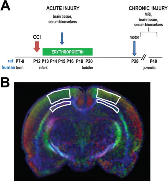
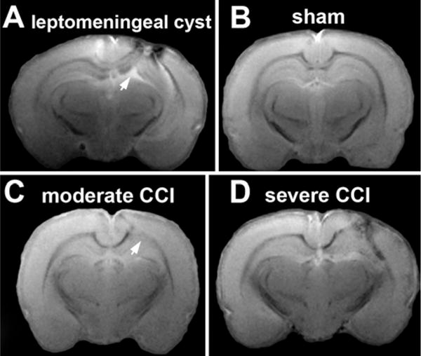
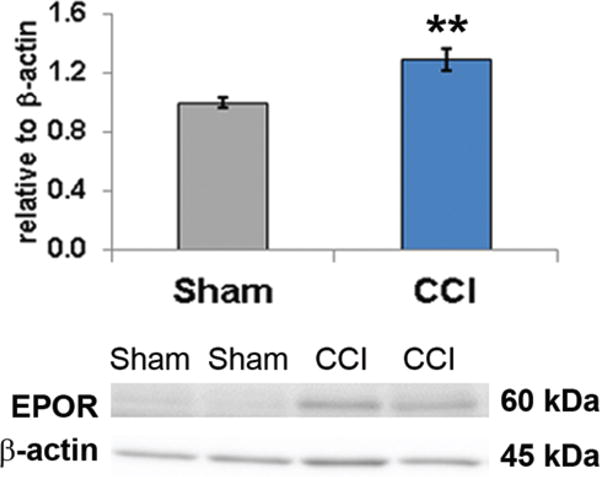
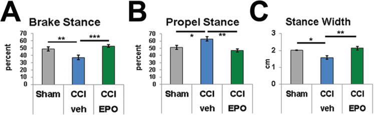
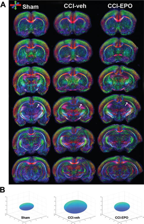
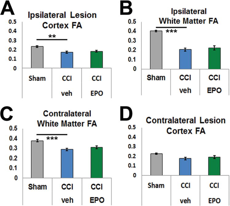
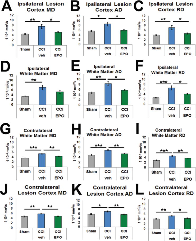
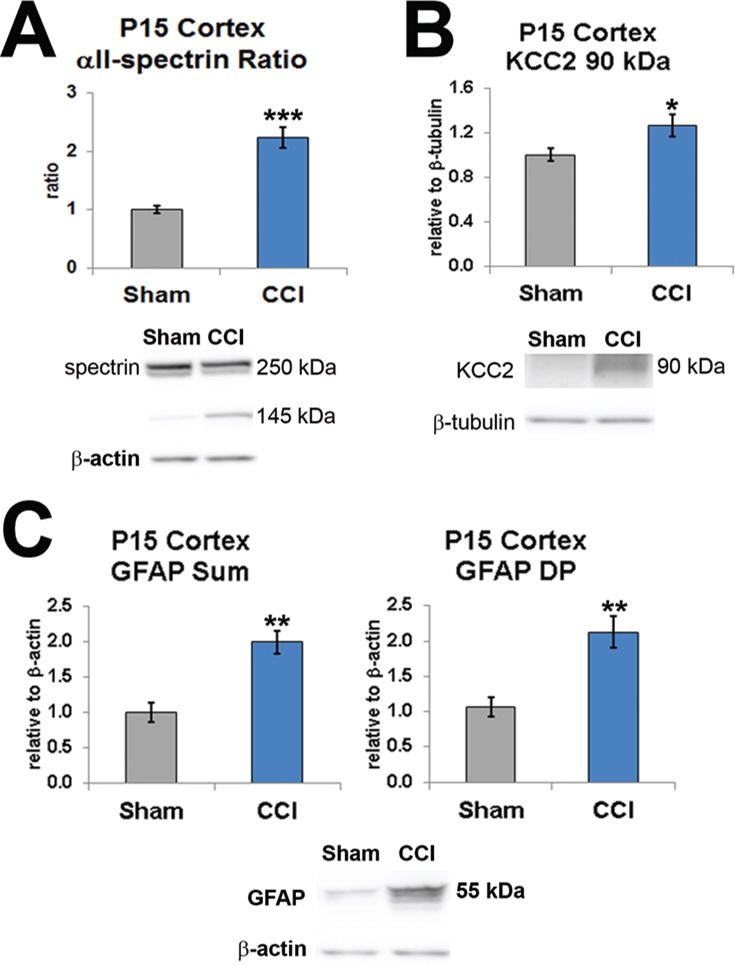
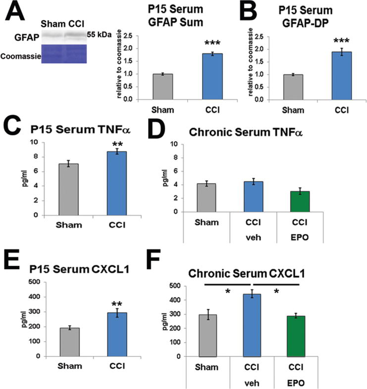
OBJECTIVE Traumatic brain injury (TBI) is a leading cause of death and severe morbidity for otherwise healthy full-term infants around the world. Currently, the primary treatment for infant TBI is supportive, as no targeted therapies exist to actively promote recovery. The developing infant brain, in particular, has a unique response to injury and the potential for repair, both of which vary with maturation. Targeted interventions and objective measures of therapeutic efficacy are needed in this special population. The authors hypothesized that MRI and serum biomarkers can be used to quantify outcomes following infantile TBI in a preclinical rat model and that the potential efficacy of the neuro-reparative agent erythropoietin (EPO) in promoting recovery can be tested using these biomarkers as surrogates for functional outcomes. METHODS With institutional approval, a controlled cortical impact (CCI) was delivered to postnatal Day (P)12 rats of both sexes (76 rats). On postinjury Day (PID)1, the 49 CCI rats designated for chronic studies were randomized to EPO (3000 U/kg/dose, CCI-EPO, 24 rats) or vehicle (CCI-veh, 25 rats) administered intraperitoneally on PID1-4, 6, and 8. Acute injury (PID3) was evaluated with an immunoassay of injured cortex and serum, and chronic injury (PID13-28) was evaluated with digitized gait analyses, MRI, and serum immunoassay. The CCI-veh and CCI-EPO rats were compared with shams (49 rats) primarily using 2-way ANOVA with Bonferroni post hoc correction. RESULTS Following CCI, there was 4.8% mortality and 55% of injured rats exhibited convulsions. Of the injured rats designated for chronic analyses, 8.1% developed leptomeningeal cyst-like lesions verified with MRI and were excluded from further study. On PID3, Western blot showed that EPO receptor expression was increased in the injured cortex (p = 0.008). These Western blots also showed elevated ipsilateral cortex calpain degradation products for αII-spectrin (αII-SDPs; p < 0.001), potassium chloride cotransporter 2 (KCC2-DPs; p = 0.037), and glial fibrillary acidic protein (GFAP-DPs; p = 0.002), as well as serum GFAP (serum GFAP-DPs; p = 0.001). In injured rats multiplex electrochemiluminescence analyses on PID3 revealed elevated serum tumor necrosis factor alpha (TNFα p = 0.01) and chemokine (CXC) ligand 1 (CXCL1). Chronically, that is, in PID13-16 CCI-veh rats, as compared with sham rats, gait deficits were demonstrated (p = 0.033) but then were reversed (p = 0.022) with EPO treatment. Diffusion tensor MRI of the ipsilateral and contralateral cortex and white matter in PID16-23 CCI-veh rats showed widespread injury and significant abnormalities of functional anisotropy (FA), mean diffusivity (MD), axial diffusivity (AD), and radial diffusivity (RD); MD, AD, and RD improved after EPO treatment. Chronically, P13-P28 CCI-veh rats also had elevated serum CXCL1 levels, which normalized in CCI-EPO rats. CONCLUSIONS Efficient translation of emerging neuro-reparative interventions dictates the use of age-appropriate preclinical models with human clinical trial-compatible biomarkers. In the present study, the authors showed that CCI produced chronic gait deficits in P12 rats that resolved with EPO treatment and that chronic imaging and serum biomarkers correlated with this improvement.

Keywords: AD = axial diffusivity; CCI = controlled cortical impact; CXCL1 = chemokine (CXC) ligand 1; DP = degradation product; DTI = diffusion tensor imaging; EP = echo planar; EPO = erythropoietin; EPOR = EPO receptor; FA = fractional anisotropy; GFAP = glial fibrillary acidic protein; IFNγ = interferon gamma; IL = interleukin; KCC2 = potassium chloride cotransporter 2; MD = mean diffusivity; MECI = multielectrochemiluminescence; P = postnatal day; PID = postinjury day; RD = radial diffusivity; ROI = region of interest; SWI = susceptibility-weighted imaging; TBI = traumatic brain injury; TNFα = tumor necrosis factor alpha; controlled cortical impact; diffusion tensor imaging; diffusivity; erythropoietin; infant; serum biomarker; trauma; traumatic brain injury; veh = vehicle; αII-SDPs = αII-spectrin DPs.


Figures

FIG. 1
FIG. 1
A: Schematic of experimental paradigm. The P12 rat pups, approximately equal to 6-month-old human infants, underwent CCI. A subset of P15 sham and CCI-veh rats underwent studies in the acute period at 72 hours after CCI (49 rats). For chronic studies (76 rats), CCI rats were randomized to receive 3000 U/kg of EPO or vehicle on PID1, 2, 3, 4, 6, and 8 and underwent analysis beginning at P28 (PID16). B: Regions of interest used for DTI analysis for the ipsilateral and contralateral white matter (thin strips) and cortex (wide strips). Figure is available in color online only.
FIG. 2
FIG. 2
Representative SWI studies of sham and CCI brains reveal the spectrum of sustained injury from P12. A: A few CCI rats developed a cranial protuberance suggestive of a leptomeningeal cyst 6–12 days after CCI. Susceptibility-weighted imaging demonstrated deformation of the lateral ventricle toward the cortical lesion (arrow), similar to the pattern observed in human infants with a leptomeningeal cyst. B: Sham rats demonstrated no abnormalities in the cortex and sharp demarcation of the underlying subcortical white matter. C: In contrast, CCI rats demonstrated a spectrum of injury, with approximately half of the CCI rats showing moderate injury with the loss of the gray-white matter demarcation (arrow) and mild extension of hypointense signal into the overlying cortex. D: The remaining half of the CCI rats that underwent SWI had severe injury with extensive focal injury to the cortex, subcortical white matter, and dorsal hippocampus.
FIG. 3
FIG. 3
Immunoblotting of EPOR protein in microdissected cortex ipsilateral to the impact revealed that CCI induces a significant increase in EPOR expression 3 days after injury and suggests that vacant CNS EPOR should be responsive to exogenous EPO treatment days after injury. CCI = CCI-veh. **p = 0.008, 2-tailed t-test with unequal variance. Figure is available in color online only.
FIG. 4
FIG. 4
Controlled cortical impact on P12 disturbed gait patterns, and extended EPO treatment reversed gait abnormalities. A: The portion of the stance spent in the brake phase was significantly reduced in CCI-veh rats, and EPO treatment restored the brake portion to sham levels. B: Similarly, the portion spent in the propel phase was increased following CCI and normalized with EPO treatment. C: The forelimb stance width was also significantly reduced in CCI rats compared with shams and normalized with EPO treatment. *p < 0.05, **p < 0.01, ***p ≤ 0.001, 2-way ANOVA with Bonferroni post hoc correction. Figure is available in color online only.
FIG. 5
FIG. 5
Diffusion tensor imaging measures the diffusion of water in the CNS. Diffusion is constrained by structures such as myelin, axons, and cellular organelles, and injury alters the diffusivity of water. A: Color maps of representative sham, CCI-veh, and CCI-EPO rat brains. Note that the color intensity is reduced in the region of the CCI (arrows), indicative of a loss of microstructural integrity. Improvement is apparent in the CCI-EPO brain. Directionality of DTI is indicated by different colors: red, transverse; green, vertical; and blue, orthogonal plane. B: The average of each of the 3 eigenvectors (λ1, λ2, and λ3) for a ROI can be modeled by an ellipsoid to show directional changes in diffusivity after injury or repair. Ellipsoids representing the mean eigenvectors for the ipsilateral cortex for sham, CCI-veh, and CCI-EPO rats demonstrate that diffusivity increases after injury in all directions in CCI-veh rat brains and that EPO treatment reduces diffusivity to levels similar to those in shams. Figure is available in color online only.
FIG. 6
FIG. 6
Ipsilateral cortex and bilateral subcortical white matter show loss of FA, a crude measure of microstructural integrity. A: In the ipsilateral cortex, FA was significantly reduced in CCI brains. B: Similarly, FA was significantly lower in the ipsilateral subcortical white matter. C: The reduction in FA following CCI extended to the contralateral subcortical white matter. D: By contrast, the FA in the contralateral cortex was not affected. *p < 0.05, **p < 0.01, ***p ≤ 0.001, 2-way ANOVA with Bonferroni post hoc correction. Figure is available in color online only.
FIG. 7
FIG. 7
Detailed analyses of DTI eigenvectors demonstrated significant efficacy of extended EPO treatment following P12 CCI, providing MRI biomarkers of treatment responsiveness. Mean diffusivity describes the average of the 3 eigenvectors depicting the ellipsoid-shaped extent of water diffusion, whereas AD and RD provide specific detail regarding directionality. The longest, primary eigenvector denotes AD, whereas RD is the mean of the 2 smaller eigenvectors. In CCI-veh brains, MD was elevated in the ipsilateral lesion cortex (A) and subcortical white matter (D). More specifically, AD was elevated in both the lesion cortex (B) and the subcortical white matter (E). Similarly, RD was also increased in both areas (C and F). Most importantly, CCI-EPO brains showed significant restoration of AD and RD in both brain regions. As expected, the magnitude of the increase in diffusivity after injury was less prominent in the contralateral subcortical white matter and cortex (G–L). The efficacy of EPO treatment was similar to that in the ipsilateral hemisphere, indicating that after injury extended EPO treatment imparts widespread microstructural restoration. *p < 0.05, **p < 0.01, ***p ≤ 0.001, 2-way ANOVA with Bonferroni post hoc correction. Figure is available in color online only.
FIG. 8
FIG. 8
Excess cortical calpain activity at P15 degrades essential neuromolecules. A: The ratio of αII-SDPs, a neuron-specific cytoskeletal protein, was significantly elevated at PID3. B: Calpain DPs for KCC2, a molecule important for neuronal function, were also significantly increased at PID3. C: The astrocyte intermediate GFAP increases proportionally with gliosis and is also degraded by calpain. After CCI, the sum total of GFAP levels (left) and the GFAP-DPs (right) were both significantly increased. *p < 0.05, **p < 0.01, ***p ≤ 0.001, 2-tailed t-test with unequal variance. Figure is available in color online only.
FIG. 9
FIG. 9
Serum biomarkers reflect CNS injury patterns. A: Serum GFAP levels were assayed with immunoblotting using Coomassie staining as a loading control, and the sum total of full-length and DPs (approximately 48 kD) was significantly elevated in serum 3 days after injury. B: Similarly, GFAP-DP levels were significantly increased at P15. C: Serum TNFα levels were significantly increased at P15. D: Serum TNFα levels normalized by the chronic phase. E: Serum CXCL1 (known as CINC in the rat) levels were also significantly increased at PID3. F: Serum CXCL1 levels remained elevated in the chronic phase and, importantly, were normalized by an extended course of EPO treatment. CCI = CCI-veh. *p < 0.05, **p < 0.01, ***p ≤ 0.001, 2-tailed t-test with unequal variance (A–C, E) and 2-way ANOVA with Bonferroni post hoc correction (D and F). Figure is available in color online only.

References

    1. Adelson PD, Dixon CE, Robichaud P, Kochanek PM. Motor and cognitive functional deficits following diffuse traumatic brain injury in the immature rat. J Neurotrauma. 1997;14:99–108. - PubMed
    1. Adelson PD, Fellows-Mayle W, Kochanek PM, Dixon CE. Morris water maze function and histologic characterization of two age-at-injury experimental models of controlled cortical impact in the immature rat. Childs Nerv Syst. 2013;29:43–53. - PubMed
    1. Adelson PD, Robichaud P, Hamilton RL, Kochanek PM. A model of diffuse traumatic brain injury in the immature rat. J Neurosurg. 1996;85:877–884. - PubMed
    1. Aloizos S, Evodia E, Gourgiotis S, Isaia EC, Seretis C, Baltopoulos GJ. Neuroprotective effects of erythropoietin in patients with severe closed brain injury. Turk Neurosurg. 2015;25:552–558. - PubMed
    1. Anblagan D, Bastin ME, Sparrow S, Piyasena C, Pataky R, Moore EJ, et al. Tract shape modeling detects changes associated with preterm birth and neuroprotective treatment effects. Neuroimage Clin. 2015;8:51–58. - PMC - PubMed

MeSH terms