Review
. 2016 Jul;5(4):408-28.
doi: 10.1002/wdev.222. Epub 2016 Feb 19.

The rise of the starlet sea anemone Nematostella vectensis as a model system to investigate development and regeneration

Affiliations
Review

The rise of the starlet sea anemone Nematostella vectensis as a model system to investigate development and regeneration

Michael J Layden et al. Wiley Interdiscip Rev Dev Biol. 2016 Jul.

Abstract

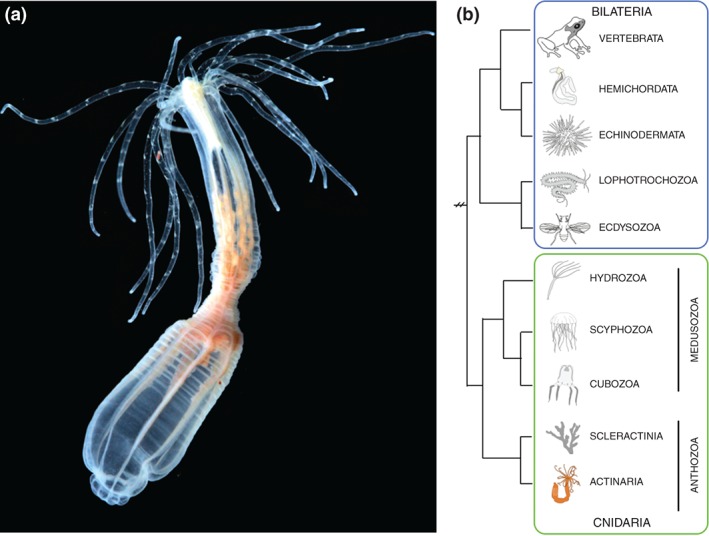
Reverse genetics and next-generation sequencing unlocked a new era in biology. It is now possible to identify an animal(s) with the unique biology most relevant to a particular question and rapidly generate tools to functionally dissect that biology. This review highlights the rise of one such novel model system, the starlet sea anemone Nematostella vectensis. Nematostella is a cnidarian (corals, jellyfish, hydras, sea anemones, etc.) animal that was originally targeted by EvoDevo researchers looking to identify a cnidarian animal to which the development of bilaterians (insects, worms, echinoderms, vertebrates, mollusks, etc.) could be compared. Studies in Nematostella have accomplished this goal and informed our understanding of the evolution of key bilaterian features. However, Nematostella is now going beyond its intended utility with potential as a model to better understand other areas such as regenerative biology, EcoDevo, or stress response. This review intends to highlight key EvoDevo insights from Nematostella that guide our understanding about the evolution of axial patterning mechanisms, mesoderm, and nervous systems in bilaterians, as well as to discuss briefly the potential of Nematostella as a model to better understand the relationship between development and regeneration. Lastly, the sum of research to date in Nematostella has generated a variety of tools that aided the rise of Nematostella to a viable model system. We provide a catalogue of current resources and techniques available to facilitate investigators interested in incorporating Nematostella into their research. WIREs Dev Biol 2016, 5:408-428. doi: 10.1002/wdev.222 For further resources related to this article, please visit the WIREs website.


Figures

Figure 1
Figure 1
Nematostella vectensis is an anthozoan cnidarian sea anemone. (a) Image of adult Nematostella polyp. Image taken by Eric Röttinger. (b) Phylogeny showing the sister relationship of cnidarians to bilaterians and Nematostella's position within the cnidarians.
Figure 2
Figure 2
Development and morphology of Nematostella. (a) Schematic of Nematostella developmental stages. Modified from Ormestad et al. Ref 90. Orange represents presumptive endoderm in blastula and the endoderm in all subsequent stages. The outer ectoderm is white, and the aboral ectodermal domain is in light blue. (b) DIC image taken by Aldine Amiel of juvenile polyp. The mouth (oral opening) is indicated by (Or), the tentacles by (tent), the pharynx by (Ph), and the mesenteries by (mes). Oral is up in all images.
Figure 3
Figure 3
Gene expression during axial patterning. Localization of transcripts (in italic) and proteins involved in the patterning of the oral–aboral (a) and directive (b) axis, respectively. The developmental stages are indicated above each cartoon. Note that not all details of the expression patterns are captured. In (a), the expression domain of wnt4 is broader than that of wntA and wnt1. frizzled5/8 is at planula stage strongly expressed at the aboral pole (green) and weakly in a broader aboral domain (pink). hoxF/anthox1 is at planula stage expressed in the small aboral pole domain (green) and in individual cells within the broader aboral domain (pink). It is not clear whether the expression domains depicted in blue, yellow, and pink are directly abutting each other. In the zygote, Disheveled protein is localized at the cortex and around the nucleus. Disheveled and Strabismus protein remain preferentially localized to the animal/blastoporal region at least until gastrula stage. In (b), BMP5‐8 is at gastrula stage co‐expressed in the ectoderm with bmp2/4, chordin, and rgm (i.e., the domain in light green). pSmad1/5/8 staining forms a gradient with highest levels on the side opposite to the bmp expressing side and low levels on the bmp expressing side. See main text for references.
Figure 4
Figure 4
(GRN) Co‐expression domains within the animal hemisphere of the Nematostella blastula. (a, b) Diagram illustrating four co‐expression domains within the animal hemisphere defined by differential spatial expression of the genes indicated in (b). (c) Examples of genes expression in either the central ring (NvsnailA), the central domain (NvfoxB), the central domain + ring (NvashB), or the external ring (Nvwnt4). In situ hybridization expression profiles are shown in either lateral view (a, top row of c) or animal view (b, bottom row of c) to distinguish expression domains. (Reprinted with permission from Ref 40. Copyright 2012 PLoS Genetics)
Figure 5
Figure 5
Morphology of Nematostella nervous system in juvenile polyps. (a, b) 3D reconstruction of confocal Z‐series acquired for two Nematostella transgenic lines that label the nervous system (a) NvElav::mOrange and (b) NvLWamide::mCherry. (c) Schematic of juvenile polyp nervous system highlights key structures and provides examples of neuronal morphologies observed in transgenic animals.
Figure 6
Figure 6
Oral regeneration of juvenile Nematostella takes place in 6 days. After sub‐pharyngeal bisection, missing oral part of the polyps regenerate within 6 days to reform a fully functional organism. Animals were fixed and stained with phalloidin (green) to show f‐actin filaments and propidium iodide (red) to visualize the nuclei. All images are lateral views with the oral part to the top.
Figure 7
Figure 7
Diagram summarizing the morphological and cellular events underlying Nematostella oral regeneration. The table below the illustration provides definitions for the various regeneration steps. *, mouth opening; ten, tentacles; ten bud, tentacle bud; pha, pharynx; pha lip, pharyngeal lip; mes, mesenteries. (Reprinted with permission from Ref 32. Copyright 2015 International Journal of Molecular Sciences)

References

FURTHER READING

    1. Rigo‐Watermeier T et al. Functional conservation of Nematostella Wnts in canonical and noncanonical Wnt‐signaling. Biol Open 2012, 1:43–51. - PMC - PubMed

References

    1. Dunn CW, Hejnol A, Matus MQ, Pang K, Browne WE, Smith SA, Seaver E, Rouse GW, Obst M. Broad phylogenomic sampling improves resolution of the animal tree of life. Nature 2008, 452:745–749. - PubMed
    1. Ryan JF, Pang K, Schnitzler CE, Nguyen A, Moreland RT, Simmons DK, Koch BJ, Francis WR, Havlak P, NISC Comparative Sequencing Program, et al. The genome of the ctenophore Mnemiopsis leidyi and its implications for cell type evolution. Science 2013, 342:1242592. - PMC - PubMed
    1. Hejnol A, Obst M, Stamatakis A, Ott M, Rouse GW, Edgecombe GD, Martinez P, Baguna J, Bailly X, Jondelius U, et al. Assessing the root of bilaterian animals with scalable phylogenomic methods. Proc R Soc B Biol Sci 2009, 276:4261–4270. - PMC - PubMed
    1. Hand C, Uhlinger KR. The culture, sexual and asexual reproduction, and growth of the sea anemone Nematostella vectensis . Biol Bull 1992, 182:169–176. - PubMed
    1. Houliston E, Momose T, Manuel M. Clytia hemisphaerica: a jellyfish cousin joins the laboratory. Trends Genet 2015, 26:159–167. - PubMed

Publication types

LinkOut - more resources