. 2016 Feb;72(Pt 2):254-65.
doi: 10.1107/S2059798315024237. Epub 2016 Jan 28.

Three-dimensional structures of two heavily N-glycosylated Aspergillus sp. family GH3 β-D-glucosidases

Affiliations

Three-dimensional structures of two heavily N-glycosylated Aspergillus sp. family GH3 β-D-glucosidases

Jon Agirre et al. Acta Crystallogr D Struct Biol. 2016 Feb.

Abstract

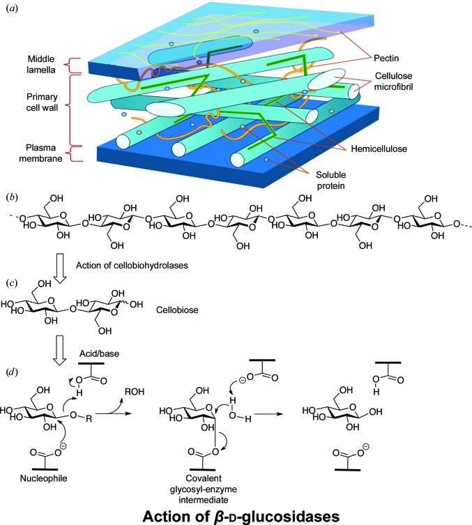
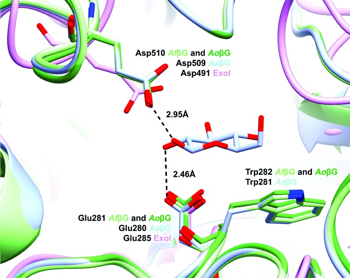
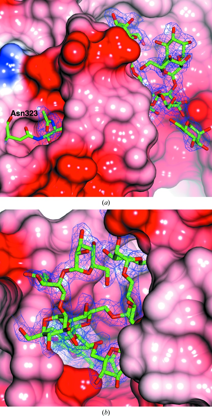
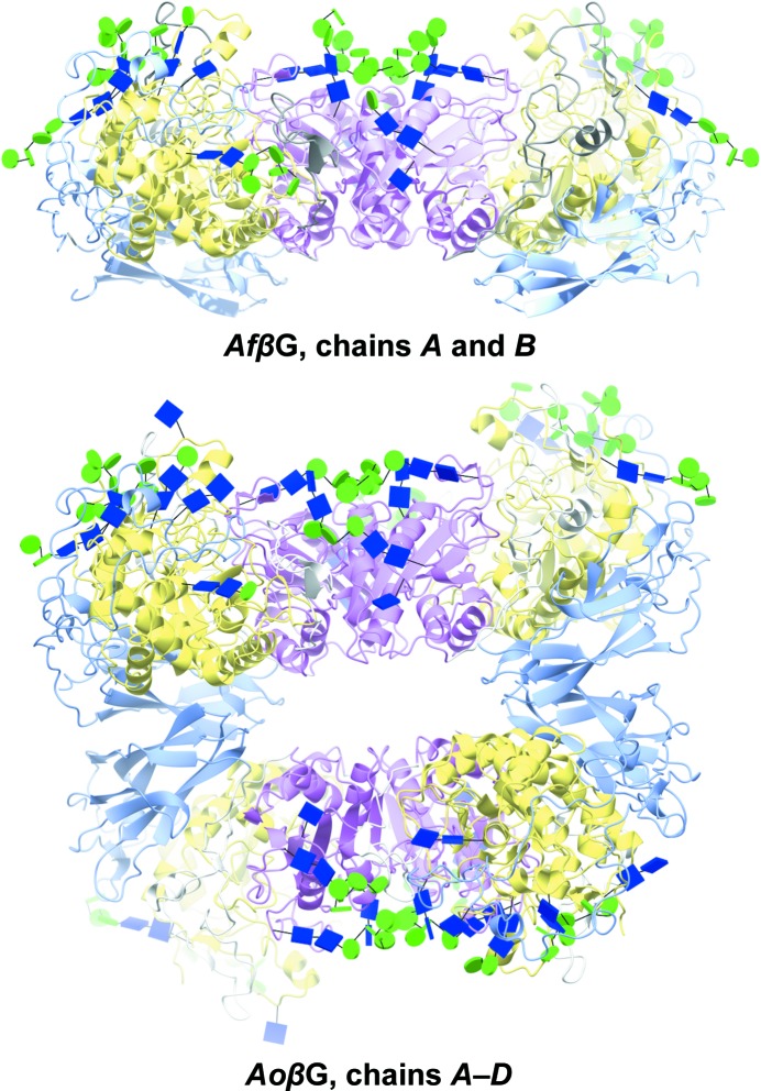
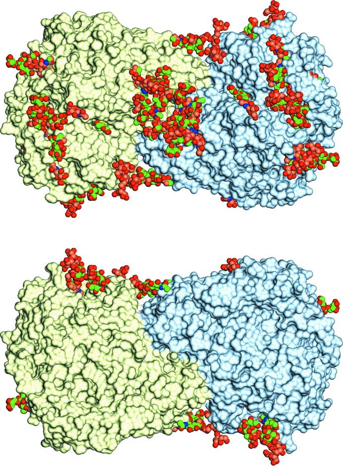
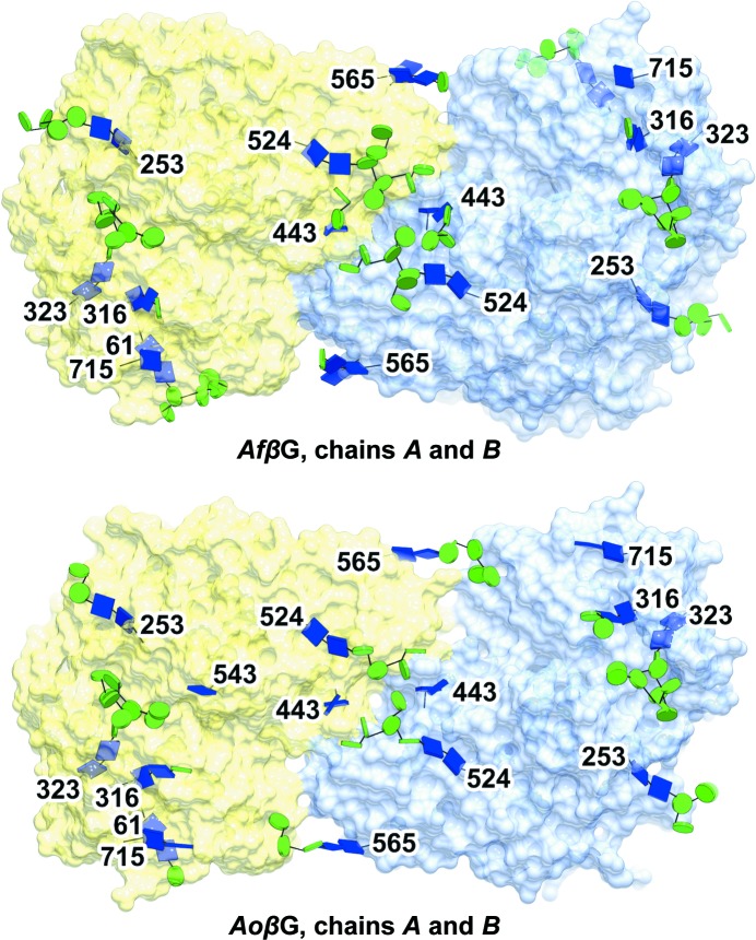
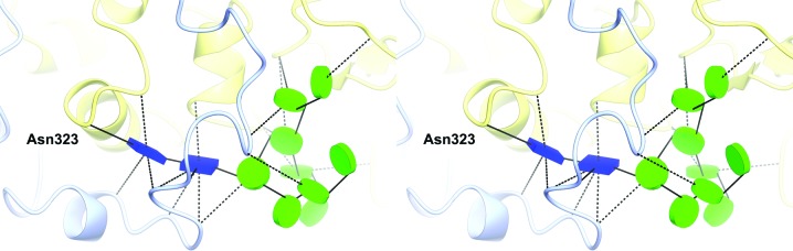
The industrial conversion of cellulosic plant biomass into useful products such as biofuels is a major societal goal. These technologies harness diverse plant degrading enzymes, classical exo- and endo-acting cellulases and, increasingly, cellulose-active lytic polysaccharide monooxygenases, to deconstruct the recalcitrant β-D-linked polysaccharide. A major drawback with this process is that the exo-acting cellobiohydrolases suffer from severe inhibition from their cellobiose product. β-D-Glucosidases are therefore important for liberating glucose from cellobiose and thereby relieving limiting product inhibition. Here, the three-dimensional structures of two industrially important family GH3 β-D-glucosidases from Aspergillus fumigatus and A. oryzae, solved by molecular replacement and refined at 1.95 Å resolution, are reported. Both enzymes, which share 78% sequence identity, display a three-domain structure with the catalytic domain at the interface, as originally shown for barley β-D-glucan exohydrolase, the first three-dimensional structure solved from glycoside hydrolase family GH3. Both enzymes show extensive N-glycosylation, with only a few external sites being truncated to a single GlcNAc molecule. Those glycans N-linked to the core of the structure are identified purely as high-mannose trees, and establish multiple hydrogen bonds between their sugar components and adjacent protein side chains. The extensive glycans pose special problems for crystallographic refinement, and new techniques and protocols were developed especially for this work. These protocols ensured that all of the D-pyranosides in the glycosylation trees were modelled in the preferred minimum-energy (4)C1 chair conformation and should be of general application to refinements of other crystal structures containing O- or N-glycosylation. The Aspergillus GH3 structures, in light of other recent three-dimensional structures, provide insight into fungal β-D-glucosidases and provide a platform on which to inform and inspire new generations of variant enzymes for industrial application.

Keywords: N-glycan; biofuels; cellulose degradation; glucosidase; glycoblocks.


Figures

Figure 1
Figure 1
Cellulose: structure and breakdown. (a) Generic representation of the plant cell wall (taken from https://commons.wikimedia.org/wiki/File:Plant_cell_wall_diagram.svg). (b) Structure of a single β-1,4-d-glucan chain. (c) Structure of the disaccharide cellobiose generated by the action of cellobiohydrolases. (d) Mechanism of a family GH3 retaining β-d-glucosidase; hydrolysis occurs via a covalent glycosyl-enzyme intermediate.
Figure 2
Figure 2
Superposition of the active sites of the enzymes. The catalytic residues proposed for ExoI (PDB entry 1ex1), Asp491 and Glu285 (Thongpoo et al., 2013 ▸), are shown superposed on AfβG, AoβG and AaβG (PDB entry 4iig). The structural figures were all produced using CCP4mg (McNicholas et al., 2011 ▸).
Figure 3
Figure 3
The electron density for the glycosylation tree attached to Asn323 in AfβG shown from two different perspectives. In (a) the first part of the tree is buried within a pocket of the protein. In (b) Asn323 is at the base of the pocket. There is well ordered density for all of the sugars. The maximum-likelihood map was contoured at the 1σ level.
Figure 4
Figure 4
Three-dimensional fold, domain organization and asymmetric unit packing of AfβG and AoβG. Both enzymes have three domains (A, yellow; B, pink; C, light blue), with a dimer being the preferred biological arrangement. AoβG has two dimers in the asymmetric unit, with all of the sugars facing opposite sides. The sugars are shown as glycoblocks, with blue squares for N-acetyl-β-d-glucosamine and green circles for d-mannopyranose.
Figure 5
Figure 5
The glycosylation sites in AfβG and AaβG with the sugars shown in space-filling representation, with the C atoms coloured brown for AaβG and green for AfβG. The surface is that of AfβG coloured by chain. The two views are from opposite sides of the dimer and emphasize how the glycosylation trees are all located on just one side.
Figure 6
Figure 6
N-Glycosylation across AfβG and AoβG. In both enzymes the abundant N-glycans all lie on one side of the molecular surface. Blue square, N-acetyl-β-d-glucosamine. Green circle, d-mannopyranose. Chain A, yellow. Chain B, light blue. The sugars are shown in the same representation as in Fig. 4 ▸.
Figure 7
Figure 7
Schematic stereoview of the longest glycan in AfβG and its interactions. The glycan N-linked to Asn323 is a complete high-mannose tree (11 sugars) that establishes numerous hydrogen bonds to adjacent residues across domain A (yellow) and domain C (light blue). This glycan is very similar in AoβG and AaβG.
Figure 8
Figure 8
GlcNAc–Asn linkages in the two energy minima as found in the AfβG structure. (a) Stereoview of all superposed GlcNAc–Asn sites for chain A. For clarity reasons, Asn residues in other rotamer forms are omitted, and only one representative GlcNAc is shown for the two conformations (1101, with blue C atoms, and 1401, with yellow C atoms). (b) Nag1101–Asn253 as a representative of the most frequent, lowest-energy linkage conformation. (c) Nag1401–Asn443 in the secondary energy minimum described by Imberty & Perez (1995 ▸), with Trp431 taking part in CH–π stacking interactions with the apolar face of Nag1401. The electron-density maps shown here were calculated from 2mF oDF c coefficients and contoured at 2σ. As they both show similar values (∼180°), ψN torsions are not depicted in (b) and (c).

Similar articles

Cited by

References

    1. Agirre, J., Davies, G., Wilson, K. & Cowtan, K. (2015). Nature Chem. Biol. 11, 303. - PubMed
    1. Agirre, J., Iglesias-Fernandez, J., Rovira, C., Davies, G., Wilson, K. & Cowtan, K. (2015). Nature Struct. Mol. Biol. 22, 833–834. - PubMed
    1. Agrawal, R., Satlewal, A., Gaur, R., Mathur, A., Kumar, R., Gupta, R. P. & Tuli, D. K. (2015). Biochem. Eng. J. 102, 54–61.
    1. Ask, M., Olofsson, K., Di Felice, T., Ruohonen, L., Penttilä, M., Lidén, G. & Olsson, L. (2012). Process Biochem. 47, 1452–1459.
    1. Bacik, J.-P., Whitworth, G. E., Stubbs, K. A., Vocadlo, D. J. & Mark, B. L. (2012). Chem. Biol. 19, 1471–1482. - PubMed

Publication types