. 2016 Mar 22;7(12):14112-24.
doi: 10.18632/oncotarget.7415.

Expression of GADS enhances FLT3-induced mitogenic signaling

Affiliations

Expression of GADS enhances FLT3-induced mitogenic signaling

Rohit A Chougule et al. Oncotarget. .

Abstract

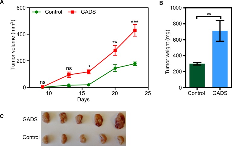
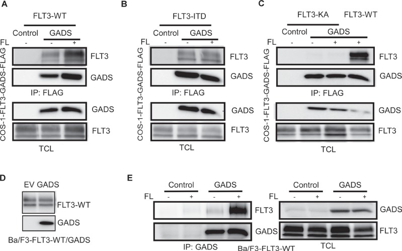
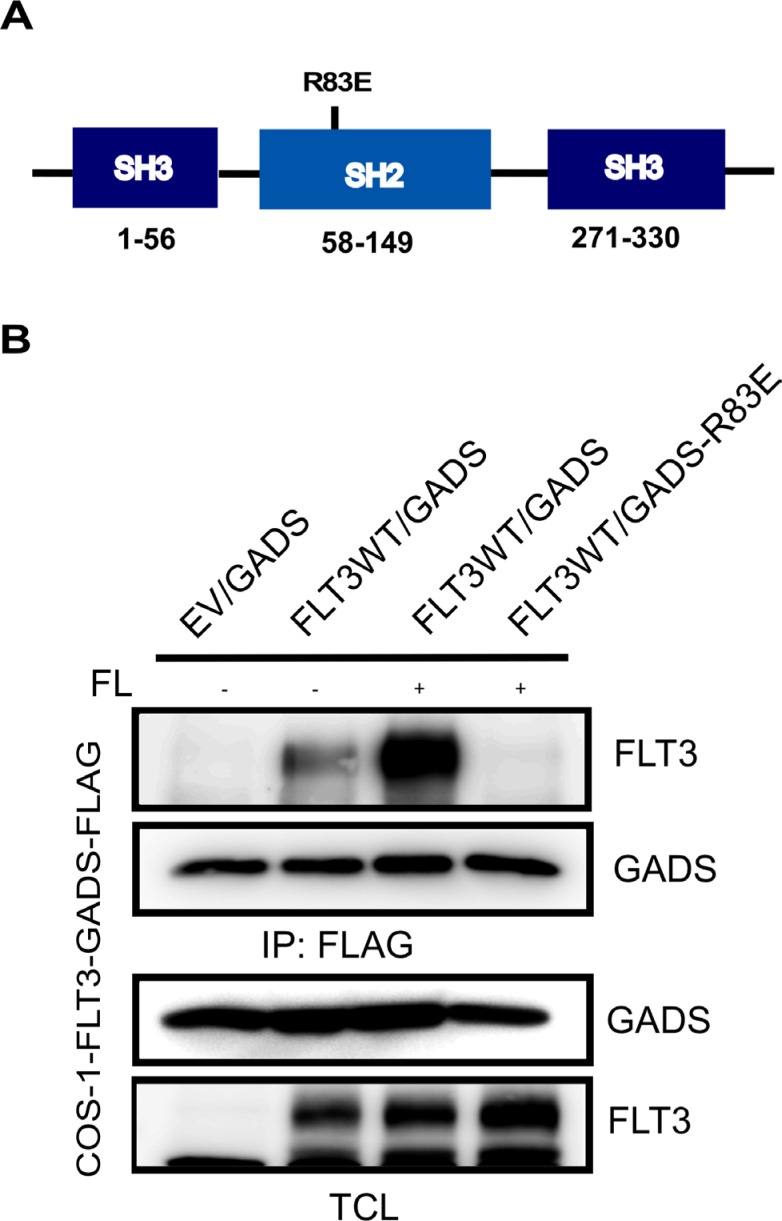
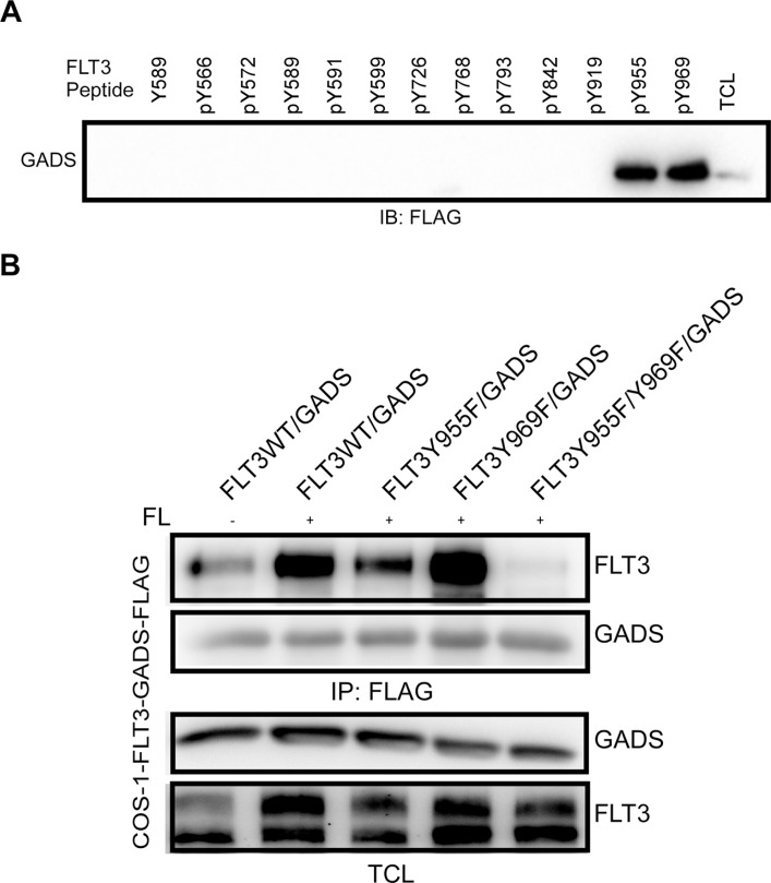
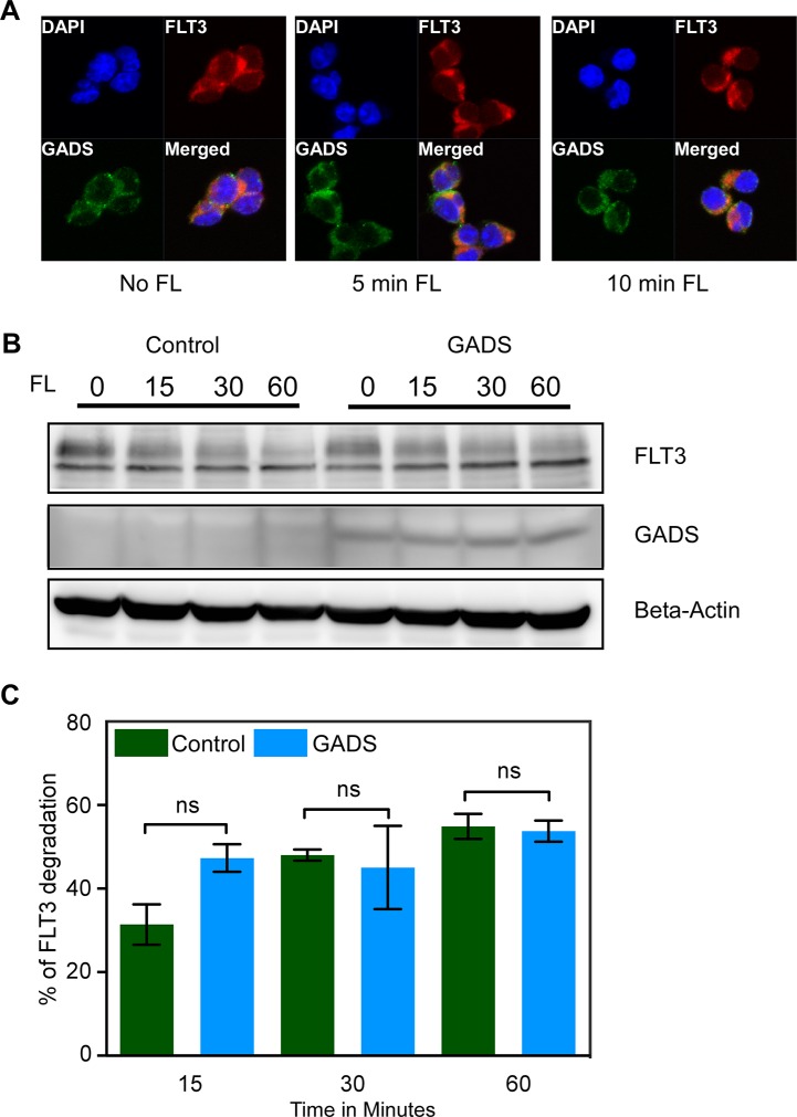
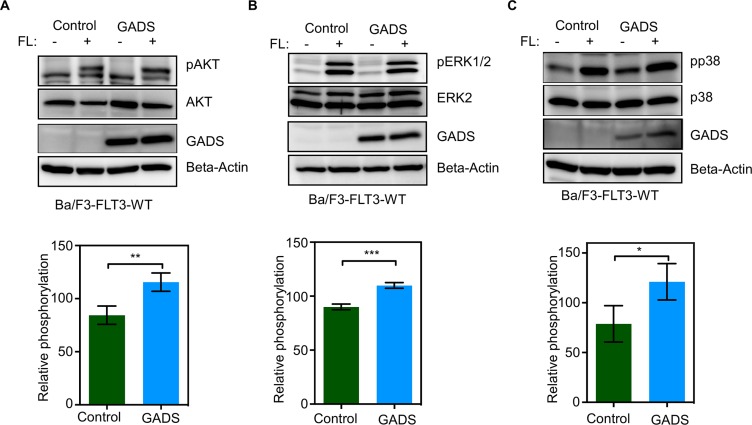
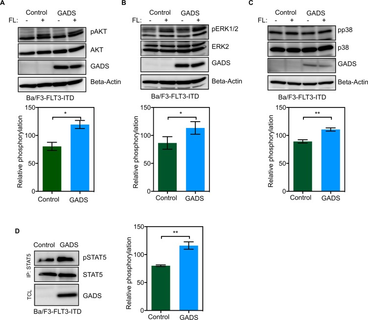
GADS is a member of a family of SH2 and SH3 domain-containing adaptors that functions in tyrosine kinase-mediated signaling cascades. Its expression is largely restricted to hematopoietic tissues and cell lines. Therefore, GADS is mainly involved in leukocyte-specific protein tyrosine kinase signaling. GADS is known to interact with tyrosine-phosphorylated SHC, BCR-ABL and KIT. The SH2 domain of GADS has a similar binding specificity to that of GRB2 but its SH3 domain displays a different binding specificity, and thus it is involved in other downstream signaling pathways than GRB2. In the present study, we examined the role of GADS in FLT3 signaling. FLT3 is a type III receptor tyrosine kinase, which is mutated in more than 30% of acute myeloid leukemia (AML) and the most common mutations is the internal tandem duplication (ITD) mutations. We observed that expression of GADS enhanced oncogenic FLT3-ITD-induced cell proliferation and colony formation in vitro. In a mouse xenograft model, GADS accelerated FLT3-ITD-dependent tumor formation. Furthermore, expression of GADS induced a transcriptional program leading to upregulation of MYC and mTORC1 target genes. GADS localizes to the cell membrane and strongly binds to ligand-stimulated wild-type FLT3 or is constitutively associated with the oncogenic mutant FLT3-ITD. We mapped the binding sites in FLT3 to pY955 and pY969 which overlaps with the GRB2 binding sites. Expression of GADS enhanced FLT3-mediated phosphorylation of AKT, ERK1/2, p38 and STAT5. Taken together, our data suggests that GADS is an important downstream component of FLT3 signaling and expression of GADS potentiates FLT3-mediated mitogenic signaling.

Keywords: AML; FLT3-ITD; GRAP2; RTK; STAT5.


Conflict of interest statement

CONFLICTS OF INTEREST

The authors declare no conflicts of interest.

Figures

Figure 1
Figure 1. GADS expression significantly contributed to cell proliferation and colony formation
Ba/F3/FLT3-ITD cells stably transfected with GADS or empty vector were washed three times with RPMI-1640 to remove IL3. (A) An equal amount of cells were lysed and the lysate was used to check FLT3 and GADS expression. (B) FLT3-ITD-dependent cell proliferation in the presence or absence of GADS expression was measured after 48 hours using PrestoBlue cell viability assay. (CD) Around 500 cells were seeded in semi-solid medium followed by counting colonies after seven days.
Figure 2
Figure 2. GADS enhances tumor growth in a mouse xenograft model
Cells were washed three times with cold PBS to remove IL3. (AC). Cells expressing GADS or empty vector were xenografted into mice and tumor growth was monitored for 23 days. Tumor volume (A) was measured twice a week and tumor weight (B) was measured after sacrificing the animals.
Figure 3
Figure 3. Gene set enrichment analysis (GSEA) showed enrichment of oncogenic pathways in GADS expressing cells
Gene expression data from microarray analysis using GADS expressing cells display enrichment in MYC target genes (A) and MTORC1 target genes (B).
Figure 4
Figure 4. GADS binds with FLT3 in response to ligand stimulation
COS1 cells were transfected with plasmids expressing FLAG-tagged GADS or empty vector and FLT3-WT (A) or FLT3-ITD (B) or FLT3-K644A/FLT3-WT (C). FLT3-K644A is a kinase-dead mutant of FLT3. Cells were stimulated or not with 100 ng/ml FL for five minutes before lysis. Lysates were subjected to anti-FLAG immunoprecipitation followed by Western blotting analysis (D) Expression of FLT3-WT and GADS were verified by Western blotting using anti-FLT3 and anti-FLAG antibodies. (E) Ba/F3 cells expressing FLT3-WT and empty vector or GADS were stimulated for 5 minutes. Cells were lysed and were subjected to immunoprecipitation with an anti-GADS antibody.
Figure 5
Figure 5. The GADS-SH2 domain associated with FLT3
(A) Schematic representation of GADS domains. (B) COS1 cells were transfected with plasmids expressing wild-type FLT3 and FLAG-tagged GADS or GADS-R83E. Cells were stimulated or not with 100 ng/ml FL for five minutes before lysis. Lysates were subjected to anti-FLAG immunoprecipitation followed by Western blotting analysis.
Figure 6
Figure 6. GADS associates with FLT3 through Y955 and Y969 residues
(A) Phosphopeptides corresponding to the known or predicted tyrosine phosphorylation sites in FLT3 were used to pull down GADS from cell lysates. (B) COS1 cells were transfected with plasmids expressing FLAG-tagged GADS or FLT3 mutants. Cells were stimulated or not with 100 ng/ml FL for five minutes before lysis. Lysates were subjected to anti-FLAG immunoprecipitation followed by Western blotting analysis.
Figure 7
Figure 7. GADS localized with FLT3 to the cell surface but did not alter FLT3 stability
(A) Ba/F3-FLT3/Empty vector and Ba/F3-FLT3/GADS cells were stimulated with 100 ng/ml FL for different time points before lysis. Cells were fixed, permeabilized, blocked and stained using PE-conjugated anti-FLT3 and Alexa Fluor 647-conjugated anti-FLAG antibody and then analyzed by confocal microscopy. (B) Ba/F3-FLT3/Empty vector and Ba/F3-FLT3/GADS cells were stimulated with 100 ng/ml FL for different time points before lysis. Lysates were subjected to Western blotting analysis. (C) Multiple blots were quantified using ImageJ and values were analyzed using GraphPad. Ns, not significant.
Figure 8
Figure 8. GADS expression enhances FLT3-induced signaling
(AC) Ba/F3-FLT3/Empty vector and Ba/F3-FLT3/GADS cells were serum- and cytokine-starved for four hours and stimulated with 100 ng/ml FL for five minutes before lysis. Lysates were subjected to Western blotting analysis. Multiple blots were quantified using ImageJ and values were analyzed using GraphPad and one representive blot is shown. **P < 0.01, ***P < 0.001.
Figure 9
Figure 9. GADS expression enhances FLT3-ITD-induced STAT5 signaling
(AD) Ba/F3-FLT3/Empty vector and Ba/F3-FLT3/GADS cells were serum- and cytokine-starved for four hours and stimulated with 100 ng/ml FL for five minutes before lysis. Lysates were subjected to Western blotting analysis. Multiple blots were quantified using ImageJ and values were analyzed using GraphPad. *P < 0.05, **P < 0.01.

Similar articles

Cited by

References

    1. Kabir NN, Rönnstrand L, Kazi JU. FLT3 mutations in patients with childhood acute lymphoblastic leukemia (ALL) Med Oncol. 2013;30:462. - PubMed
    1. Masson K, Rönnstrand L. Oncogenic signaling from the hematopoietic growth factor receptors c-Kit and Flt3. Cell Signal. 2009;21:1717–1726. - PubMed
    1. Lyman SD, James L, Vanden Bos T, de Vries P, Brasel K, Gliniak B, Hollingsworth LT, Picha KS, McKenna HJ, Splett RR, et al. Molecular cloning of a ligand for the flt3/flk-2 tyrosine kinase receptor: a proliferative factor for primitive hematopoietic cells. Cell. 1993;75:1157–1167. - PubMed
    1. Lyman SD, James L, Johnson L, Brasel K, de Vries P, Escobar SS, Downey H, Splett RR, Beckmann MP, McKenna HJ. Cloning of the human homologue of the murine flt3 ligand: a growth factor for early hematopoietic progenitor cells. Blood. 1994;83:2795–2801. - PubMed
    1. Hannum C, Culpepper J, Campbell D, McClanahan T, Zurawski S, Bazan JF, Kastelein R, Hudak S, Wagner J, Mattson J, et al. Ligand for FLT3/FLK2 receptor tyrosine kinase regulates growth of haematopoietic stem cells and is encoded by variant RNAs. Nature. 1994;368:643–648. - PubMed

MeSH terms