. 2016 Mar 22;7(12):14781-90.
doi: 10.18632/oncotarget.7438.

Tumoral indoleamine 2, 3-dioxygenase 1 is regulated by monocytes and T lymphocytes collaboration in hepatocellular carcinoma

Affiliations

Tumoral indoleamine 2, 3-dioxygenase 1 is regulated by monocytes and T lymphocytes collaboration in hepatocellular carcinoma

Qiyi Zhao et al. Oncotarget. .

Abstract

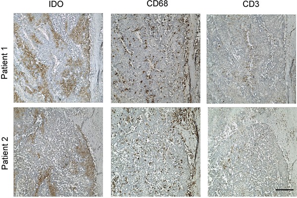
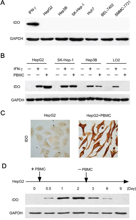
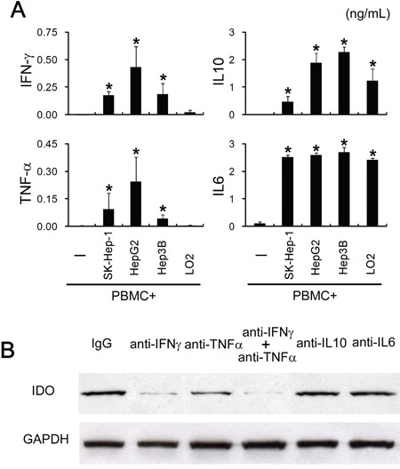
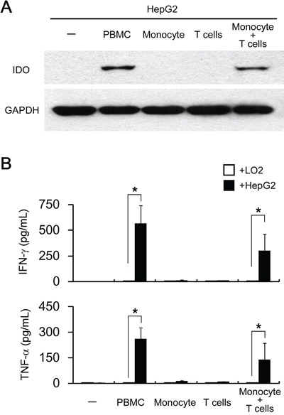
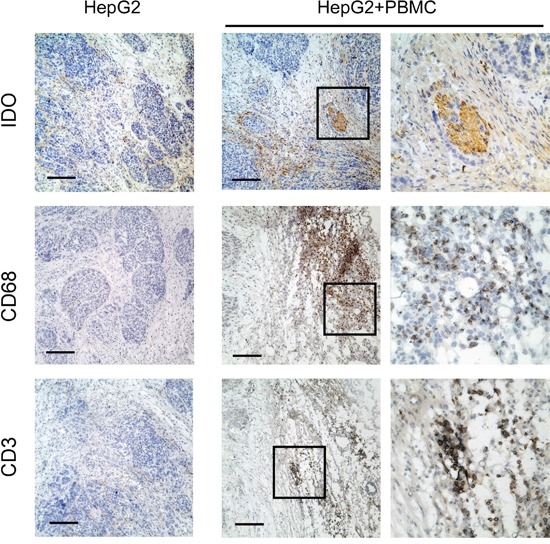
Indoleamine 2, 3-Dioxygenase 1 (IDO1) in cancer cells plays a critical role in tumor immunosuppression. However, the precise mechanisms regulating tumoral IDO1 expression in tumor milieus remain unclear. Here, we reported that IDO1 expression in tumor cells of hepatocelluar carcinomas (HCC), displayed a discrete rather than uniform pattern. In vitro culture, human hepatoma cell lines did not constitutively express IDO1. Interestingly, co-culture with peripheral blood mononuclear cells (PBMC) significantly induced and maintained IDO1 expression in these tumor cells, predominantly through IFN-γ. Mechanistically, we showed that IDO1 expression in tumor cells was only induced when co-cultured with both T lymphocytes and monocytes. Moreover, the cooperation between T lymphocytes and monocytes played an indispensable role on the tumoral IDO1 expression in immunocompromised mice. Taken together, our data supported the notion that IDO1 expression in tumor cells might serve as a counter-regulatory mechanism regulated by immune system, and provided new insights into the collaborative action of different inflammatory cells in tumor immunosuppression.

Keywords: IDO1; T cells; monocytes; tumor cells.


Conflict of interest statement

CONFLICTS OF INTEREST

The authors declare no conflicts of interest.

Figures

Figure 1
Figure 1. Discontinuous tumoral IDO1 expression in human hepatoma samples
Paraffin-embedded hepatocelluar carcinoma samples were stained for Ab against IDO1. Positive signals appear in brown (DAB staining). One of five representative areas is shown. Scale bar, 200 μm.
Figure 2
Figure 2. Immune cells contributed to the induction and maintenance of IDO1 in human hepatoma cells
A. Human hepatoma (HepG2, SK-Hep-1, Hep3B, Huh7, SMMC-7721 and BEL-7402) cell lines did not constitutively express IDO1 in vitro. The IFN-γ treated HepG2 served as a positive control. B. Tumor cells (HepG2, SK-Hep-1, Hep3B) or normal liver cells (L02) were cultured with PBMC at a ratio of 1: 3 or IFN-γ (100 IU/ml) as IDO1-positive control for 2 days. For the coculture groups, PBMC were wash away before protein extraction. The expression of IDO1 protein in the attached cells was detected by western blot. C. HepG2 were cultured alone or with PBMC for 2 days. IDO1 protein was detected by immunocytochemistry. One of five representative areas is shown. D. PBMC were added to HepG2 at day 0, and were washed away after 2-day coculture. The IDO1 protein in the attached cells was detected by western blot at indicated times.
Figure 3
Figure 3. Tumoral IDO1 expression was regulated by pro-inflammatory cytokines
A. PBMC were cocultured with tumor cells lines (HepG2, SK-Hep-1, Hep3B) or normal liver cells (L02), and the production of cytokine (TNF-α, IFN-γ, IL-6, IL-10) was measured by ELISA. B. HepG2 cocultured with PBMC were treated with indicated blocking Abs or control IgG. The expression of IDO1 in HepG2 was determined by western blot and ELISA.
Figure 4
Figure 4. Both T lymphocytes and monocytes are responsible for pro-inflammatory cytokines driven tumoral IDO1 expression in vitro
HepG2 (hepatoma cell line) or L02 (normal liver cell line) were cocultured with PBMC (1: 3) or with purified monocytes or/and T cells (1: 1: 2) for 2 days. A. The expression of IDO1 in HepG2 cells were determined by western blot. B. The production of IFN-γ and TNF-α was determined by ELISA. *p < 0.05 are shown.
Figure 5
Figure 5. Human T lymphocytes and monocytes elevated IDO1 in human tumor cells in a xenograft model in vivo
Human hepatoma (HepG2) was implanted subcutaneously into nonobese diabetic/severe combined immunodeficient (NOD/SCID) mice alone, or co-implantation with human PBMC (1:3) as described in materials and methods. Paraffin-embedded tumor tissues were stained for Abs against human IDO1, CD68 or CD3, respectively. Scale bar, 100 μm.

References

    1. Munn DH, Mellor AL. Indoleamine 2,3 dioxygenase and metabolic control of immune responses. Trends Immunol. 2013;34:137–143. - PMC - PubMed
    1. Munn DH, Mellor AL. IDO and tolerance to tumors. Trends Mol Med. 2004;10:15–18. - PubMed
    1. Munn DH, Mellor AL. Indoleamine 2,3-dioxygenase and tumor-induced tolerance. J Clin Invest. 2007;117:1147–1154. - PMC - PubMed
    1. Vigneron N, van Baren N, Van den Eynde BJ. Expression profile of the human IDO1 protein, a cancer drug target involved in tumoral immune resistance. Oncoimmunology. 2015;4:e1003012. - PMC - PubMed
    1. Theate I, van Baren N, Pilotte L, Moulin P, Larrieu P, Renauld JC, Herve C, Gutierrez-Roelens I, Marbaix E, Sempoux C, Van den Eynde BJ. Extensive profiling of the expression of the indoleamine 2,3-dioxygenase 1 protein in normal and tumoral human tissues. Cancer immunology research. 2015;3:161–172. - PubMed

MeSH terms

Substances