. 2016 Feb 18;61(4):563-574.
doi: 10.1016/j.molcel.2016.01.026.

Releasing Activity Disengages Cohesin's Smc3/Scc1 Interface in a Process Blocked by Acetylation

Affiliations

Releasing Activity Disengages Cohesin's Smc3/Scc1 Interface in a Process Blocked by Acetylation

Frederic Beckouët et al. Mol Cell. .

Abstract

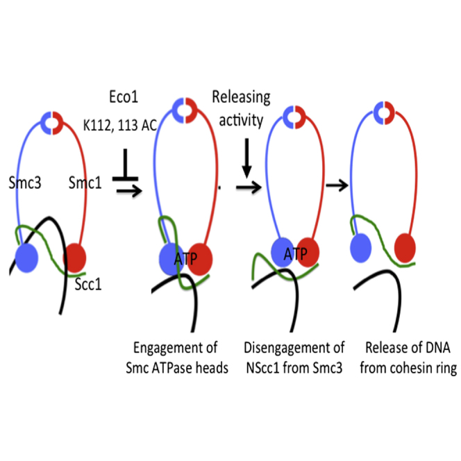
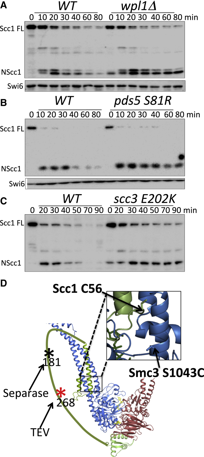
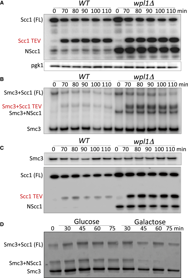
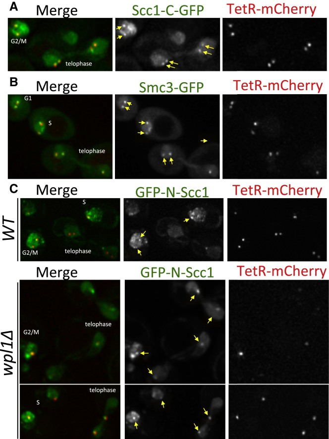
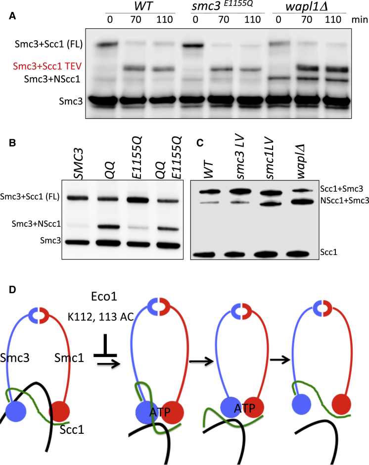
Sister chromatid cohesion conferred by entrapment of sister DNAs within a tripartite ring formed between cohesin's Scc1, Smc1, and Smc3 subunits is created during S and destroyed at anaphase through Scc1 cleavage by separase. Cohesin's association with chromosomes is controlled by opposing activities: loading by Scc2/4 complex and release by a separase-independent releasing activity as well as by cleavage. Coentrapment of sister DNAs at replication is accompanied by acetylation of Smc3 by Eco1, which blocks releasing activity and ensures that sisters remain connected. Because fusion of Smc3 to Scc1 prevents release and bypasses the requirement for Eco1, we suggested that release is mediated by disengagement of the Smc3/Scc1 interface. We show that mutations capable of bypassing Eco1 in Smc1, Smc3, Scc1, Wapl, Pds5, and Scc3 subunits reduce dissociation of N-terminal cleavage fragments of Scc1 (NScc1) from Smc3. This process involves interaction between Smc ATPase heads and is inhibited by Smc3 acetylation.


Figures

None
Graphical abstract
Figure 1
Figure 1
Stability of Scc1 Cleavage Fragments in Releasing Activity Mutants (A–C) Wild-type (K17960), wpl1Δ (K20236), pds5-S81R (K20521), and Scc3-E202K (K20526) strains expressing CDC20 from the GAL promoter were grown to logarithmic phase at 25°C in YP medium containing galactose, transferred to galactose-free media to induce metaphase arrest (time 0), and anaphase triggered by galactose readdition. Separase cleavage of Scc1 was followed by western blotting, detecting N-terminal Myc tag on Scc1. (D) Model of the ATPase domains of Smc1 and Smc3 in an engaged state driven by ATP binding. The separase cleavage site in Scc1 at position 181 is marked with a black asterisk; TEV sites at position 268 are marked with a red asterisk.
Figure 2
Figure 2
Wapl Triggers Dissociation of NScc1 from Smc3 (A) Wild-type and wpl1Δ strains K22156 (MATα SMC3(S1043C)-HA6 MYC3-SCC1(TEV268) YEp-PGAL1 TEV) and K22155 (MATα wpl1Δ SMC3(S1043C)-HA6 MYC3-SCC1(TEV268) YEp-PGAL1 TEV) grown to logarithmic phase at 25°C in YP medium containing raffinose were G2/M arrested by incubating with nocodazole for 2 hr. TEV protease was then induced by addition of galactose and cleavage of Scc1 monitored by Western blotting, detecting Myc epitopes. (B) Samples from (A) were treated with 5 mM BMOE to induce in vivo thiol specific crosslinking between Smc3 S1043C and Scc1 C56. Smc3-HA3 immunoprecipitated from whole-cell extracts was analyzed by western blotting detecting HA epitopes. (C) Smc3-HA3 was immunoprecipitated from untreated cells. Coimmunoprecipitation of Scc1 protein was analyzed by western blotting using Myc antibodies. (D) Strain K22555 (MATα SMC3(S1043C)-HA6 MYC3-SCC1(TEV268) pGAL1-10-WPL1) expressing Wapl from the GAL promoter was grown in YP medium containing raffinose at 25°C and arrested in G2/M due to incubation with nocodazole for 2 hr. One-half of the culture was incubated in the presence of glucose, while the other half was induced to express Wapl by galactose addition. BMOE-induced crosslinking between Smc3 S1043C and Scc1 C56 was analyzed by western blotting using anti-Myc antibodies.
Figure 3
Figure 3
Live-Cell Imaging NScc1’s Dissociation from Smc3 (A and B) An array of 448 tetracycline operators (TetO) was integrated between the BMH1 and PDA1 genes on the long arm of chromosome V in haploid (C, K23761 and K23764) or diploid (A, K23388; B, K23183) cells expressing a version of Scc3 fused to the Tet repressor as well as low levels of a Tet repressor protein fused to mCherry (TetR-mCherry) to mark the location of operators. Exponentially growing cells (in YPD medium at 25°C) were placed on 2.5% agarose pads made of synthetic complete medium containing glucose. Live-cell imaging was performed under a spinning disk confocal system at 25°C. The recruitment of C-terminally GFP-tagged Scc1 (A) and C-terminally GFP-tagged Smc3 (B) to the TetO arrays through Scc3-TetR fusion protein is shown (arrows). (C) The localization of N-terminally GFP-tagged Scc1 to TetO arrays in wild-type (upper panel) waplΔ cells (lower panels) is marked with arrows. We failed to detect GFP-NScc1 at the Tet operators in 20 or more late anaphase/telophase nuclei in Wpl1+ cells.
Figure 4
Figure 4
Scc3’s Highly Conserved Surface Is Essential for NScc1 Dissociation (A) Surface conservation of Scc3 orthologs projected on Z.r. Scc3 (blue, most conserved; red, least conserved) highlighting the conserved K404 (S. cerevisiae). (B) Diploid strain (MATa/α wpl1Δ eco1Δ scc3K404E) was sporulated, tetrads dissected, and selected haploid segregants with their genotypes shown. (C) Exponentially growing cells from wild-type (K22156), wpl1Δ (K22155), pds5-S81R (K20521), and scc3-E404K (K24349), all MATα SMC3(S1043C)-HA6 MYC3-SCC1(TEV268) were grown in YPD medium at 25°C and treated with 5 mM BMOE to crosslink Smc3 S1043C and Scc1 C56. Crosslinking was analyzed by western blotting using anti HA antibody. (D) HIS-tagged wild-type Scc3, Scc3K404E, and Wapl proteins were purified from E. coli using TALON resin followed by Size exclusion using Superdex 200 16/60 column. Wild-type Scc3 or the K404E mutant protein was incubated either alone or with Wapl. After separation of the proteins by gel filtration using a Superose 6 column, the peak fractions were analyzed by SDS-PAGE and Coomassie staining, the fractions containing the Scc3/Wapl complexes are highlighted with a red box. The peak profiles of Scc3, Wapl, and the Scc3 Wapl complex are shown in the right for the wild-type and the E404K mutant proteins.
Figure 5
Figure 5
Effect of smc3 and scc1 Mutations on NScc1 Release (A) Strains K24297 (MATa SMC3(S1043C)-HA6 MYC3-SCC1) and K24343 (MATa SMC3(S1043C)-HA6 MYC3-scc1M102K) growing exponentially in YPD medium at 25°C were treated with 5 mM BMOE to crosslink Smc3 S1043C with either wild-type Scc1 or Scc1 M102K. The crosslinking was analyzed by western blotting using an HA epitope-specific antibody. The structure of Smc3-Scc1NTD complex (PDB: 4UX3) is shown on the right with Scc1 M102, Smc3 R107, K112, K113, and D1189 residues marked. (B) Strains K24217 (MATa SMC3 URA3::SMC3 S1043C-PK6 MYC3-SCC1) and K24485 (MATa SMC3 URA3::SMC3 R107I S1043C-PK6, MYC3-SCC1) were treated as in (A) and crosslinking analyzed by western blotting using anti PK antibody. The data are from the same western blot, with irrelevant lanes removed. (C) Strains K24217 (MATa SMC3 URA3::SMC3 S1043C-PK6 MYC3-SCC1), K24493 (MATa SMC3 URA3::smc3 K112 K113R S1043C-PK6 MYC3-SCC1), K24495 (MATa, SMC3, URA3::smc3 K112 K113R S1043C-PK6 MYC3-SCC1 M102K), K24497 (MATa SMC3 URA3::SMC3 S1043C D1189H-PK6 MYC3-SCC1), and K24499 (MATa SMC3 URA3::smc3 K112 K113R S1043C D1189H-PK6 MYC3-SCC1) were analyzed as in (B). The data shown in the right panel are from the same blot, with irrelevant lanes removed.
Figure 6
Figure 6
Smc3 Acetylation Blocks NScc1 Dissociation (A) HOS1 or hos1Δ strains with galactose inducible WPL1, K22555 (MATa SMC3 (S1043C)-HA6 MYC3-SCC1(TEV268) pGAL1-10-WPL1) and K22810 (MATa hos1Δ SMC3(S1043C)-HA6 MYC3-SCC1(TEV268) pGAL1-10-WPL1) were grown in YP Raff medium at 25°C and arrested in nocodazole for 2 hr. Galactose was then added to induce Wapl. Samples were taken at the indicated time points to induce in vivo crosslinking with 5 mM BMOE. Crosslinking was analyzed by western blotting using anti HA antibodies. Uncrosslinked samples were also analyzed similarly (shown in Figure S1B). (B) Exponentially growing strains K24217 (MATa SMC3 URA3::SMC3 S1043C-PK6 MYC3-SCC1(TEV268)) and K24218 (MATa, SMC3 URA3::smc3K112 113Q S1043C-PK6 MYC3-SCC1(TEV268)) in YPD medium at 25°C were subjected to in vivo thiol-specific crosslinking with 5 mM BMOE. Crosslinking was analyzed by western blotting using anti PK(V5) antibody. (C) Strain K24090 containing a 2.3 kb circular minichromosome, six cysteines within the Smc1-Smc3-Scc1 interfaces, eco1ts(G211H), and galactose-inducible WPL1 gene was grown at 25°C, arrested in G1 by pheromone, and permitted to go through S phase at 37°C in the presence of nocodazole. After addition of either galactose or glucose to induce Wapl expression (or not) samples were taken at times 0 and 60 min for in vivo crosslinking with BMOE. Scc1-PK6-immunoprecipitated DNA denatured with SDS was detected by Southern blotting. Catenated monomers (CM), catenated dimers (CD). (D) Calibrated ChIP-seq profiles of Smc3 E1155Q and Smc3 E1155Q K112Q K113Q showing the number of reads at each base pair away from the CDEIII element averaged over all 16 chromosomes.
Figure 7
Figure 7
Disengagement of NScc1 from Smc3 Requires a Single Round of ATP Hydrolysis (A) Strains K23070 (MATa SMC3 URA3::SMC3 S1043C-HA6 MYC3-SCC1(TEV268) YEp-PGAL1-TEV), K23067 (MATa SMC3 URA3::smc3 S1043C E1155Q-HA6::URA3 MYC3-SCC1(TEV268) YEp-PGAL1-TEV), and K23068 (MATa SMC3 SMC3 S1043C-HA6 MYC3-SCC1(TEV268) wpl1Δ YEp-PGAL1 TEV) were treated and analyzed as described in Figure 2B. (B) Strains K24217 (MATa SMC3 URA3::SMC3 S1043C-PK6::URA3 MYC3-SCC1(TEV268)), K24218 (MATa SMC3 URA3::smc3K112 K113Q S1043C-PK6 MYC3-SCC1(TEV268)), K24219 (MATa SMC3 URA3::smc3E1155Q S1043C-PK6 MYC3-SCC1(TEV268)), and K24220 (MATa SMC3 URA3::smc3K112 K113Q E1155Q S1043C-PK6 MYC3-SCC1(TEV268)) were grown and analyzed as described in Figure 6B. (C) Exponentially growing strains K23070, K23068, K24911 (SMC3 smc3L1126V S1043C::LEU2 MYC3-SCC1(TEV268)), and K24523 (SMC3 S1043C::ADE2 MYC3-SCC1(TEV268) smc1 L1129V) were treated as described in Figure 6B and analyzed by western blotting using anti-MYC antibodies. (D) Shown is a model for how releasing activity dissociates Scc1-NTD from Smc3’s coiled coil leading to escape of entrapped DNAs in a process involving ATP-dependent engagement of SMC ATPase heads. Acetylation of Smc3 residues K112 and K113 is suggested to inhibit ATP-dependent head engagement.

Similar articles

Cited by

References

    1. Arumugam P., Gruber S., Tanaka K., Haering C.H., Mechtler K., Nasmyth K. ATP hydrolysis is required for cohesin’s association with chromosomes. Curr. Biol. 2003;13:1941–1953. - PubMed
    1. Arumugam P., Nishino T., Haering C.H., Gruber S., Nasmyth K. Cohesin’s ATPase activity is stimulated by the C-terminal Winged-Helix domain of its kleisin subunit. Curr. Biol. 2006;16:1998–2008. - PubMed
    1. Beckouët F., Hu B., Roig M.B., Sutani T., Komata M., Uluocak P., Katis V.L., Shirahige K., Nasmyth K. An Smc3 acetylation cycle is essential for establishment of sister chromatid cohesion. Mol. Cell. 2010;39:689–699. - PMC - PubMed
    1. Bürmann F., Shin H.C., Basquin J., Soh Y.M., Giménez-Oya V., Kim Y.G., Oh B.H., Gruber S. An asymmetric SMC-kleisin bridge in prokaryotic condensin. Nat. Struct. Mol. Biol. 2013;20:371–379. - PubMed
    1. Chan K.L., Roig M.B., Hu B., Beckouët F., Metson J., Nasmyth K. Cohesin’s DNA exit gate is distinct from its entrance gate and is regulated by acetylation. Cell. 2012;150:961–974. - PMC - PubMed

Publication types

MeSH terms

Substances