

New p32/gC1qR Ligands for Targeted Tumor Drug Delivery
- PMID: 26895508
- PMCID: PMC5433940
- DOI: 10.1002/cbic.201500564
New p32/gC1qR Ligands for Targeted Tumor Drug Delivery
Abstract
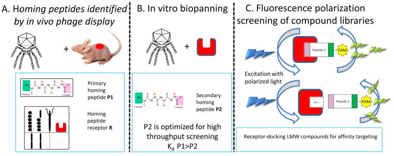
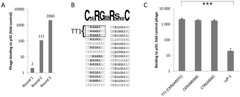
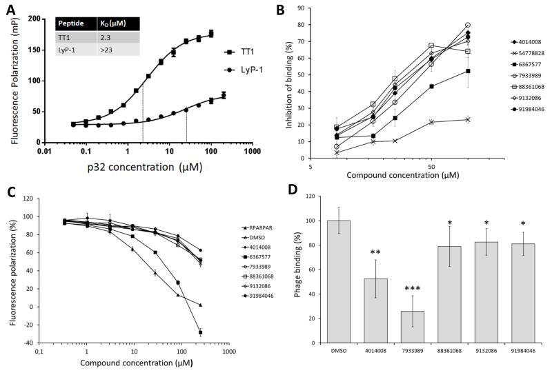
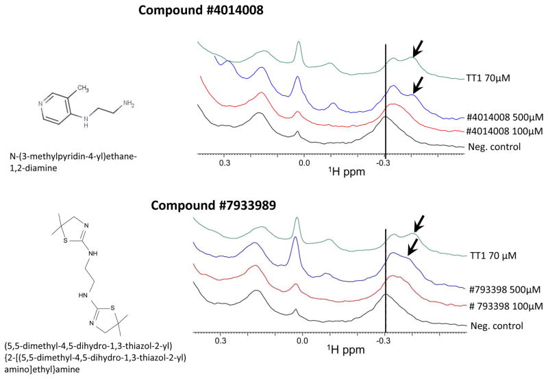
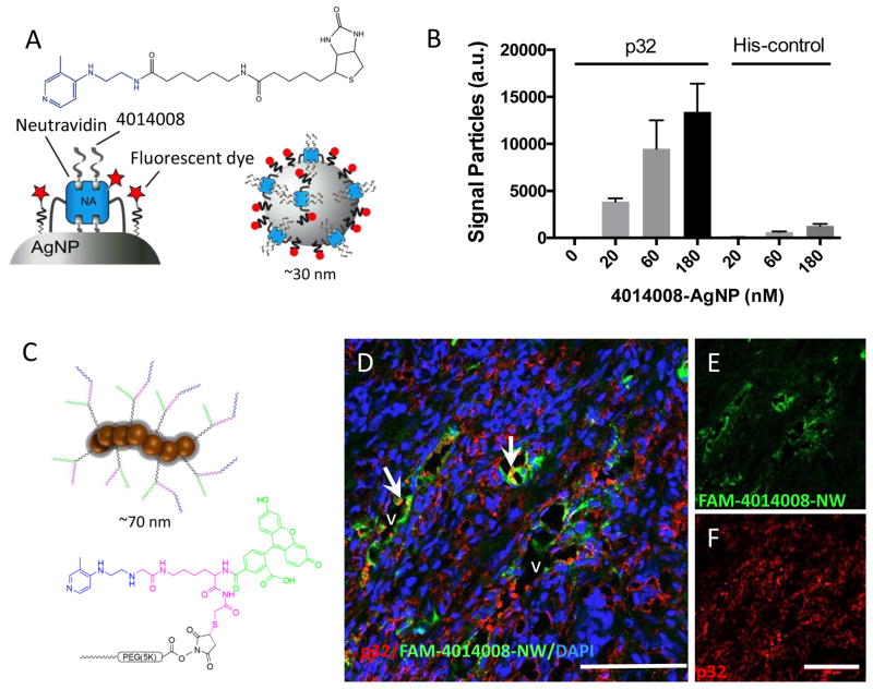
Cell surface p32, the target of LyP-1 homing peptide, is upregulated in tumors and atherosclerotic plaques and has been widely used as a receptor for systemic delivery of payloads. Here, we identified an improved LyP-1-mimicking peptide (TT1, CKRGARSTC). We used this peptide in a fluorescence polarization-based high-throughput screening of a 50,000-compound chemical library and identified a panel of compounds that bind p32 with low micromolar affinity. Among the hits identified in the screen, two compounds were shown to specifically bind to p32 in multiple assays. One of these compounds was chosen for an in vivo study. Nanoparticles surface-functionalized with this compound specifically adhered to surfaces coated with recombinant p32 and, when injected intravenously, homed to p32-expressing breast tumors in mice. This compound provides a lead for the development of p32-targeted affinity ligands that circumvent some of the limitations of peptide-based probes in guided drug delivery.
Keywords: cancer; drug delivery; high-throughput screening; nanoparticles; peptides.
© 2016 WILEY-VCH Verlag GmbH & Co. KGaA, Weinheim.
Figures
Similar articles
-
Design and In Vitro Evaluation of Bispecific Complexes and Drug Conjugates of Anticancer Peptide, LyP-1 in Human Breast Cancer.Pharm Res. 2017 Feb;34(2):352-364. doi: 10.1007/s11095-016-2066-2. Epub 2016 Nov 28. Pharm Res. 2017. PMID: 27896591
-
Targeting of p32 in peritoneal carcinomatosis with intraperitoneal linTT1 peptide-guided pro-apoptotic nanoparticles.J Control Release. 2017 Aug 28;260:142-153. doi: 10.1016/j.jconrel.2017.06.005. Epub 2017 Jun 8. J Control Release. 2017. PMID: 28603028 Free PMC article.
-
Structure Reconstruction of LyP-1: Lc(LyP-1) Coupling by Amide Bond Inspires the Brain Metastatic Tumor Targeted Drug Delivery.Mol Pharm. 2018 Feb 5;15(2):430-436. doi: 10.1021/acs.molpharmaceut.7b00801. Epub 2017 Dec 22. Mol Pharm. 2018. PMID: 29215294
-
Recent progress in LyP-1-based strategies for targeted imaging and therapy.Drug Deliv. 2019 Dec;26(1):363-375. doi: 10.1080/10717544.2019.1587047. Drug Deliv. 2019. PMID: 30905205 Free PMC article. Review.
-
Research Progress on Cyclic-Peptide Functionalized Nanoparticles for Tumor-Penetrating Delivery.Int J Nanomedicine. 2024 Nov 26;19:12633-12652. doi: 10.2147/IJN.S487303. eCollection 2024. Int J Nanomedicine. 2024. PMID: 39624118 Free PMC article. Review.
Cited by
-
LyP-1-fMWNTs enhanced targeted delivery of MBD1siRNA to pancreatic cancer cells.J Cell Mol Med. 2020 Mar;24(5):2891-2900. doi: 10.1111/jcmm.14864. Epub 2020 Jan 22. J Cell Mol Med. 2020. PMID: 31968405 Free PMC article.
-
Tumor penetrating peptides for improved drug delivery.Adv Drug Deliv Rev. 2017 Feb;110-111:3-12. doi: 10.1016/j.addr.2016.03.008. Epub 2016 Apr 1. Adv Drug Deliv Rev. 2017. PMID: 27040947 Free PMC article. Review.
-
Core vs. surface labelling of mesoporous silica nanoparticles: advancing the understanding of nanoparticle fate and design of labelling strategies.Nanoscale Adv. 2022 Mar 1;4(9):2098-2106. doi: 10.1039/d1na00719j. eCollection 2022 May 3. Nanoscale Adv. 2022. PMID: 36133445 Free PMC article.
-
Anti gC1qR/p32/HABP1 Antibody Therapy Decreases Tumor Growth in an Orthotopic Murine Xenotransplant Model of Triple Negative Breast Cancer.Antibodies (Basel). 2020 Oct 6;9(4):51. doi: 10.3390/antib9040051. Antibodies (Basel). 2020. PMID: 33036212 Free PMC article.
-
Neuropilins in the Context of Tumor Vasculature.Int J Mol Sci. 2019 Feb 1;20(3):639. doi: 10.3390/ijms20030639. Int J Mol Sci. 2019. PMID: 30717262 Free PMC article. Review.
References
-
- Laakkonen P, Porkka K, Hoffman J, Ruoslahti E. A tumor-homing peptide with a targeting specificity related to lymphatic vessels. Nat Med. 2002;8:751–755. - PubMed
Publication types
MeSH terms
Substances
Grants and funding
LinkOut - more resources
Full Text Sources
Other Literature Sources
Medical
Research Materials
Miscellaneous
