. 2016 Apr 15;310(8):L733-46.
doi: 10.1152/ajplung.00419.2015. Epub 2016 Feb 19.

Enhanced resolution of experimental ARDS through IL-4-mediated lung macrophage reprogramming

Affiliations

Enhanced resolution of experimental ARDS through IL-4-mediated lung macrophage reprogramming

F R D'Alessio et al. Am J Physiol Lung Cell Mol Physiol. .

Abstract

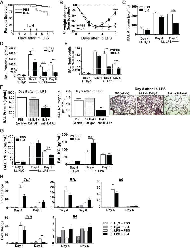
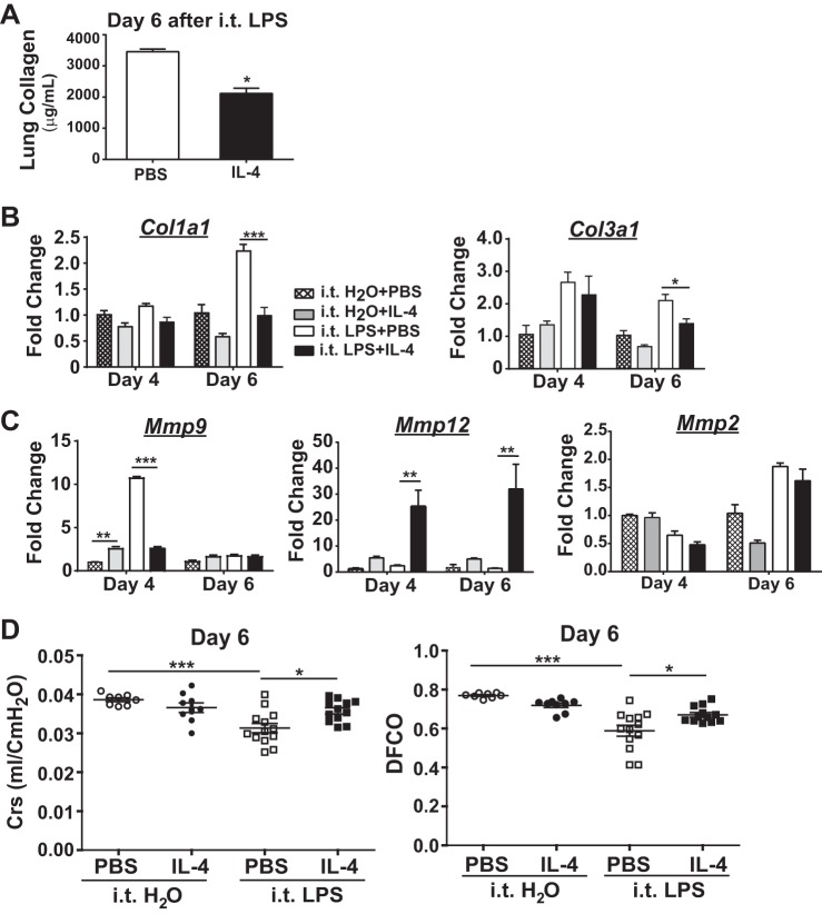
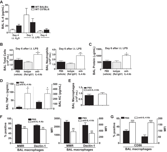
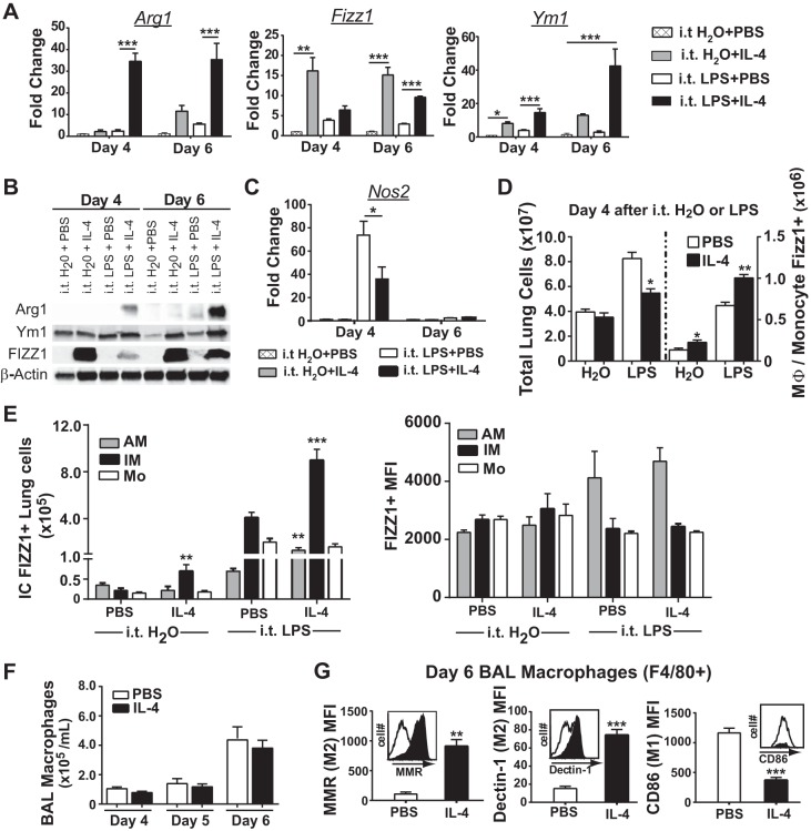
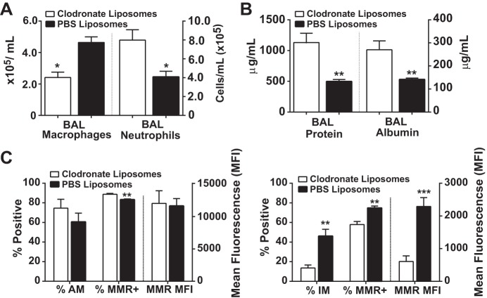
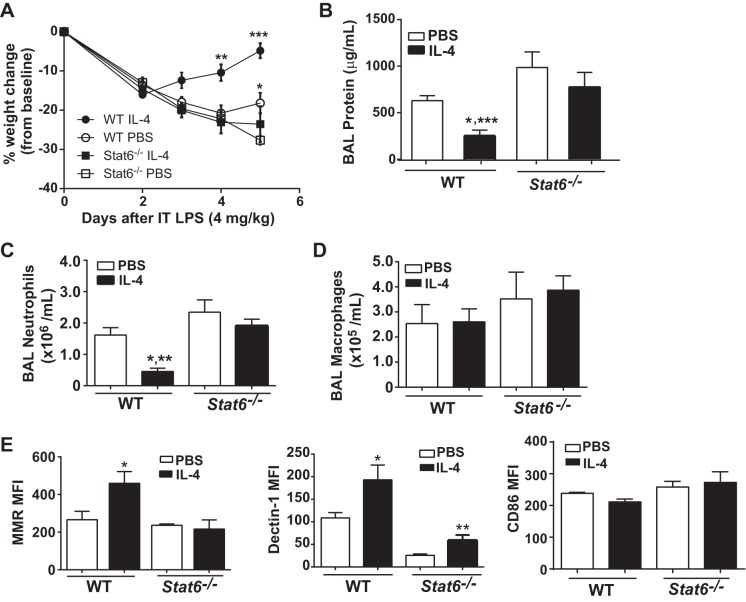
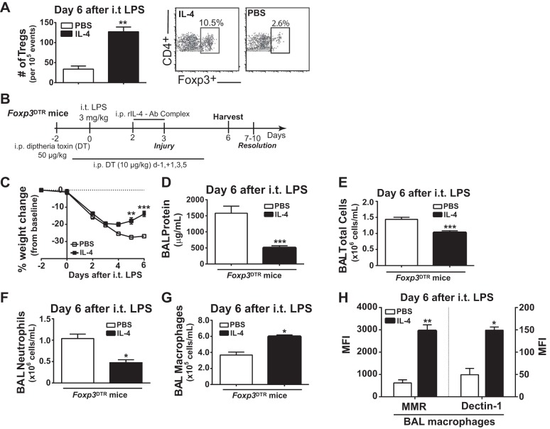
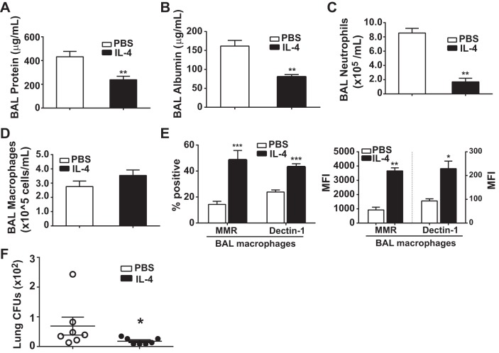
Despite intense investigation, acute respiratory distress syndrome (ARDS) remains an enormous clinical problem for which no specific therapies currently exist. In this study, we used intratracheal lipopolysaccharide or Pseudomonas bacteria administration to model experimental acute lung injury (ALI) and to further understand mediators of the resolution phase of ARDS. Recent work demonstrates macrophages transition from a predominant proinflammatory M1 phenotype during acute inflammation to an anti-inflammatory M2 phenotype with ALI resolution. We tested the hypothesis that IL-4, a potent inducer of M2-specific protein expression, would accelerate ALI resolution and lung repair through reprogramming of endogenous inflammatory macrophages. In fact, IL-4 treatment was found to offer dramatic benefits following delayed administration to mice subjected to experimental ALI, including increased survival, accelerated resolution of lung injury, and improved lung function. Expression of the M2 proteins Arg1, FIZZ1, and Ym1 was increased in lung tissues following IL-4 treatment, and among macrophages, FIZZ1 was most prominently upregulated in the interstitial subpopulation. A similar trend was observed for the expression of macrophage mannose receptor (MMR) and Dectin-1 on the surface of alveolar macrophages following IL-4 administration. Macrophage depletion or STAT6 deficiency abrogated the therapeutic effect of IL-4. Collectively, these data demonstrate that IL-4-mediated therapeutic macrophage reprogramming can accelerate resolution and lung repair despite delayed use following experimental ALI. IL-4 or other therapies that target late-phase, proresolution pathways may hold promise for the treatment of human ARDS.

Keywords: ARDS; acute lung injury; interleukin-4; macrophage; resolution.


Figures

Fig. 1.
Fig. 1.
Wild-type (WT; C57BL/6) mice treated with IL-4 demonstrate improved survival and accelerated acute lung injury (ALI) resolution. A: Kaplan-Meier survival curves for mice exposed to intratracheal (i.t.) LPS (4 mg/kg) and treated with IL-4 or PBS (n = 27–30, P = 0.006 by Mantel-Cox). B: daily percent weight change from baseline for each group following intratracheal LPS (n = 6–10, ***P < 0.001 by repeated-measures ANOVA). Bronchoalveolar lavage (BAL) albumin (C) and BAL protein (D) values, as well as BAL neutrophil (E) numbers at day 4 in intratracheal H2O-exposed mice, or at days 4–6 in intratracheal LPS-exposed mice (n = 4 for intratracheal H2O groups, 6–10 for intratracheal LPS groups, *P < 0.05, **P < 0.01 by one-way ANOVA). F: following intratracheal LPS, mice received either PBS (vehicle), heat-inactivated (h.i.) IL-4 + Rat IgG1 (protein control), or IL-4 + anti-IL-4 Ab (active IL-4) on days 2–4. BAL protein and BAL neutrophils were quantified at day 5 after intratracheal LPS (n = 7 per group, *P < 0.05, **P < 0.01 by one-way ANOVA). Representative hematoxylin and eosin stain of the lung at day 5 after intratracheal LPS in all 3 groups at ×20 magnification. G: BAL TNF-α and KC after intratracheal H2O or LPS (**P < 0.01 by t-test, n = 4 for intratracheal H2O groups, 5–6 for LPS groups). H: gene expression changes for Tnf, Il1b, Il6, Il10, and Il4 in lung tissues following intratracheal LPS or H2O in IL-4- or PBS-treated mice (n = 3–6, *P < 0.05, **P < 0.01 by one-way ANOVA).
Fig. 2.
Fig. 2.
Fibroproliferative changes are reduced in WT (C57BL/6) mice treated with IL-4 after ALI. A: lung collagen at day 6 following intratracheal (i.t.) LPS (n = 5–6, *P < 0.05). Gene expression for the collagen genes Col1a1 and Col3a1 (B) and the matrix metalloproteinase (MMP) genes Mmp9, Mmp12, and Mmp2 (C) quantified in lung tissue and normalized to Actinb at days 4 and 6 after intratracheal LPS or intratracheal H2O (n = 3–5, *P < 0.05, **P < 0.01, ***P < 0.001 by one-way ANOVA). D: dynamic lung compliance (Crs; ml/cmH2O) and diffusing capacity (DFCO) after intratracheal LPS or intratracheal H2O (n = 8–12, *P < 0.05, ***P < 0.001 by one-way ANOVA).
Fig. 3.
Fig. 3.
Following intratracheal LPS exposure, blocking IL-4 delays endogenous ALI resolution. A: BAL IL-4 levels were quantified following intratracheal LPS exposure to WT C57BL/6 and BALB/c mice (n = 4). Following intratracheal LPS (5 mg/kg) exposure to BALB/c mice, mice received either PBS (vehicle), Rat IgG1 (isotype antibody), or anti-IL-4 Ab (blocking antibody) on days 1–5. BAL total cell count and neutrophils (B) as well as BAL protein (C) were quantified at day 6 after intratracheal LPS (n = 5–6, *P < 0.05, **P < 0.01 by one-way ANOVA). BAL TNF-α (D) and BAL macrophages (E) were also quantified at day 6 after intratracheal LPS in PBS- and anti-IL-4 Ab-exposed groups (n = 4, *P < 0.05 by t-test). Percent positive and MFI expression of the M2 macrophage markers MMR and Dectin-1 (F) and the M1 marker CD86 (G) among F4/80+ macrophages (n = 4–5, **P < 0.01 and ***P < 0.001 by t-test). MFI, mean fluorescence intensity.
Fig. 4.
Fig. 4.
Lung macrophages prominently express M2 proteins following IL-4-treatment in intratracheal LPS-exposed WT mice. A: mRNA expression for the M2 markers Arg1, Fizz1, and Ym1 quantified in the whole lung at days 4 and 6 among intratracheal H2O- or LPS-exposed mice with IL-4 or PBS treatment (n = 3–6, *P < 0.05, **P < 0.01, and ***P < 0.001 by one-way ANOVA). B: whole lung tissue immunoblots for Arg1, Ym1, FIZZ1, and β-actin after intratracheal LPS or H2O. C: mRNA expression of the M1 marker Nos2 quantified in the whole lung of mice at days 4 and 6 among the 4 groups (n = 3–6, *P < 0.05 by one-way ANOVA). D: total lung cells (left) and the number of lung monocytes and macrophages that expressed intracellular FIZZ1 using flow cytometry (right) at day 4 after intratracheal H2O or intratracheal LPS (n = 4–5, *P < 0.05, and **P < 0.01 by one-way ANOVA). E: intracellular (IC) FIZZ1 expression quantified by the number of positive cells and MFI for monocyte (Mo; Ly6c+CD11b+CD64low/−), interstitial macrophage (IM; CD64+CD11b+F4/80+), and alveolar macrophage (AM; CD64+SiglecF+CD11b) populations in the whole lung of each group (n = 4–5, **P < 0.01 and ***P < 0.001 by t-test comparing H2O or LPS groups for AM, IM, or Mo populations). F: BAL macrophage numbers in intratracheal LPS-exposed mice followed by IL-4 or PBS (n = 6–10). G: M2 markers MMR and Dectin-1, and the M1 marker CD86, quantified on the surface of F4/80+ macrophages at day 6 after intratracheal LPS. Representative flow histogram plots of the number of cells (y-axis) and fluorescence intensity (x-axis, log scale) for each marker are shown (n = 4–5, **P < 0.01 and ***P < 0.001 by t-test).
Fig. 5.
Fig. 5.
Macrophage depletion mitigates IL-4 benefits on ALI resolution. A: BAL macrophage and BAL neutrophil number at day 5 after intratracheal LPS and IL-4 in mice receiving either liposomal clodronate (macrophage depleted) or PBS liposomes (control) (n = 4–5, *P < 0.05 by t-test). B: BAL protein and BAL albumin at day 5 quantified in both groups following IL-4 therapy (n = 4–5, **P < 0.01 by t-test). C: percent depletion of alveolar (AM; F4/80+CD11c+) and interstitial (IM; F4/80+CD11b+) macrophage subpopulations, as well as the percentage and MFI of MMR expression among these cells (n = 4–5, *P < 0.05, **P < 0.01, and ***P < 0.001 by t-test).
Fig. 6.
Fig. 6.
IL-4 does not accelerate ALI resolution in Stat6−/− mice. Weight change (A), BAL protein (B), neutrophils (C), and macrophages (D) quantified in WT and Stat6−/− mice with IL-4 or PBS treatment on day 5 after intratracheal LPS (n = 7–10. For BAL protein,*P < 0.05 compared with WT sham and Stat6−/− IL-4 and ***P < 0.001 compared with Stat6−/− PBS by t-test. For BAL neutrophils, *P < 0.05 to WT PBS, and **P < 0.01 compared with Stat6−/− by t-test). E: among BAL macrophages (F4/80+), MFI for mannose receptor (MMR), Dectin-1, and CD86 quantified by flow cytometry (n = 4–5, *P < 0.05 and **P < 0.01 by t-test).
Fig. 7.
Fig. 7.
Regulatory T cells (Tregs) are not necessary for IL-4 to accelerate ALI resolution. A: alveolar Tregs were enumerated at day 6 following intratracheal LPS in PBS or IL-4 treated mice (n = 4–5, **P < 0.01 by t-test), following identification by dual positive expression of CD4 and Foxp3 using flow cytometry. B: experimental design for Treg depletion in Foxp3DTR mice using intraperitoneal injection of diphtheria toxin (DT). All Foxp3DTR mice received DT to deplete Tregs and either IL-4 or PBS treatment following intratracheal LPS. C: Daily weight difference compared with baseline (day 2) expressed as percent change (n = 7–8, **P < 0.01, ***P < 0.001 by repeated-measures ANOVA). At day 6 after intratracheal LPS, ALI markers including total protein (D), cell count (E), neutrophils (F), and macrophages (G) were quantified following BAL in IL-4 and PBS-treated Foxp3DTR mice (n = 8, *P < 0.05, ***P < 0.001 by t-test). H: among BAL macrophages (F4/80+), the MFI for MMR and Dectin-1 (n = 4, *P < 0.05, **P < 0.01 by t-test).
Fig. 8.
Fig. 8.
IL-4 accelerates ALI resolution following lung Pseudomonas (PAO1) challenge. BAL protein (A), albumin (B), neutrophils (C), and macrophages (D) were quantified (n = 6, **P < 0.01 by t-test.) at day 4 in WT mice exposed to intratracheal PAO1 [∼2 × 10⁶ colony-forming units (CFUs)] followed by IL-4 or PBS on days 2 and 3. E: among BAL macrophages (F4/80+), the percent positive and MFI for the M2 markers MMR and Dectin-1 (n = 6, *P < 0.05, **P < 0.01, ***P < 0.001 by t-test). F: lung CFUs were quantified at day 4 after WT mice were exposed to intratracheal PAO1 (∼1.5 × 10⁶ CFUs) followed by IL-4 or PBS on days 2 and 3 (n = 7, *P < 0.05 by t-test).

References

    1. Acute Respiratory Distress Syndrome Network. Ventilation with lower tidal volumes compared with traditional tidal volumes for acute lung injury, and the acute respiratory distress syndrome. The Acute Respiratory Distress Syndrome Network. N Engl J Med 342: 1301–1308, 2000. - PubMed
    1. Aggarwal NR, D'Alessio FR, Tsushima K, Files DC, Damarla M, Sidhaye VK, Fraig MM, Polotsky VY, King LS. Moderate oxygen augments lipopolysaccharide-induced lung injury in mice. Am J Physiol Lung Cell Mol Physiol 298: L371–L381, 2010. - PMC - PubMed
    1. Aggarwal NR, King LS, D'Alessio FR. Diverse macrophage populations mediate acute lung inflammation and resolution. Am J Physiol Lung Cell Mol Physiol 306: L709–L725, 2014. - PMC - PubMed
    1. Aggarwal NR, Tsushima K, Eto Y, Tripathi A, Mandke P, Mock JR, Garibaldi BT, Singer BD, Sidhaye VK, Horton MR, King LS, D'Alessio FR. Immunological priming requires regulatory T cells and IL-10-producing macrophages to accelerate resolution from severe lung inflammation. J Immunol 192: 4453–4464, 2014. - PMC - PubMed
    1. Bosmann M, Russkamp NF, Ward PA. Fingerprinting of the TLR4-induced acute inflammatory response. Exp Mol Pathol 93: 319–323, 2012. - PMC - PubMed

MeSH terms