. 2016 Feb 19:47:33.
doi: 10.1186/s13567-016-0318-0.

Virus replicon particles expressing porcine reproductive and respiratory syndrome virus proteins elicit immune priming but do not confer protection from viremia in pigs

Affiliations

Virus replicon particles expressing porcine reproductive and respiratory syndrome virus proteins elicit immune priming but do not confer protection from viremia in pigs

Melanie Eck et al. Vet Res. .

Abstract

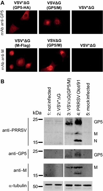
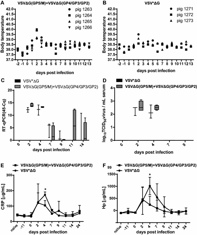
Porcine reproductive and respiratory syndrome virus (PRRSV) is the causative agent of one of the most devastating and economically significant viral disease of pigs worldwide. The vaccines currently available on the market elicit only limited protection. Recombinant vesicular stomatitis virus (VSV) replicon particles (VRP) have been used successfully to induce protection against influenza A virus (IAV) in chickens and bluetongue virus in sheep. In this study, VSV VRP expressing the PRRSV envelope proteins GP5, M, GP4, GP3, GP2 and the nucleocapsid protein N, individually or in combination, were generated and evaluated as a potential vector vaccine against PRRSV infection. High level expression of the recombinant PRRSV proteins was demonstrated in cell culture. However, none of the PRRSV antigens expressed from VRP, with the exception of the N protein, did induce any detectable antibody response in pigs before challenge infection with PRRSV. After challenge however, the antibody responses against GP5, GP4 and GP3 appeared in average 2 weeks earlier than in pigs vaccinated with the empty control VRP. No reduction of viremia was observed in the vaccinated group compared with the control group. When pigs were co-vaccinated with VRP expressing IAV antigens and VRP expressing PRRSV glycoproteins, only antibody responses to the IAV antigens were detectable. These data show that the VSV replicon vector can induce immune responses to heterologous proteins in pigs, but that the PRRSV envelope proteins expressed from VSV VRP are poorly immunogenic. Nevertheless, they prime the immune system for significantly earlier B-cell responses following PRRSV challenge infection.


Figures

Figure 1
Figure 1
Characterization of VRP-mediated expression of GP5 and M. A Immunofluorescence analysis of MARC-145 cells 6 h after infection with VSV*ΔG(GP5-HA), VSV*ΔG(M-Flag), VSVΔG(GP5/M) or with the VSV*ΔG control VRP. In the top panels, expression of GP5 is detected using the anti-GP5 mAb 3AH9. In the bottom panels, expression of M is detected using the anti-M mAb 11E10C7. B Western blot analysis of lysates from non-infected cells (lane 1) or cells infected with VSV*ΔG (lane 2), VSVΔG(GP5/M) (lane 3), PRRSV Olot/91 (lane 4) or mock (lane 5). The proteins were separated by SDS-PAGE under reducing (+βME) conditions and blotted onto a nitrocellulose membrane. The GP5 and M proteins were detected with the anti-GP5 and anti-M mAbs and with a porcine anti-PRRSV serum that reacted also with N. α-tubulin served as loading control using the anti-α-tubulin mAb. Protein molecular weight (kDa) standards are indicated.
Figure 2
Figure 2
Characterization of VRP-mediated expression of GP3 and GP4. Immunofluorescence analysis of MARC-145 cells 6 h after infection with VSV*ΔG(GP3-Flag), VSV*ΔG(GP4-HA), VSVΔG(GP4/GP3/GP2) or with the VSV*ΔG control VRP. Expression of GP3 (top panels) and GP4 (bottom panels) were detected with the anti-GP3 mAb VII2D and with a rabbit anti-GP4 serum, respectively.
Figure 3
Figure 3
Characterization of VRP-mediated expression of the GP3 ectodomain. A Immunofluorescence analysis of MARC-145 cells 6 h after infection with VSV*ΔG(GP3ecto-Myc) or with the VSV*ΔG control. Expression of GP3ecto is detected with a rabbit anti-Myc serum (top panels) and with the anti-GP3 mAb VII2D (bottom panels). B Western blot analysis of lysates (ly) and supernatants (sn) from cells infected with VSV*ΔG(GP3ecto-Myc) separated by SDS-PAGE under non-reducing (-βME) conditions, using the anti-Myc serum for GP3ecto-Myc detection (arrowhead). Protein molecular weight (kDa) standards are indicated.
Figure 4
Figure 4
Characterization of VRP-mediated expression of N. A Immunofluorescence analysis of MARC-145 cells 6 h after infection with VSV*ΔG(N) or with the VSV*ΔG control. The expression of N is detected with the anti-N mAb 13E2. B Western blot analysis of lysates from non-infected cells (lane 1) or cells infected with VSV*ΔG (lane 2), VSVΔ*G(N) (lane 3), PRRSV Olot/91 (lane 4) or mock (lane 5). The proteins were separated by SDS-PAGE under non-reducing (-βME) conditions and blotted onto a nitrocellulose membrane. N antigen was detected with the anti-N mAb as above. Protein molecular weight (kDa) standards are indicated.
Figure 5
Figure 5
Body temperatures, virus load and acute phase proteins after challenge infection of pigs vaccinated with VSVΔG(GP5/M) or with VSV*ΔG as control. Two groups of 5 SPF pigs each were vaccinated three times with 28 and 35 days interval, with 107 PFU/pig of VSVΔG(GP5/M) or of VSV*ΔG as mock control, respectively. 4 weeks after the 3rd vaccination, the pigs were infected intranasally with 106 PFU/pig of the homologous Olot/91 virus. The body temperatures A, B are shown for the individual animals. Blood was collected at the indicated days after infection. PRRSV load in serum was determined by real-time RT-qPCR (C) and by virus titration in MARC-145 cells (D), and the values expressed in 45-Cq and TCID50/mL serum (detection limit 1.7 log10 TCID50/mL), respectively. The serum concentrations (µg/mL) of the porcine acute phase proteins CRP (E) and Hp (F) were determined by ELISA. The data of panels CF represent the mean of five pigs per group, with error bars representing the standard deviation (SD).
Figure 6
Figure 6
Body temperatures, virus load and acute phase proteins after challenge infection of pigs vaccinated with a mixture of VSVΔG(GP5/M) and VSVΔG(GP4/GP3/GP2) or with VSV*ΔG as control. Two groups of 4 and 3 SPF pigs were vaccinated two times at 28 days interval with a mixture of 107 PFU/pig of the VSVΔG(GP5/M) and VSVΔG(GP4/GP3/GP2) VRP or with the VSV*ΔG mock control, respectively. 26 days after the 2nd vaccination, the pigs were infected intranasally with 106 PFU/pig of the homologous Olot/91 virus. The body temperatures A, B are shown for the individual animals. Blood was collected at the indicated days post infection. PRRSV load in serum was determined by real-time RT-qPCR (C) and by virus titration in MARC-145 cells (D), and the values expressed in 45-Cq and TCID50/mL serum (detection limit 1.7 log10 TCID50/mL), respectively. The serum concentrations (µg/mL) of the porcine acute phase proteins CRP (E) and Hp (F) were determined by ELISA. The data of panels CF represent the mean of four and three pigs per group respectively, with error bars representing the SD and asterisks showing significant differences (p < 0.05).

Similar articles

Cited by

References

    1. Karniychuk UU, Nauwynck HJ. Pathogenesis and prevention of placental and transplacental porcine reproductive and respiratory syndrome virus infection. Vet Res. 2013;44:95. doi: 10.1186/1297-9716-44-95. - DOI - PMC - PubMed
    1. Rossow KD. Porcine reproductive and respiratory syndrome. Vet Pathol. 1998;35:1–20. doi: 10.1177/030098589803500101. - DOI - PubMed
    1. Lunney JK, Benfield DA, Rowland RR. Porcine reproductive and respiratory syndrome virus: an update on an emerging and re-emerging viral disease of swine. Virus Res. 2010;154:1–6. doi: 10.1016/j.virusres.2010.10.009. - DOI - PMC - PubMed
    1. Neumann EJ, Kliebenstein JB, Johnson CD, Mabry JW, Bush EJ, Seitzinger AH, Green AL, Zimmerman JJ. Assessment of the economic impact of porcine reproductive and respiratory syndrome on swine production in the United States. J Am Vet Med Assoc. 2005;227:385–392. doi: 10.2460/javma.2005.227.385. - DOI - PubMed
    1. An TQ, Tian ZJ, Xiao Y, Li R, Peng JM, Wei TC, Zhang Y, Zhou YJ, Tong GZ. Origin of highly pathogenic porcine reproductive and respiratory syndrome virus, China. Emerg Infect Dis. 2010;16:365–367. doi: 10.3201/eid1602.090005. - DOI - PMC - PubMed

Publication types

MeSH terms