. 2016 Apr 22;291(17):9133-47.
doi: 10.1074/jbc.M115.694075. Epub 2016 Feb 19.

Regulator of G Protein Signaling 7 (RGS7) Can Exist in a Homo-oligomeric Form That Is Regulated by Gαo and R7-binding Protein

Affiliations

Regulator of G Protein Signaling 7 (RGS7) Can Exist in a Homo-oligomeric Form That Is Regulated by Gαo and R7-binding Protein

Junior Tayou et al. J Biol Chem. .

Abstract

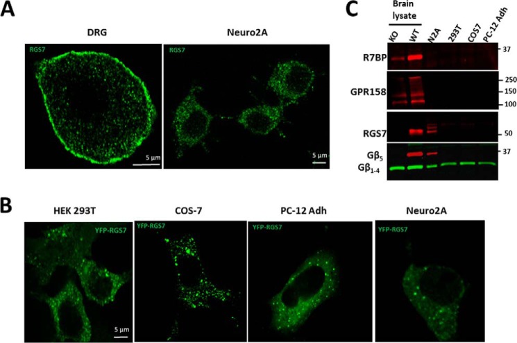
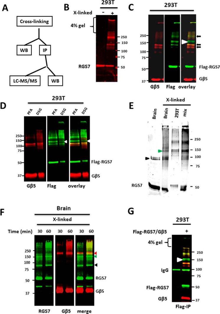
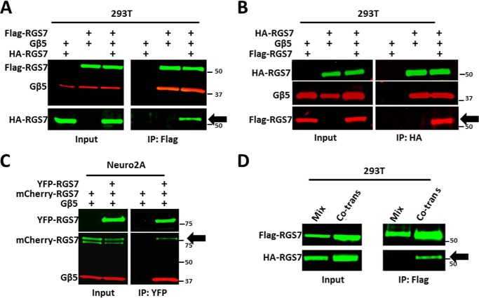
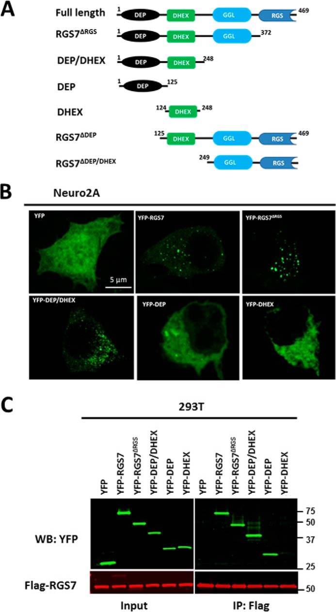
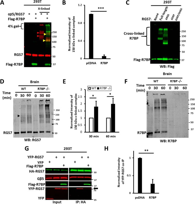
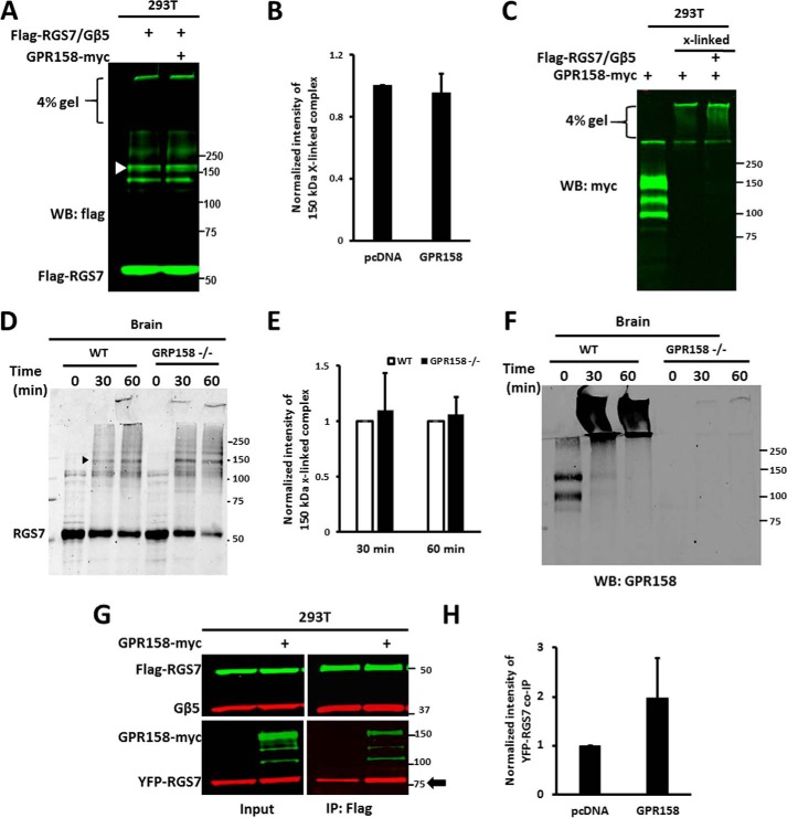
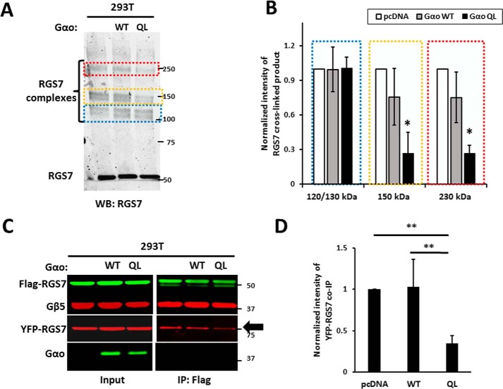
RGS (regulator of G protein signaling) proteins of the R7 subfamily (RGS6, -7, -9, and -11) are highly expressed in neurons where they regulate many physiological processes. R7 RGS proteins contain several distinct domains and form obligatory dimers with the atypical Gβ subunit, Gβ5 They also interact with other proteins such as R7-binding protein, R9-anchoring protein, and the orphan receptors GPR158 and GPR179. These interactions facilitate plasma membrane targeting and stability of R7 proteins and modulate their activity. Here, we investigated RGS7 complexes using in situ chemical cross-linking. We found that in mouse brain and transfected cells cross-linking causes formation of distinct RGS7 complexes. One of the products had the apparent molecular mass of ∼150 kDa on SDS-PAGE and did not contain Gβ5 Mass spectrometry analysis showed no other proteins to be present within the 150-kDa complex in the amount close to stoichiometric with RGS7. This finding suggested that RGS7 could form a homo-oligomer. Indeed, co-immunoprecipitation of differentially tagged RGS7 constructs, with or without chemical cross-linking, demonstrated RGS7 self-association. RGS7-RGS7 interaction required the DEP domain but not the RGS and DHEX domains or the Gβ5 subunit. Using transfected cells and knock-out mice, we demonstrated that R7-binding protein had a strong inhibitory effect on homo-oligomerization of RGS7. In contrast, our data indicated that GPR158 could bind to the RGS7 homo-oligomer without causing its dissociation. Co-expression of constitutively active Gαo prevented the RGS7-RGS7 interaction. These results reveal the existence of RGS protein homo-oligomers and show regulation of their assembly by R7 RGS-binding partners.

Keywords: G protein; G protein-coupled receptor (GPCR); R7-binding protein (R7BP); immunoprecipitation; oligomer; protein cross-linking; regulator of G protein signaling (RGS); subcellular localization.


Figures

FIGURE 1.
FIGURE 1.
Subcellular localization of endogenous and overexpressed RGS7. A, adult mouse day 6 in vitro DRG neurons (left panel) or N2A cells (right panel) were stained with anti-RGS7 antibodies as described under “Experimental Procedures.” B, indicated cell lines were transfected with plasmids expressing YFP-RGS7 and Gβ5, and YFP was directly imaged by fluorescence microscopy. C, lysates from indicated cell lines were analyzed by Western blot for R7BP, GPR158, RGS7, Gβ5, and G protein β subunit (1–4) expression. Wild-type and Gβ5 knock-out mouse brains were used as a control. Shown are representative immunoblots from two experiments.
FIGURE 2.
FIGURE 2.
R7BP or GPR158 cause redistribution of RGS7 from cytoplasmic granules to plasma membrane. HEK293T cells were co-transfected with taggedRGS7 and Gβ5, with or without FLAG-R7BP (A and B) or GPR158-Myc (A and C) plasmids. A, YFP-RGS7 was immunoprecipitated from the cell lysate, and the eluates were probed for RGS7, Gβ5, and R7BP. B, immunoprecipitated YFP-RGS7 complex was probed for RGS7, Gβ5, and GPR158. C, cells expressing YFP-RGS7 and Gβ5 (top row), or these proteins together with FLAG-R7BP (middle row), or GPR158-Myc (bottom row) were analyzed by confocal microscopy. YFP-RGS7 was detected by epifluorescence, and R7BP or GPR158 were detected using antibodies against FLAG or Myc tag, respectively. D, quantification of the result in C. Regions of interest corresponding to plasma membrane (cell periphery) and cytosol were selected within a cell. Fluorescence intensities within these regions were measured using Leica LAS AF Lite image analysis software. The data show the means ± S.D. of the plasma membrane to cytosol ratio of the YFP-RGS7 fluorescence. ***, p <0.001. For every condition, we analyzed 20–25 cells in each of three independent transfection experiments. The ratio of 1:1 in the absence of R7BP and GPR158 represents the fluorescence distribution for cytosol-localized proteins.
FIGURE 3.
FIGURE 3.
Chemical cross-linking of RGS7 complexes in live cells. A, flow chart representing our cross-linking, Western blot (WB), immunoprecipitation (IP), and mass-spectrometry (LC-MS/MS) experiments. B, HEK293T cells expressing RGS7 and Gβ5 were incubated with 1% PFA for 30 min and analyzed by immunoblot with anti-RGS7 antibodies, as described under “Experimental Procedures.” Left lane shows a representative Western blot of the control cells (untreated with PFA) probed with anti-RGS7 antibody. Right lane, lysate from cells treated with PFA. C, immunoblot analysis of lysates from cells expressing FLAG-RGS7 and Gβ5 probed with antibodies against FLAG (green) and Gβ5 (red). Black arrows indicate the cross-linked complexes containing both RGS7 and Gβ5. White arrowhead at ∼150-kDa denotes an RGS7 complex that does not contain Gβ5. D, HEK293T cells were transfected with FLAG-RGS7 and Gβ5 and cross-linked with either PFA or DSG. The resulting products were analyzed by Western blot to compare the patterns of cross-linked proteins using antibodies against FLAG (green) and Gβ5 (red). E, cross-linking of endogenous RGS7 in mouse brain was done using PFA as described under “Experimental Procedures.” To compare the apparent molecular weight of the cross-linked RGS7 products from brain and transfected HEK293T cells, the samples were resolved on 8% gel. The arrowhead points to the band that is identical in electrophoretic mobility. When the two samples were mixed (mix), the intensity of this band increased. F, mouse brain slices were treated with PFA for 30 or 60 min. The proteins were resolved on a 10% gel and analyzed by Western blot using anti-RGS7 and anti-Gβ5 antibodies. Because both primary antibodies were from rabbit, we used the following method. First, the immunoblot was stained using anti-RGS7 antibodies and scanned using the Odyssey instrument (green). The filter was then stripped and probed with anti-Gβ5 antibodies (red). The two images were merged to compare the exact position of RGS7 and Gβ5-positive bands. Red arrowhead denotes the protein cross-linking product that contains both RGS7 and Gβ5. Green arrowhead points to the 150-kDa RGS7 complex. Black arrowhead shows a nonspecific band revealed by the RGS7 antibody in the brain extracts. G, immunoprecipitation of the cross-linked RGS7 complexes from transfected HEK293T cells using anti-FLAG antibody. The eluates from the beads were analyzed by immunoblot and by mass spectrometry (see text and Table 1). Left lane, lysates from untransfected cells were used as a negative control. Shown are representative immunoblots from three (D, E, and F) or more than five (B, C, and G) independent experiments.
FIGURE 4.
FIGURE 4.
Homo-oligomerization of RGS7. A, HEK293T cells were co-transfected with HA-RGS7 and Gβ5, FLAG-RGS7 and Gβ5, or all the three constructs. Cell lysates were subjected to IP using anti-FLAG antibody, as described under “Experimental Procedures.” The eluates from the resin were analyzed using anti-FLAG, HA, and Gβ5 antibodies. B, reciprocal IP using anti-HA beads. Cell transfection and lysis were done as in A; immunoprecipitation was performed using anti-HA antibodies, and the bound material was probed for the presence of FLAG-tagged RGS7. C, mCherry-RGS7 was co-immunoprecipitated with YFP-RGS7 from N2A cell lysates. The experiment was performed essentially as in A or B. D, two separate cell cultures were transfected with either FLAG-RGS7/Gβ5 or HA-RGS7/Gβ5, harvested, and lysed. The two lysates were mixed (mix) prior to co-IP with immobilized anti-FLAG antibody. In parallel, FLAG-RGS7, HA-RGS7, and Gβ5 plasmids were co-transfected (co-trans), and cell lysates were analyzed by co-IP and immunoblot.
FIGURE 5.
FIGURE 5.
5 is required for stability but not for homo-oligomerization of RGS7. A, HEK293T cells expressing FLAG-, YFP-, or mCherry-tagged RGS7 with or without Gβ5 were lysed and analyzed by Western blot using antibodies against RGS7, Gβ5, and actin. The white arrow shows FLAG-RGS7 expressed in the absence of Gβ5. B, cells on coverslips were transfected with YFP-RGS7 with or without CFP-Gβ5, then fixed, and YFP and CFP direct fluorescence was detected using a confocal microscope (×63 objective lens). C, lysates from cells expressing YFP-RGS7 and mCherry-RGS7 with or without Gβ5 were subjected to immunoprecipitation using immobilized YFP antibody. The eluates were analyzed by Western blot using YFP, Gβ5, and mCherry antibodies. The black arrow points to the co-immunoprecipitated mCherry-RGS7.
FIGURE 6.
FIGURE 6.
DEP domain is essential for oligomerization of RGS7. A, RGS7 constructs used in the study. B, N2A cells were transfected with plasmids encoding Gβ5 and the indicated YFP-tagged deletion mutants of RGS7. After 24 h, cells were fixed, and YFP epifluorescence was detected by confocal microscopy. C, HEK293T cells expressing FLAG-RGS7, Gβ5 together with YFP, or the indicated YFP-tagged RGS7 mutants were lysed, and the lysates were subjected to immunoprecipitation using anti-FLAG antibody. The eluates from the beads were analyzed by immunoblot using anti-GFP and anti-FLAG antibodies. WB, Western blot.
FIGURE 7.
FIGURE 7.
R7BP causes dissociation of RGS7 homo-oligomer. A, HEK293T cells expressing RGS7 and Gβ5, with or without FLAG-R7BP, were subjected to cross-linking as described in the legend to Fig. 3. The lysates were analyzed by immunoblot using a combination of anti-FLAG (green) and RGS7 (red) antibodies. Red arrowheads denote the 150-kDa and 230 RGS7 complexes. Note that in the presence of R7BP these bands disappear. Instead, there are two new bands of ∼110 and ∼200 kDa (yellow arrowheads) revealed with both anti-RGS7 and anti-FLAG antibodies. B, quantification of the 150-kDa band intensity in A. Blots were scanned and analyzed as described under “Experimental Procedures.” The fluorescence intensity determined for the 150-kDa band was normalized to the RGS7 band in the non-cross-linked control sample. The value obtained for 150-kDa band without R7BP (pcDNA) was set to the arbitrary unit of 1.0. Data show means ± S.D. from four independent transfection experiments. ***, p <0.001. C, cells were transfected with FLAG-R7BP and Gβ5 together with or without (none) full-length RGS7 or its deletion mutants. They were subjected to cross-linking with PFA, and the lysates were analyzed by immunoblot using anti-FLAG antibody to detect R7BP. WB, Western blot. D, cross-linking of endogenous RGS7 in the brain tissue from wild-type (WT) and R7BP knock-out (R7BP−/−) mice. Cross-linking with PFA was performed for 30 or 60 min as described under “Experimental Procedures” and the legend to Fig. 3E; shown is a representative immunoblot probed with anti-RGS7 antibody. E, quantification of data represented in D. The intensity of the 150-kDa band (black arrow) was normalized to the non-cross-linked RGS7, and the resulting value for WT mouse was set to 1.0. The data show the means ± S.D. from four independent experiments performed with the tissues from three individual WT (open bars) and three R7BP−/− (black bars) animals. *, p <0.05. The knock-out of R7BP resulted in a 1.8–2.0-fold increase in the 150-kDa band. F, pattern of cross-linked endogenous R7BP revealed by anti-R7BP antibody. G, HEK293T cells were co-transfected with HA-RGS7, YFP-RGS7, and Gβ5, with or without FLAG-R7BP. Cell lysates were immunoprecipitated with anti-HA antibody. The inputs and eluates from IP were probed with the indicated antibodies. The black arrow pointing to YFP-RGS7 highlights the reduction of the interaction between HA-RGS7 and YFP-RGS7 in the presence of R7BP. The RGS7, Gβ5, and R7BP plasmids used for transfection were added in the ratio of 4:1:5, respectively. H, quantification of the data shown in G. YFP-RGS7 band intensity in co-IP eluate was normalized to the intensity of the HA-RGS7 band. The results are expressed as means ± S.D. (n = 4, **, p <0.01).
FIGURE 8.
FIGURE 8.
Effect of GPR158 on RGS7 self-association. A, HEK293T cells expressing FLAG-RGS7/Gβ5 with or without GPR158-Myc were treated with PFA. The lysates were analyzed by immunoblot using anti-FLAG antibodies. White arrowhead points to the 150-kDa cross-linked RGS7 product. WB, Western blot. B, 150-kDa band intensity compared between cells with and without GPR158, means ± S.D., n = 3. C, same samples as in A were probed with anti-Myc antibody. D, cross-linking of endogenous RGS7 in the brain tissue from wild-type (WT) and GPR158−/− mice. Cross-linking was performed for 30 or 60 min; shown is a representative immunoblot probed with anti-RGS7 antibody. E, data from D was quantified as described in the legend to Fig. 7E. F, cross-linked brain samples probed with the antibody against GPR158. G, FLAG-RGS7 was immunoprecipitated from HEK293T cells expressing FLAG-RGS7, Gβ5, YFP-RGS7 with or without GPR158Myc. The lysates were analyzed by immunoblot using specific antibodies. The ratio of plasmid DNA in the transfection was 4:1:5 (RGS7, Gβ5, and GPR158, respectively). H, quantification of the results in G. YFP-RGS7 band (co-IP) was normalized to FLAG-RGS7 (IP) as described in the legend to Fig. 7. The observed increase in RGS7 self-association in the presence of GPR158 was not statistically significant (n = 4, p = 0.095).
FIGURE 9.
FIGURE 9.
Effect of Gαo subunit on RGS7 homo-oligomerization. A, HEK293T cells transfected with RGS7, Gβ5 with or without wild-type Gαo or its constitutively active Q205L (QL) mutant. The ratio of plasmid DNA in transfection was 4:1:5 (RGS7, Gβ5, Gαo, or pcDNA, respectively). After PFA-induced cross-linking, the cell lysates were analyzed by immunoblot using RGS7 antibody. The areas denoted by the colored broken lines highlight the positions of the 120/130-kDa Gβ5-RGS7 conjugate, the 150-kDa RGS7, and 250-kDa Gβ5-RGS7 oligomers. WB, Western blot. B, quantification of the data in A. The normalized band intensity in the control (pcDNA) was set to 1.0. Data are means ± S.D., n = 3. *, p <0.05. C, lysates from cells expressing FLAG-RGS7, YFP-RGS7, Gβ5, with or without wild-type or mutant Gαo were subjected to immunoprecipitation using FLAG antibody. The eluates were analyzed by immunoblot using FLAG, YFP, Gβ5, and Gαo antibodies. D, quantification of data shown in C. YFP-RGS7 co-IP intensity was normalized to FLAG-RGS7 IP. The results are expressed as means ± S.D.; **, p <0.01.

Similar articles

Cited by

References

    1. Neer E. J. (1995) Heterotrimeric G proteins: organizers of transmembrane signals. Cell 80, 249–257 - PubMed
    1. Cabrera-Vera T. M., Vanhauwe J., Thomas T. O., Medkova M., Preininger A., Mazzoni M. R., and Hamm H. E. (2003) Insights into G protein structure, function, and regulation. Endocr. Rev. 24, 765–781 - PubMed
    1. Clapham D. E., and Neer E. J. (1997) G protein βγ subunits. Annu. Rev. Pharmacol. Toxicol. 37, 167–203 - PubMed
    1. Dohlman H. G., Apaniesk D., Chen Y., Song J., and Nusskern D. (1995) Inhibition of G-protein signaling by dominant gain-of-function mutations in Sst2p, a pheromone desensitization factor in Saccharomyces cerevisiae. Mol. Cell. Biol. 15, 3635–3643 - PMC - PubMed
    1. Lan K. L., Zhong H., Nanamori M., and Neubig R. R. (2000) Rapid kinetics of regulator of G-protein signaling (RGS)-mediated Gαi and Gαo deactivation. Gα specificity of RGS4 and RGS7. J. Biol. Chem. 275, 33497–33503 - PubMed

Publication types

MeSH terms